博 士 学 位 論 文
Role of glutathione peroxidase 4 (GPx4) for oxidative
homeostasis in vascular and eye tissues, and the effects of
vitamin E on cytotoxicity caused by lipid oxidation in loss
of GPx4 expression
博 士 学 位 論 文
Role of glutathione peroxidase 4 (GPx4) for oxidative
homeostasis in vascular and eye tissues, and the effects of
vitamin E on cytotoxicity caused by lipid oxidation in loss
of GPx4 expression
平成 28 年 11 月 21 日
(英文題目)
Role of glutathione peroxidase 4 (GPx4) for oxidative
homeostasis in vascular and eye tissues, and the effects of
vitamin E on cytotoxicity caused by lipid oxidation in loss
of GPx4 expression
Osamu Sakai
(Advisor: Prof. Shigeru Ueshima)
(和文題目)
血管及び眼組織における抗酸化酵素グルタチオンペ
ルオキシダーゼ 4 (GPx4)の役割及び GPx4 発現低下
による過酸化脂質障害に対する
ビタミン E の効果
酒井 修
(指導:上嶋 繁 教授)
Table of Contents
1. Preface……….1
2. Role of GPx4 for oxidative homeostasis in human vascular endothelial cells, and t h e c o m p e n s a t o r y a c t i v i t y o f b r o w n r i c e o n G P x 4 a b l a t i o n condition……….………..5
2.1 Introduction………...……….5
2.2 Materials and methods………....7
2.3 Results………10
2.4 Discussion………..16
2.5 Summary………18
3. GPx4 is an essentialfor survival and protection against oxidative stress in corneal epithelial cells, and the effects of vitamin E on cell damage induced by GPx4 depletion……….….20
3.1 Introduction………...20
3.2 Materials and methods………..21
3.3 Results………...26
3.4 Discussion……….36
3.5 Summary………...38
4. Role of GPx4 in glutamate-induced oxytosis in the retina………....40
4.1 Introduction………...40
4.2 Materials and methods………..41
4.3 Results………...46
4.5 Summary………...56
5. Role of GPx4in conjunctival epithelial cells………...……...58
5.1 Introduction………...58
5.2 Materials and methods………..59
5.3 Results………63
5.4 Discussion………..70
5.5 Summary………72
6. Summary and perspectives……….74
7. Abbreviations………..79
8. Acknowledgments………...80
9. References………81
1
1. Preface
The eyes are constantly exposed to sunlight, and oxidative stress is implicated in several ocular diseases including pterygium, dry eye, conjunctivochalasis, atopic keratoconjunctivitis, age-related macular degeneration, glaucoma, and diabetic retinopathy.1-7 Oxidative stress may cause or aggravate ocular injury resulting in decreased visual acuity or even vision loss. On the other hand, major vascular risk factors, such as excessive dietary, fat intake, smoking or alcohol consumption, increase the oxidative stress on the arterial endothelium, 8-10 and oxidative stress is thought to contribute to the pathogenesis of many vascular diseases, including atherosclerosis, hypertension and coronary artery disease.11-13 Therefore, the aggravation of oxidative stress in blood vessels could help evaluate the risk for development of these vascular disease.
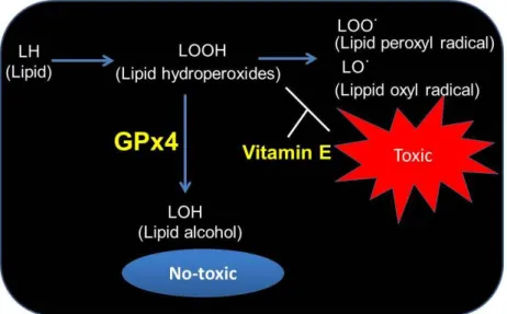
Redox homeostasis is maintained by various antioxidant enzymes (superoxide dismutase (SOD), catalase, and glutathione peroxidase (GPx), etc.)14-17 and dietary antioxidants (vitamins, carotenoids, lutein, and glutathione, etc.) (Figure 1).18-20 Lack of antioxidant enzymes and dietary antioxidants cause oxidative damage to biomolecules (lipids, proteins, DNA), eventually leading to many eye and vascular diseases (Figure 1). Among oxidative stresses, lipid oxidation is especially known to be implicated in a variety of pathophysiologic processes of various diseases.12,21-23 Oxidized lipids are highly reactive and lead to DNA fragmentation and protein modification.24,25 Byproducts of lipid peroxidation such as 4-hydroxynonenal (4-HNE) have been identified in ocular and vascular diseases such as dry eye, glaucoma, diabetic retinopathy, atherosclerosis, and hypertension,21,22,26-28 and is known to induce cell
2
damage such as cell death and growth inhibition.29,30 Therefore, lipid oxidation is a key contributor to the progression and perhaps to the origin of ocular and vascular diseases, and a decrease of lipid peroxidation products may be beneficial for various pathological conditions. However, despite the importance of the defense mechanism against lipid peroxidation, the importance of specific antioxidant enzymes and dietary antioxidants in vascular and eye tissue is not well defined.
Figure 1. Balance of oxidative stress and ant-oxidative system
Glutathione peroxidase 4 (GPx4) has a high preference for lipid hydroperoxides and directly reduces peroxidized phospholipids in cellular membranes (Figure 2).31,32 GPx4 is one of the eight GPx isozymes found in mammals, and is ubiquitously expressed.33 Knockout mice of GPx4 die at embryonic day 8,34 and loss of GPx4 results in lipid oxidation leading to cell death in various cells such as cancer cells, neurons, and T cells.35-38 GPx4 is essential for maintaining tissue homeostasis by preventing cell demise and tissue damage. Moreover, GPx4 is considered to be a central regulator of ferroptosis, which is mediated by lipid peroxidation. The process of ferroptosis is
3
triggered by the iron-dependent accumulation of lipid peroxides,37,38 and is also distinct from apoptosis, and necrosis.39 Whereas, the overexpression of GPx4 confers protection against oxidative stress-mediated injury.40,41 Therefore, GPx4 is thought to be important for cell protection from lipid oxidative stress.
In contrast, many dietary antioxidants have been identified,18-20 and vitamin E is one of the best known antioxidants.42 Vitamin E is the major lipid-soluble antioxidant, and is essential to protect the tissue against lipid oxidative damage.43-44 In addition, it has been reported vitamin E acts in conjunction with GPx4 to inhibit lipid peroxidation, and cell death under GPx4 depletion was rescued by vitamin E in several cells (Figure 2).36,45,46 Thus, vitamin E can potentially compensate for the lack of GPx4. Moreover, it is known that sources of vitamin E include nuts, olive oil, and brown rice, etc.47-49 Thus, these foods may act as a highly efficient back-up system for GPx4 in the prevention of lipid peroxidation processes.
Figure 2. Regulation of redox signaling, lipid hydroperoxides, as intracellular messengers by GPx4 and vitamin E.
4
As discussed above, lipid oxidation is involved in the pathology of ocular and vascular diseases, and antioxidative enzymes and dietary antioxidants play an important role in defense of oxidant stress. However, the essentiality of antioxidants, which are enzymes and dietary antioxidants, has remained unclear in eye and blood vessel. In the present study, I elucidated the importance of GPx4 in ocular (cornea, retina and conjunctiva) and vascular. Furthermore, I also assessed the effects of vitamin E, which is one of the dietary antioxidants, on cell damage by downregulation of GPx4.
5
2. Role of GPx4 for oxidative homeostasis in human vascular
endothelial cells, and the compensatory activity of brown rice on
GPx4 ablation condition
2.1 Introduction
Endothelial dysfunction has been identified in patients with several vascular diseases, including atherosclerosis, diabetes mellitus, and hypercholesterolemia.50-52 The accumulation of byproducts of oxidative metabolism has been observed in the blood vessels of patients with vascular diseases,11,13 and oxidative stress is thought to cause endothelial dysfunction. Antioxidants, including various agents such as antioxidant enzymes (glutathione peroxidase, superoxide dismutase, and catalase), and dietary antioxidants (carotenoids, glutathione, vitamin C, and vitamin E), play an important role in the cellular protection cascade against oxidative damage, and lack of antioxidants cause endothelial dysfunction in the vascular system. However, the importance of antioxidant enzyme and dietary antioxidants have not been fully understood in vascular endothelial cells.
Among oxidative stresses, lipid peroxidation in particularly is known to be implicated in a number of pathophysiologic processes,21-23 and byproducts of lipid peroxidation such as 4-HNE induces cell damage such as apoptosis and growth inhibition.29,30 Moreover, increase of 4-HNE has been observed in a variety of vascular diseases such as atherosclerosis.26 GPx4 is one of eight glutathione peroxidases in mammals,33,34 and has been proposed to play an important role in the reduction of lipid peroxides and protect cells from lipid hydroperoxides.33-41 Knockout mice of GPx4 die at embryonic day 8,34 and loss of GPx4 results in lipid peroxidation leading to cell death in many cells.34-38 Therefore, GPx4 is thought to be crucial for cell protection from lipid
6
oxidative stress in various cells.
In addition, the ablation of GPx4 induced ferroptotic cell death, and GPx4 is thought to regulate the ferroptosis.37-39 Ferroptosis is a recently recognized form of regulated cell death.37-39 The process of ferroptosis is triggered by the iron-dependent accumulation of lipid peroxides.37-39 Ferroptotic cell death could not be prevented by chemical or genetic inhibitors of apoptosis53,54 or inhibitors of necroptosis,55,39 suggesting that ferroptotic cell death was distinct from apoptosis, and necrosis. Moreover, ferroptosis has been implicated in multiple physiological and pathological processes, including cancer cell death, neurotoxicity, neurodegenerative diseases, and T-cell immunity,37-39 although ferroptosis is not known to be involved in vascular diseases. Loss of GPx4 is reported to induce cell damage in vascular endothelial cells in endothelial-specific GPx4 knockout mice,46 but the regulatory mechanism of the cell death is not well defined in vascular endothelial cells.
On the other hand, many dietary antioxidants have been identified,18-20 and vitamin E is known to be the major lipid-soluble antioxidant.42-43 Vitamin E is essential to protect tissue against oxidative damage induced by lipid peroxidation.43,44 In addition, it has been reported that vitamin E acts in conjunction with GPx4 to inhibit lipid peroxidation, and cell death under GPx4 depletion was rescued by vitamin E in some cells.36,46 Thus, vitamin E can potentially compensate for the lack of GPx4.
Rice is one of the main foods in the diet of most populations. Brown rice is more nutritious than white rice, and is known to be an anti-oxidant rich food.47 Therefore, brown rice is thought to play an important role in the concentration of antioxidants ingested daily. The highest amount of vitamin E is contained in brown rice,47 and brown rice has been reported to possess a high antioxidant capacity, which reduces lipid
7
peroxidation.56 Thus, brown rice may rescue cell damage induced by loss of GPx4. In the present study, I clarified the importance of GPx4 and the implication of ferroptosis on cell death induced by GPx4 loss in vascular endothelial cells, using the siRNA knockdown technique. In addition, I examined the effect of brown rice extract on conditional ablation of GPx4.
2.2 Material and methods
2.2.1 Human Vascular Endothelial Cell Culture and siRNA transfection
In this study, human umbilical vein endothelial cells (HUVECs) (Lonza) were cultured in EGM-2 (Lonza) containing vascular endothelial growth factor (VEGF), basic fibroblast growth factor (bFGF), insulin-like growth factor-1 (IGF-1), epidermal growth factor (EGF), hydrocortisone, heparin, gentamicin sulfate amphotericin and 2% fetal bovine serum (FBS) under 5% CO2 at 37°C. Cells at 20-30% confluence were
transfected with 25 nM siRNA that specifically knockdown GPx4 and scrambled control siRNA (Invitrogen) using lipofectamine RNAiMAX (Invitrogen) according to the manufacturer's instructions. Experiments were performed on cells below passage 8. Morphology of transfected cells was assessed with an inverted phase-contrast microscope.
2.2.2 RT-PCR
Twenty four hours after transfection with GPx4 siRNA or scrambled control siRNA, total RNA was extracted from cells by using Nucleo Spin RNA XS (Takara Bio). Complementary DNA was prepared by master mix with genomic DNA remover (ReverTra Ace qPCR RT with gDNA Remover; Toyobo). Real-time RT-PCR was
8
carried out with 7500 Real-PCR System (Applied Biosystems) using SYBR Premix Ex Taq II (Takara Bio). The values for each gene were normalized to the level of GAPDH. The primer sequences used in RT-PCR were as follows: human GAPDH (Fwd, 5-TTGATTTTGGAGGGATCTCG-3 and Rev, 5-AACTTTGGCATTGTGGAAGG-3), human GPx4 (Fwd, 5-GCACATGGTTAACCTGGACA-3, Rev, 5-CTGCTTCCCG AACTGGTTAC-3).
2.2.3 Western Blot Analysis
Twenty four hours after transfection of GPx4 siRNA or scrambled control siRNA, the proteins were extracted from cells. SDS-PAGE of cellular proteins was performed on gel (Mini-PROTEAN TGX Any kD; Bio-Rad Laboratories) with tris-glycine- SDS running buffer (Bio-Rad Laboratories) for 30 min at 250 V. Proteins were transferred to polyvinylidene fluoride (PVDF) membranes (Millipore Corp) at 100 V for 60 minutes at ice-cold temperature using tris-glycine buffer, and then incubated in blocking buffer made of 5% non-fat milk in phosphate-buffered saline with 0.1% Tween-20. Membranes were then probed with antibodies to GAPDH (Santa Cruz Biotechnology and GPx4 (Santa Cruz Biotechnology). Binding of secondary antibodies, conjugated to horseradish peroxidase, was visualized with chemiluminescent substrate (Pierce Biotechnology).
2.2.4 Activity of apoptosis
Activation of apoptosis was examined by immunoblotting for caspase 3 and poly (ADP-ribose) polymerase (PARP). Three days after transfection with siRNA, immunoblotting was conducted using antibodies to caspase 3 (Cell Signaling
9
Technology), PARP (Cell Signaling Technology), and GAPDH (Santa Cruz Biotechnology) as described above. Cells treated with 1 µM staurosporine were also used as a positive control for caspase activity.
2.2.5 Determination of lipid peroxidation
Accumulations of peroxidized lipids were assessed by immunohistochemical detection of 4-HNE. Four days after transfection with GPx4 siRNA or scrambled control siRNA, cells were fixed with 4% paraformaldehyde for 15 min, washed three times with phosphate-buffered saline (PBS), and permeabilized with a 0.1% Triton X-100 solution containing 5% goat serum in PBS. Permeabilized cells were washed three times with PBS containing 5% goat serum and incubated with anti-4-HNE antibodies (JaICA) overnight at 4°C. Cells were then washed three times with PBS. Alexa 488-conjugated anti-mouse IgG secondary antibodies (Thermo Scientific,) were applied for 1 h at room temperature and washed three times with PBS. Fluorescent images were observed using a fluorescence microscope (Keyence). The fluorescence intensities were semi-quantified using Image J software (http://imagej.nih.gov/ij/; provided in the public domain by the National Institutes of Health, Bethesda, MD, USA).
2.2.6 Cytotoxicity assay
Cytotoxicity assay was performed usinglactate dehydrogenase (LDH) cytotoxicity detection kit (Takara Bio). After 4 days of transfection with GPx4 siRNA or scrambled control siRNA, LDH activity in the extracellular medium and cell lysate was measured
10
according to the manufacturer’s instructions; and then extracellular LDH activity was calculated as percentage of the total LDH activity.
2.2.7 Cell Proliferation assay
Proliferation of cells treated with GPx4 or scrambled control siRNA was assessed using WST-8 assay (Dojindo Molecular Tech) at 4 days after transfection. The WST-8 assay was performed according to the manufacturer’s instructions.
2.2.8 The treatment of α-tocopherol, brown rice extracts, ferrostatins-1, and Z-VAD-FMK in HUVEC treated with GPx4 siRNA
α-tocopherol, the major form of vitamin E, is an important lipid-soluble antioxidant.42 Brown rice contains several vitamins such as vitamin E,47 and has the highest antioxidant activity.56 Ferrostatin-1 is the specific ferroptosis inhibitor.39 Z-VAD-FMK is the specific caspase inhibitor.57
α-tocopherol (Sigma) was dissolved in methanol. Brown rice (Agriculture, Kindai University) was mixed with methanol and kept for one overnight at 37°C. Extract from the rice bran was separated from the residue by a centrifugation at 1000×g for 10 min. Ferrostatin-1 (Sigma) and Z-VAD-FMK (Sigma) were dissolved in DMSO.
To examine the effect of α-tocopherol, brown rice extract, and ferrostatin-1 on vascular endothelial cells treated with GPx4 siRNA, they were cultured with α-tocopherol (10 µM), extract (0.1mg/mL) from brown rice, ferrostatin-1 (10 µM), and Z-VAD-FMK (1 µM) 1 day after transfection.
11
Data are expressed as mean ± standard error of the mean (SEM). Values were analyzed statistically using Student’s t-test or Dunnett’s test. P < 0.05 was considered statistically significant.
2.3 Results
2.3.1 Knockdown of GPx4 using siRNA in HUVECs
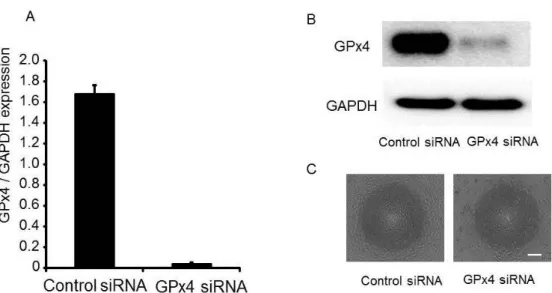
HUVECs were transfected with siRNA specifically silencing GPx4 or scrambled control siRNA. The messenger RNA expression was evaluated by real-time RT-PCR. After 24 hours of transfection, mRNA expression of GPx4 was downregulated by more than 98% (Figure 3A). Moreover, a significant reduction in GPx4 protein levels was observed in 25 nM GPx4 siRNA-treated cells as compared to control (Figure 3B).
I examined the morphological characteristics of HUVECs. Control siRNA-treated cells showed to be compact, uniform, and cobblestone appearance in shape (Figure 3C). Conversely, GPx4 siRNA-treated cells exhibited signs of cell damage including spheroid structures (Figure 3C).
Figure 3. Knockdown of GPx4 siRNA in HUVEC. (A) Knockdown efficiency evaluated by mRNA levels (n = 4). (B) Knockdown efficiency evaluated by protein levels using
12
immunoblot analysis. (C) Phase contrast morphology of HUVEC transfected with siRNA of scramble control and GPx4 24 hour after transfection. Scale bar, 50 µm.
2.3.2 Effect of GPx4 knockdown on lipid peroxidation
Lipid peroxidation induced by oxidants and oxidative stress, generates a huge variety of lipid peroxidation products, including ketones, alkanes and aldehydes, such as malondialdehyde (MDA) and 4-HNE.21,29,30 To evaluate lipid peroxidation, I performed immunostaining of 4-HNE. After 4 days of transfection, knockdown of GPx4 significantly increased the level of lipid oxidation (Figure 4A, B).
Figure 4. Determination of lipid peroxidation. (A) Detection of 4-HNE by fluorescence microscopy using 4-HNE antibodies. (B) The fluorescence intensities of 4-HNE were quantified using ImageJ (NIH) (n =4-5). ##P < 0.01 relative to control siRNA group (Student’s t-test). **P < 0.01 relative to GPx4 siRNA group (Dunnet-test). α-Toc=α-tocopherol.
2.3.3 Effect of GPx4 knockdown on cytotoxicity and proliferation
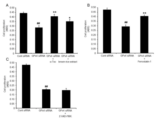
To test the cytotoxicity, I assessed LDH activity. The LDH activity assay showed that GPx4 siRNA-treated HUVEC released a significantly higher level of LDH than control siRNA treated-cells (Figure 5A, B, C). Next, the effect of GPx4 knockdown on proliferation was evaluated via a WST-8 assay. HUVECs were treated with GPx4
13
siRNA or scrambled control siRNA. At 4 days of transfection, GPx4 silencing induced a significant reduction in the cellular proliferation (Figure 6A, B, C), suggesting that GPx4 was essential for growth of HUVECs.
Figure 5. LDH from HUVEC treated with control or GPx4 siRNA 4 days after transfection. (A) α-tocopherol (α-Toc) and brown rice extract prevented the LDH release induced by GPx4 knockdown (n =4). ##P < 0.01 relative to control siRNA group (Student’s t-test).
**
P < 0.01 relative to GPx4 siRNA group (Dunnet-test). (B) Ferrostatin-1 prevented the LDH release induced by GPx4 knockdown (n =4). (C) Z-VAD-FMK did not prevented the LDH release induced by GPx4 knockdown (n =4). ##P < 0.01 relative to control siRNA group (Student’s t-test). *P < 0.05 relative to GPx4 siRNA group (Student’s t-test).
2.3.4 α-tocopherol and brown rice extracts rescued cytotoxic effects of GPx4 knockdown
α-tocopherol and brown rice extracts significantly prevented the LDH release and the delay of cell growth caused by knockdown of GPx4 siRNA (Figure 5A, 6A). In addition, increase of 4-HNE was rescued by treatment with α-tocopherol and brown rice
14
extracts (Figure 4A, B), suggesting that cell damage induced by GPx4 ablation is involved in lipid peroxidation.
Figure 6. Proliferation of HUVEC treated with GPx4 siRNA. Proliferation was evaluated by WST-8 assay at 4 days after transfection. (A) α-tocopherol (α-Toc) and brown rice extract ameliorated suppression of proliferation by GPx4 knockdown (n =4). ##P < 0.01 relative to control siRNA group (Student’s t-test). **P < 0.01 and *P < 0.05 relative to GPx4 siRNA group (Dunnet-test). (B) Ferrostatin-1 ameliorated suppression of proliferation by GPx4 knockdown (n =4). (C) Z-VAD-FMK did not ameliorated suppression of proliferation by GPx4 knockdown. ##P < 0.01 relative to control siRNA group (Student’s t-test). **P < 0.01 relative to GPx4 siRNA group (Student’s t-test).
2.3.5 Action mechanism of cytotoxicity induced by GPx4 knockdown on vascular endothelial cells.
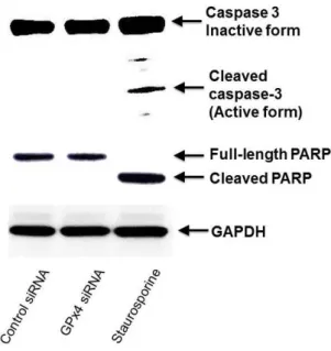
Caspase 3 and PARP are well-known to be implicated in apoptosis, and is activated by self proteolysis or cleavage by another caspase such as active caspase-8. We
15
examined the cleavage of caspase 3 and PARP as the indicator of apoptosis activation by immunoblotting.
Cleaved caspase-3 and PARP were not detected in not only control siRNA but also GPx4siRNA cells, though staurosporine induced the cleavage of caspase 3 and PARP (Figure 7).
Ferrostatin-1 partially ameliorated the increase in LDH and the decrease in cell growth activity caused by GPx4 knockdown (Figure 5B, 6B), suggesting that cell damage induced by GPx4 ablation is involved in ferroptosis. On the other hand, Z-VAD-FMK, caspase inhibitor, did not rescue the cytotoxicity induced GPx4 ablation (Figure 5C, 6C).
Figure 7. The influence of GPx4 knockdown on caspase-3 activation in HUVEC. The activation of caspase-3 was detected by immunoblot analysis using specific antibodies for caspase-3 and PARP.
16 2.4 Discussion
Major findings of the present study are the following: (1) GPx4 is an essential antioxidant enzyme for maintaining redox homeostasis and proliferation in human vascular endothelial cells. (2) Cytotoxicity induced by loss of GPx4 is implicated in ferroptosis but not caspase dependent apoptosis. (3) Thesupplementation of brown rice, which contains a high level of vitamin E, canrescue cell damage induced by lack of GPx4 in vascular endothelial cells, and plays an important role in supporting antioxidant function in lacking GPx4 cells.
Maintaining cellular redox balance is important for cell survival and tissue homeostasis. Many antioxidant enzymes have been reported to control redox balance and ameliorate the damaging effects of oxidative stress in vascular endothelial cell.58,59 However, the importance of antioxidant enzymes and dietary antioxidants remains to be fully clarified in vascular endothelial cells. In the present study, I observed that GPx4 knockdown caused cell death and delay of proliferation in HUVEC. These results suggested that GPx4 is a key antioxidant enzyme in regulating not only cell survival but also proliferation in vascular endothelial cells. Lipid peroxidation induces ferroptosis, and recently GPx4 is considered to be a central regulator of ferroptosis, which is mediated by lipid peroxidation.37-39,55 Ferroptosis is identified as a novel form of iron-dependent cell death different from apoptosis or necrosis, and has been implicated in various pathologies of cell death such as cancer. 37-39 However, the implication of ferroptosis on cell death induced by GPx4 loss in vascular endothelial cell has remained unknown. My data showed ferrostatin-1, an inhibitor of ferroptosis, prevented cell death induced by GPx4 ablation in HUVEC. These results suggest that cell death induced by GPx4 ablation is involved in ferroptosis in vascular endothelial cells. On the other hand,
17
GPx4 knockdown did not induce the activation of caspase 3. Furthermore, caspase inhibitor did not rescue cell death. Some reports showed the cell death by loss of GPx4 was not implicated in caspase activation, my results were in line with the results of previous reports.36-38 Therefore, these results suggest that GPx4 may be a regulator of ferroptosis in vascular endothelial cells. However, the death caused by loss of GPx4 is also reported to be involved in necroptosis in other cells.60 The mechanism of the cell death mediated by GPx4 may be different among cell types, and further investigation is also required to fully understand the mechanism of the cell death.
Increase in lipid peroxidation is also known to damage the vascular endothelial cells, and to be associated with diseases like atherosclerosis, cardiovascular disease, and hypercholesterolemia.26,61,62 In addition, byproducts of lipid hydroperoxide is identified in patients of vascular diseases.26,61,62 4-HNE is a major common byproduct of lipid peroxidation, and is a highly toxic molecule.26,28,29 My studies found that 4-HNE was significantly elevated in GPx4 knockdown, suggesting that GPx4 controls the production of lipid hydroperoxides in vascular endothelial cells.
Vitamin E is known to function as a lipid-soluble antioxidant that eliminates peroxyl radicals and prevents the propagation of lipid peroxidation. Brown rice contains higher amounts of vitamin E.47 Vitamin E has been considered to be the major antioxidant in rice bran.56 In addition, brown rice is the rich source of γ-oryzanol and phytic acid, and γ-oryzanol and phytic acid has antioxidant properties.47
Thus, not only vitamin E but also γ-oryzanol and phytic acid in brown rice may contribute to prevent the lipid oxidation.
From my results, I observed that addition of vitamin E and extract of brown rice prevented the cell damage and the delay of proliferation induced by Gpx4 ablation.
18
Thus, it is thought that vitamin E rich food, such as brown rice, has a protective effect on lipid peroxidation, acting as an effective backup system for GPx4 in vascular endothelial cells.
Downregulation of the activities or expressions of antioxidant enzymes has been observed in some pathologies.16,63 Several studies showed decreased expression of antioxidant enzymes with aging,64,65 and GPx4 expression was also reported to be decreased in aged rats.66 Aging may increase onset risk of vascular diseases, which are implicated in GPx4 down regulation. Thus, dietary intake of brown rice, vitamin E rich food, may help to prevent vascular diseases involved in lipid peroxidation with aging.
In conclusion, my data demonstrated that GPx4 is an essential antioxidant enzyme to maintain redox state and protect vascular endothelial cells from oxidative stress. These findings encourage a further investigation of GPx4 as a novel therapeutic target for vascular endothelial disorders. Cell death in the GPx4-deficient vascular endothelial cells is implicated in ferroptosis but not apoptosis, and I found that GPx4 is a regulator of ferroptosis in vascular endothelial cells. Furthermore, brown rice can compensate for GPx4 loss by protecting cells against lipid peroxidation. My data also suggest that supplementation of brown rice, and vitamin E rich foods, is useful for the pathologies of vascular diseases driven by lipid peroxidation.
2.5 Summary
Purpose: To elucidate the importance of GPx4 in human vascular endothelial cells, and
the compensatory activity of brown rice on GPx4 ablation condition.
Methods: Human umbilical vein endothelial cell (HUVEC) was used. Cells were
19
through LDH activity. Lipid peroxidation immunostained for 4-HNE, and cell proliferation (WST-8) were conducted. In addition, rescue effects of brown rice extract and α-tocopherol against the adverse effects of deficient GPx4 expression were examined.
Results: Knockdown of GPx4, remarkably induced cytotoxicity in HUVEC. Cell death
was induced through GPx4 knockdown. The proliferation of GPx4 siRNA-transfected cells were delayed compared with control siRNA-transfected cells. α-tocopherol and brown rice extract ameliorated lipid peroxidation, cytotoxicity, and delay of proliferation induced by GPx4 knockdown. Furthermore, ferrostatin-1, inhibitor of ferroptosis, also prevented the cytotoxicity, and the delay of proliferation.
Conclusions: GPx4 is an essential antioxidant enzyme for protecting lipid peroxidation,
and may be as a regulator of ferroptosis in vascular endothelial cells. Furthermore, vitamin E rich food, such as brown rice, can compensate for GPx4 loss by protecting cells against lipid peroxidation.
20
3. GPx4 is an essential for survival and protection against oxidative
stress in corneal epithelial cells, and the effects of vitamin E on cell
damage induced by GPx4 depletion
3.1 Introduction
The cornea is constantly exposed to environmental insults, and oxidative stress from these insults is considered to be implicated in corneal diseases.1-3
Redox homeostasis is maintained by various antioxidant enzymes including catalase, SOD, and GPx.15,17,18 Downregulation of the activities or expressions of antioxidant enzymes has been observed in some pathologies.16,63 Abnormal accumulation of byproducts produced because of oxidative stress has been identified in corneal tissue and in tear fluid of the patients with corneal diseases, such as dry eye, conjunctivochalasis, and atopic keratoconjunctivitis,1-3 as well as in animal models for pathologies involving corneal epithelium.66-68 However, despite the importance of the defense mechanism against oxidative stress, which has been widely accepted, the importance of specific antioxidant enzymes in corneal epithelial cells is not fully understood.
GPx4 is one of the eight GPx isozymes found in mammals.33 It is ubiquitously expressed34 and has a unique substrate specificity that directly reduces peroxidized lipids in cell membrane.33 Lipid peroxidation is implicated in a variety of pathophysiological processes,28,29,30 and byproducts of lipid peroxidation, such as 4-HNE, are known to induce cell damage, including growth inhibition and cell death.29,30 Conventional GPx4 knockout mice die at embryonic day 8.34 Loss of GPx4 results in lipid peroxidation leading to cell death,35,36 whereas the overexpression of GPx4 confers protection against oxidative stress-mediated injury.40, 41
21
In the present study, I elucidated the importance of GPx4 in corneal epithelial cells
in vitro and in vivo.
3.2 Materials and methods
3.2.1 Cell culture and transfection of siRNA
Human corneal epithelial cells (HCEC, SV40-T Ag-immortalized human corneal epithelial cell line) that was established by Araki-Sasaki et al.69 was cultured in Dulbecco's modified Eagle medium (DMEM)/F12 medium with 10% heat-inactivated fetal bovine serum (Invitrogen) and 100 U penicillin plus 100 µg/ml streptomycin under 5% CO2 at 37°C.
Cells were transfected with 25 nM siRNA for catalase, GPx1, GPx4, SOD1, SOD2, or scramble control siRNA (Invitrogen) using lipofectamine RNAiMAX (Invitrogen) following the manufacturer's instruction. Morphology of transfected cells was assessed with an inverted phase-contrast microscope. In some experiments, α-tocopherol (10 µM) and ferrostatin-1 (10 µM) was added after 24 h of GPx4 siRNA transfections.
3.2.2 Real-time RT-PCR
Two days after transfection with siRNA, total RNA of the cells was isolated using Isogen (Nippon Gene) according to the manufacturer’s instructions. For the in vivo studies, total RNA was isolated from microsurgically dissected mouse cornea in the same manner. Subsequently, RNA was reverse-transcribed into cDNA by ReverTra Ace® qPCR RT Master Mix with gDNA Remover (Toyobo). Quantitative real-time PCR was carried out with thermal cycler dice (Takara) using Platinum SYBR Green qPCR SuperMix-UDG (Invitrogen). The levels of GAPDH were used as inner control.
22
The sequences of the primers used in the real-time RT-PCR were as follows: human GAPDH (Fwd, 5-TTGATTTTGGAGGGATCTCG-3- and Rev, 5-AACTT TGGCATTGTGGAAGG-3), human catalase (Fwd, 5-GCCTGGGACCCAATT ATCTT-3, Rev, 5-GAATCTCCGCACTTCTCCAG-3), human GPx1 (Fwd, 5-CTCTTCGAGAAGTGCGAGGT-3, Rev, 5-TCGATGTCAATGGTCTGGAA-3), human GPx4 (Fwd, 5-GCACATGGTTAACCTGGACA-3, Rev, 5-CTGCTTC CCGAACTGGTTAC-3), human SOD1(Fwd, 5-TGGCCGATGT GTCTATTGAA-3, Rev, 5-GGGCCTCAGACTACATCCAA-3), human SOD2 (Fwd, 5-TTGGCC AAGGGAGATGTTAC-3, Rev,5- AGTCACGTTTGATGGCTTCC-3), mouse GAPDH (Fwd, 5- CACATTGGGGGTAGGAACAC -3 and Rev, 5- AACTTTGGCA TTGTGGAAGG -3), and mouse GPx4 (Fwd, 5- CGCGATGATTGGCGCT -3 and Rev, 5- CACACGAAACCCTGTACTTATCC -3).
3.2.3 Immunoblotting
For in vitro experiment, cells after 2 days of transfection with siRNA were used. For
in vivo experiment, the dissected mouse corneas were used. Proteins were extracted
from the cells and mouse corneas using LIPA buffer. SDS-PAGE of the proteins was performed on Mini-PROTEAN TGX Any kD gel (Bio-Rad Laboratories) with tris-glycine-SDS running buffer (Bio-Rad Laboratories). Immunoblot analysis was performed by electrotransferring proteins from the gels onto polyvinylidene fluoride (PVDF) membranes (Millipore, Billerica) at 100 V for 60 minutes at ice-cold temperature using tris-glycine buffer. The membranes were probed with antibodies to GAPDH (Santa Cruz Biotechnology), catalase (Santa Cruz Biotechnology), GPx1 (Cell Signaling Technology), GPx4 (Cayman), SOD1 (Santa Cruz Biotechnology), or SOD2
23
(GeneTex). Binding of secondary antibodies, conjugated to alkaline phosphatase or to horseradish peroxidase, was visualized with BCIP/NBT substrate (Bio-Rad Laboratories) or chemiluminescent substrate (Pierce).
3.2.4 Caspase activity
Activation of caspase was examined by immunoblotting for caspase 3. Three days after transfection with siRNA, immunoblotting was conducted using antibodies to caspase 3 (Cell Signaling Technology) and GAPDH (Santa Cruz Biotechnology) as described above. Cells treated with 1 µM staurosporine were also used as a positive control for caspase activity.
3.2.5 Cytotoxicity assay
Membrane breakage and cell death were quantified using release of LDH into the culture medium. Three days after transfection with siRNA, cytotoxicity by the knockdown of SOD1, SOD2 catalase, GPx1, or GPx4 was evaluated using LDH cytotoxicity detection kit (Takara). LDH activity was measured in the extracellular medium and in the cell lysate according to the manufacturer’s instructions, and then extracellular LDH activity was calculated as percentage of the total LDH activity.
3.2.6 Determination of lipid peroxidation
Accumulations of peroxidized lipids were assessed by immunohistochemical detection of 4-HNE. After 3 days of transfection with siRNA, cells were fixed with 4% paraformaldehyde for 15 min, washed three times with PBS, and permeabilized with 0.1% of Triton X-100 solution containing 5% goat serum in PBS. Permeabilized cells
24
were washed three times with PBS containing 5% goat serum, incubated with anti-4-HNE antibodies (JaICA) for 1 day at 4°C. Then, cells were washed again three times with PBS. Alexa 488-conjugated anti-mouse IgG secondary antibodies (Invitrogen) were applied, the sample left at room temperature for 1 hour, and excess antibodies were removed by washing cells three times with PBS. Fluorescent images were observed with a fluorescence microscope (Keyence). The fluorescence intensities of the dots stained with 4-HNE were quantified using Image J software.
3.2.7 Determination of reactive oxygen species (ROS)
Production of ROS was determined using an oxidationsensitive fluorescent probe, 2′7′-dichlorofluorescin diacetate (DCFH-DA). Cells treated with GPx4 or control siRNA at 4 days after transfection were incubated with 100 µM DCFH-DA (Invitrogen) for 30 minutes, and rinsedwith proliferation medium. Then, the fluorescence was analyzed at 485-/535-nm excitation.
3.2.8 Annexin V and propidium iodide (PI) staining
Annexin V/PI staining was performed using the FITC Annexin V Apoptosis Detection Kit (BD Bioscience). Three days after transfection with siRNA, cells were stained by FITC-conjugated Annexin and PI for 15 min at room temperature, and rinsed with PBS. Fluorescent images were obtained with a fluorescence microscope (Keyence).
3.2.9 AIF translocation
25
been shown to translocate from mitochondria to nucleus when cell death is induced.70 Localization of AIF was evaluated by immunostaining using anti-AIF antibodies (Santa Cruz Biotechnology) after 3 days of transfection with siRNA. Nucleus was stained with DAPI. Fluorescent images were obtained with a fluorescence microscope (Keyence).
3.2.10 Cell viability assay
Cellular viability was assessed using WST-8 assay (Dojindo) at 0, 1, 3, and 5 days after siRNA transfection, following the manufacturer’s instructions.
3.2.11 In vitro wound closure assay
In vitro wound closure assay was performed based on the previous literature.68
HCEC cells were seeded onto a 24-well cell culture plate, in which a 7-mm-diameter circular seal was affixed to the bottom of each well, and cultured for 24 hours. Next, the cells were transfected with siRNA. Two days after transfection, affixed seals were removed from the bottom of each well to generate cell-free areas of the same size. The cells were cultured for an additional 48 hours. Then, the plates were washed two times using PBS, and the cells were fixed with 10% formalin neutral buffer solution. The fixed cells were washed three times using PBS, and stained with 0.05% toluidine blue solution. The bottom of each of the stained experimental wells was photographed, and the remaining wound area size was measured using Image J software.
3.2.12 Corneal Epithelial Wound Healing in mice
I used GPx4+/+ and GPx4+/− mice with C57BL/6 background.71 Animals were maintained in ordinary animal cages under constant 12-h light/dark cycles. Food and
26
water were available ad libitum. All animal experiments were performed in accordance with the Association for Research in Vision and Ophthalmology (ARVO) Statement for the Use of Animals in Ophthalmic and Vision Research and the NIH Guiding Principles in the Care and Use of Animals (DHEW Publication, NIH 80-23), and were approved by the Institutional Animal Research Committee of the University of Tokyo.
Mice were anesthetized by intramuscular injection of a mixture of ketamine and xylazine. Paper filters (2 mm diameter) soaked in n-heptanol were attached to the center of each corneal surface for 1 minute to remove corneal epithelia, and then the treated eyes were washed with saline. The epithelial defect was stained with 1% fluorescein solution and photographed at 0, 6, 12, 18, 24, 30, 36, 42, and 48 h after epithelial debridement. The area of the epithelial defect was measured on photographs using Image J software.
3.2.13 Statistical Analysis
Data were presented as mean ± standard error mean (SEM). Statistical analysis was performed with 2-tail Student’s t-test or one-way analysis of variance (ANOVA) followed by Tukey’s test. P < 0.05 was considered statistically significant.
3.3 Results
3.3.1 Knockdown of antioxidant enzymes
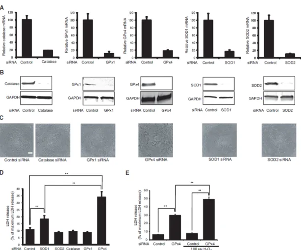
Human corneal epithelial cells were transfected with siRNA specifically silencing catalase, GPx1, GPx4, SOD1, or SOD2. Two days after transfection, mRNA (Figure 8A) and protein (Figure 8B) levels were measured through real-time RT-PCR and immunoblotting. The mRNA levels of all the antioxidant enzymes were downregulated
27
by >80%. In addition, a significant downregulation in the protein levels of each antioxidant enzyme was also confirmed.
I examined the morphological characteristics of corneal epithelial cells treated with each targeted siRNA 3 days after transfection (Figure 8C). Cells transfected with control siRNA appeared to be compact, uniform, and cobblestone pavement in shape. The shape of the cells transfected with catalase, GPx1, SOD1, or SOD2 was similar to that of cells transfected with control siRNA. Conversely, cells transfected with GPx4 siRNA exhibited signs of cell damage including spheroid structures.
Cytotoxicity was evaluated by measuring LDH activity. Knockdown of catalase, GPx1, and SOD2 did not affect LDH activity (Figure 8D). Knockdown of GPx4 and SOD1 significantly increased the activity of LDH. However, the LDH activity of GPx4 knockdown was significantly higher than that of SOD1 knockdown.
To further clarify the protective effect of GPx4 under oxidative stress conditions, I investigated the effect of GPx4 knockdown on cytotoxicity enhanced by hydrogen peroxide (Figure 8E). LDH activity of the cells transfected with control siRNA was not influenced by the addition 100 µM hydrogen peroxide. Conversely, LDH activity of the cells transfected with GPx4 siRNA significantly increased after treatment with 100 µM hydrogen peroxide. Knockdown of GPx4 enhanced cytotoxicity under mild oxidative stress, suggesting an important role for GPx4 against oxidative stress.
28
Figure 8. Knockdown of different antioxidant enzymes using siRNA in corneal epithelial cells. (A) Knockdown efficiency evaluated by mRNA levels. Mean + standard error mean (SEM; n = 3–4). (B) Knockdown efficiency evaluated by protein levels using immunoblot analysis. Reproducibility was confirmed in triplicate. (C) Phase contrast morphology of corneal epithelial cells transfected with siRNA of scramble control, catalase, GPx1, GPx4, SOD1, or SOD2 at day 3 after transfection. Scale bar, 50 µm. (D) LDH release from corneal epithelial cells three days after transfection with siRNA for scramble control, catalase, GPx1, GPx4, SOD1, or SOD2. Mean SEM (n = 4). **p < 0.01 using Tukey’s test. (E) Knockdown of GPx4 enhanced LDH release induced by H2O2. Data are mean + SEM (n = 4). **p < 0.01 using Tukey’s test.
3.3.2 α-tocopherol rescued cytotoxic effects of GPx4 knockdown
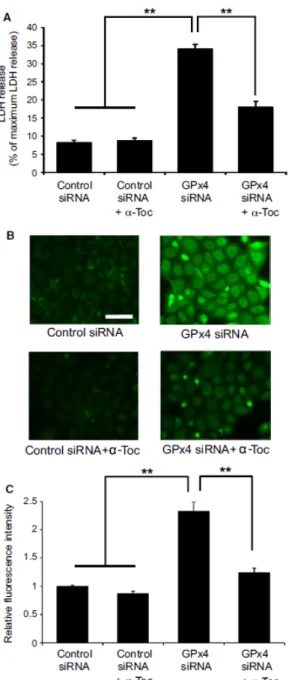
α-tocopherol has been reported to confer protection against cytotoxicity and cell death induced by GPx4 deficiency,36 which I subsequently tested in corneal epithelial cells in vitro. My results show that α-tocopherol significantly prevented LDH release
29
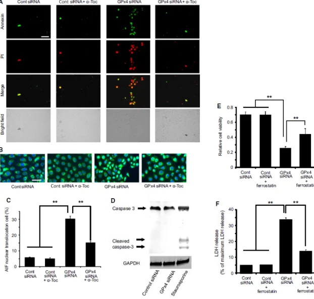
from cells transfected with GPx4 siRNA (Figure 9A). Next, I evaluated lipid hydroperoxide generation using immunostaining for 4-HNE and total intracellular ROS using DCFH-DA. Results show that both 4-HNE and total ROS were significantly elevated in cells transfected with GPx4 siRNA, which was rescued by treatment with α-tocopherol (Figure 9B, C, D). Figure 3 shows the cell death induced by GPx4 knockdown. Annexin V and PI staining indicated that most of the dead cells were annexin V positive with PI staining after 3 days of GPx4 silencing while the number of cells with either Annexin V or PI staining only was relatively small. In addition, the cell death was rescued by α-tocopherol treatment (Figure 10A).
Next, I investigated possible mechanisms for the cell death by GPx4. The percentage of cells with AIF translocation to the nucleus increased in cells transfected with GPx4 siRNA (Figure 10B, C). Furthermore, α-tocopherol prevented the AIF translocation induced by GPx4 knockdown (Figure 10B, C). In contrast, cleaved caspase-3, implicated in caspase-dependent apoptosis, was not detected in cells transfected with control siRNA or GPx4 siRNA (Figure 10D), while staurosporine treatment(positive control) led to the activation of caspase 3. I further examined the implication of ferroptotic mechanism using ferrostatin-1, an inhibitor of ferroptosis. Ferrostatin-1 partially ameliorated the decrease in cell viability (Figure 10E) and the increase in LDH activity (Figure 10F) caused by GPx4 knockdown.
30
Figure 9. α-tocopherol rescues cytotoxic effects of GPx4 knockdown in corneal epithelial cells. (A) α-tocopherol prevented the LDH release induced by GPx4 knockdown (n = 4). **p < 0.01 using Tukey’s test. (B) Accumulation of 4-HNE was evaluated by immunofluorescence. Scale bar, 50 µm. (C) Fluorescence intensities for 4-HNE were quantified using Image J (n = 8–9). **p < 0.01 using Tukey’s test. (D) Total intracellular ROS was quantified using DCFH-DA (n = 4). **p< 0.01 and *p < 0.05 using Tukey’s test.
31
Figure 10. Cell death caused by GPx4 knockdown in corneal epithelial cells. (A) Representative image of annexin V and PI staining. Majority of the staining was annexin V positive with or without PI staining. Scale bar, 50 mm. (B, C) Nuclear translocation of AIF (green) induced by GPx4 knockdown was evaluated in the total number of cells. DAPI was used for nuclear staining (n = 6–10). **p < 0.01 using Tukey’s test. Scale bar, 50 mm. (D) Caspase-3 and cleaved caspase-3 (active form) were immunoblotted for cells transfected with siRNA for scramble control or GPx4. Staurosporin (1 µM) served as a positive control. Reproducibility was confirmed in triplicate. (E) Effect of ferrostatin-1 (10 µM) to rescue the decreased cell viability induced by GPx4 knockdown. **p <
32
0.01 using Tukey’s test. (F) Effect of ferrostatin-1 (10 µM) to rescue the increased LDH activity induced by GPx4 knockdown. **p < 0.01 using Tukey’s test.
3.3.3 Effects of GPx4 knockdown on cell viability
I examined the effects of GPx4 knockdown on corneal epithelial cell growth. First, I evaluated cell viability using WST-8 assay. There was no significant difference in cell viability between cells transfected with GPx4 and control siRNA up to one day after transfection (Figure 11A). However, at 3 and 5 days after transfection, the viability of GPx4 siRNA-transfected cells was significantly lower than that of control siRNA-transfected cells (Figure 11A), suggesting that GPx4 is essential for growth of corneal epithelial cells.
Next, I examined the effects of GPx4 knockdown on the wound closure system of corneal epithelial cells in vitro. Two days after wound creation, a significant delay in the wound closure was observed in the cells treated with GPx4 siRNA, and α-tocopherol ameliorated the delay caused by GPx4 knockdown (Figure 11B, C).
33
Figure 11. Wound healing model of corneal epithelial cells in vitro. (A) Viability was evaluated by WST-8 assay at day 0, 1, 3, 5, and 7 after transfection (n = 5). **p < 0.01 by Student’s t-test. (B, C) Cell viability and migration was evaluated in wound healing model in vitro. Remaining wound area (% of each initial area) at 48 h after wound creation was compared (n = 4). *p<0.05 and **p < 0.01 using Tukey’s test.
34
3.3.4 Corneal epithelial wound healing in GPx4+/+ and GPx4+/− mice
I confirmed the decreased expression of GPx4 in both the mRNA and protein level in the cornea of GPx4+/− mice compared to that of GPx4+/+ mice (Figure 12A, B). In line with the decreased GPx4 expression, lipid peroxidation levels in the cornea of GPx4+/− mice were significantly higher than those in the cornea of GPx4+/+ mice (Figure 12C, D). Then, I examined corneal epithelial wound healing in GPx4+/− mice and GPx4+/+ mice after topical exposure to n-heptanol. At 18, 24, 30, and 36 h after n-heptanol treatment, the remaining epithelial defect area in GPx4+/− mice was larger than that in GPx4+/+ mice (Figure 12E, F). The epithelial defect was resurfaced in all the GPx4+/+ mice by 36 h after exposure to n-heptanol, whereas even at 42 h the defect was not completely resurfaced in GPx4+/− mice.
35
Figure 12. Corneal epithelial wound healing in GPx4+/− and GPx4+/+ mice. (A) GPx4 mRNA levels in the cornea of GPx4+/− and GPx4+/+ mice (n = 5–6). **p < 0.01 by Student’s t-test. (B) GPx4 protein levels were determined using western blot.
36
Reproducibility was confirmed in triplicate. (C) Accumulation of 4-HNE was evaluated by immunofluorescence. Scale bar, 100 µm. (D) Fluorescence intensities for 4-HNE were quantified using Image J (n = 3). *p < 0.05 by Student’s t-test. (E) Representative photographs of corneal epithelial. Wound healing in GPx4+/− and GPx4+/+ mice. Green areas represented fluorescein-stained wounded areas (F) The remaining area size of the wounds (% of each initial wound area) was compared between GPx4+/− and GPx4+/+ mice (n =10–12). **p<0.01 and *p<0.05 using Student’s t-test.
3.4 Discussion
The major contribution of the present study is that I show that GPx4 is by itself an important antioxidant enzyme for maintaining redox homeostasis and wound healing in corneal epithelial cells. Decreased expression of GPx4 led to cytotoxicity by oxidative stress, caspase-independentcell death with nuclear translocation of AIF, and decreased viability and wound healing in corneal epithelial cells. I confirmed that α-tocopherol could potentially compensate for the lack of GPx4 in corneal epithelial cells.
Oxidative stress and antioxidant system have been intensively discussed in the cornea pathologies.68.72,73 However, the importance of a specific antioxidant enzyme has not been fully understood. Degeneration and dysfunction of lacrimal glands leading to age-related dry eye signs has only been reported in mice deficient of SOD1.74 In the present study, I silenced the expression of various antioxidant enzymes in corneal epithelial cells and found that GPx4 deficiency led to a significant increase in cytotoxicity compared to the silencing of other antioxidant enzymes. Although the remnant expression levels of each antioxidant enzymes after knockdown might be
37
slightly different, the results might suggest the paramount importance of GPx4 as a defense mechanism in the corneal epithelium. In fact, even in the GPx4-haplodeficient mice, a significant delay in epithelial wound repair was observed in vivo.
It is known that byproducts of lipid hydroperoxide cause cell death and inhibition of cell proliferation,29,30 and are considered to be implicated in pathologies of corneal diseases such as atopic keratoconjunctivitis and dry eye.63,67 4-HNE is a major product generated during lipid peroxidation, and is a highly toxic molecule.28-30 Recently, a distinctive iron-dependent cell death, called ferroptosis has been primarily characterized in cancer cells and GPx4 is considered to be a central regulator of ferroptosis that is mediated by lipid peroxidation.38 In the present study, α-tocopherol prevented lipid peroxidation and cell death due to GPx4 deficiency, and moreover, ferrostatin-1 partially rescued decreased cell viability and increased LDH release by GPx4 knockdown. My results suggest an implication of ferroptosis in the cytotoxicity and cell death in the GPx4-deficient corneal epithelial cells. However, further investigations are necessary for the exact mechanism of cells death.
To the best of my knowledge, I firstly observed a delay in the corneal epithelial wound healing because of lack of a specific antioxidant enzyme. It has been reported that dry eye phenotypes appear in aged SOD1 knockout mice.66 The researchers observed degeneration and dysfunction of lacrimal glands that have been speculated as causes of corneal epithelial damage.66 Although my in vitro data indicated that the loss of GPx4 in corneal epithelium led to impaired viability and delayed wound healing, an implication of dysfunctional lacrimal gland was not examined in GPx4+/− mice. Another related report highlighted a delay in corneal epithelial wound healing in nuclear factor-like 2 (Nrf2) mice.68 The Nrf2 protein is a transcription factor that regulates the
38
expressions of numerous antioxidant enzymes and proteins. Therefore, the importance of the specific antioxidant enzyme was not the focus of the study, whereas the importance of Nrf2-associated antioxidant defense mechanisms was clearly delineated.
In conclusion, my data demonstrated that GPx4 is a major antioxidant enzyme that is not only crucial for maintaining redox homeostasis but also for wound healing in corneal epithelial cells. Deficient GPx4 can aggravate the corneal pathology and may highlight a new therapeutic target for corneal disorders such as dry eye and keratoconjunctivitis. In addition, α-tocopherol has a protective effect on lipid peroxidation, acting as an effective backup system for GPx4 in corneal epithelial cells.
3.5 Summary
Purpose: Oxidative stress is involved in the pathologies of corneal epithelial cells.
However, the importance of specific antioxidant enzymes in corneal epithelial cells is not fully understood. The purpose of this study is to elucidate the role of GPx4 in corneal epithelial cells. In addition, I examined the compensatory activity of vitamin E on GPx4 ablation condition.
Methods: For in vitro studies, an immortalized human corneal epithelial cell line was
used. Cells were transfected with siRNA for catalase, GPx1, GPx4, SOD1, SOD2, or scramble control siRNA. Cytotoxicity measured through LDH activity, lipid peroxidation immunostained for 4-HNE, cell proliferation, and cell death were compared between cells transfected with GPx4 siRNA or scramble control siRNA. In addition, the rescue effects of α-tocopherol and ferrostatin-1, a ferroptosis inhibitor, were examined in the cells with deficient GPx4 expression. For in vivo studies, I applied n-heptanol on the cornea of GPx4+/+ and GPx4+/− mice to create corneal epithelial wound. The epithelial defect area size was measured at 0, 6, 12, 18, 24, 30, 36, 42, and
39
48 h after epithelial wound creation.
Results: Knockdown of GPx4 strongly induced cytotoxicity and cell death in human
corneal epithelial cells. Cell death induced by GPx4 knockdown was characterized by positive staining for both annexin V and propidium iodide, nuclear translocation of AIF, and without activation of caspase 3, and was rescued by α-tocopherol and ferrostatin-1. The delayed wound healing of GPx4 siRNA-transfected cells were ameliorated by α-tocopherol in vitro. In addition, loss of one GPx4 allele was sufficient to significantly delay the healing of experimental corneal epithelial wounds in vivo.
Conclusions: GPx4 is an antioxidant enzyme that is by itself important for oxidative
homeostasis, cell survival, and wound healing in corneal epithelial cells. In addition, α-tocopherol has a protective effect on lipid peroxidation, acting as an effective backup system for GPx4 in corneal epithelial cells.
40
4. Role of GPx4 in glutamate-induced oxytosis in the retina.
4.1 Introduction
Glutamate-induced neurotoxicity has been studied for its possible role in the pathogenesis of numerous neurological disorders, including Alzheimer’s disease, Parkinson’s disease, amyotrophic lateral sclerosis, and ischemic stroke.75 Glutamate-induced toxicity may also be implicated in the ocular neurodegenerative changes in glaucoma76-79 and diabetic retinopathy.80,81 In fact, several studies have reported an increase in glutamate levels in the vitreous of patients with glaucoma76 and proliferative diabetic retinopathy.80,81 Because excess extracellular glutamate induces oxidative stress and cell death, glutamate-induced neurotoxicity is commonly called “oxytosis” 75.Treatments with antioxidants ameliorated the progression of the mouse model of glaucoma82,83 and diabetic retinopathy84 and suppressed cytotoxicity in retinal ganglion cells (RGCs) induced by N-methyl-D-aspartate (NMDA), the selective agonist for the glutamate receptor (NMDA receptor).85,86 Furthermore, treatment with an antioxidant suppressed the elevation of glutamate levels in the retinas of diabetic rats.87 In addition, several studies have suggested the importance of endogenous antioxidative defense mechanisms, including a superoxide dismutase and thioredoxins in RGCs.88,89
In glutamate-induced oxytosis, elevated levels of extracellular glutamate or increased susceptibility to extracellular glutamate can induce glutathione depletion and lipid peroxidation.75 Among antioxidant enzymes, GPx4 can directly reduce complex lipid hydroperoxides that are incorporated in biomembranes or lipoproteins.90 GPx4-deficient mice are lethal on embryonic day 8,34 and studies have identified drastic disease phenotypes of photoreceptors,35 and cerebral neurons,91 in conditional knockout mice.
41
In the present study, I evaluated the role of GPx4 in glutamate-induced oxytosis in the rat retinal precursor cell line R28 and the mouse retina.
4.2 Material and methods
4.2.1 Cell culture and transfection of siRNA
The rat retinal precursor cell line R28 was a kind gift from Dr. Yoshiaki Kiuchi (Hiroshima University, Department of Ophthalmology and Visual Sciences). R28 was established by immortalization of postnatal day 6 rat neuroretinal tissue using the psi2 replication incompetent retroviral vector.92 Unlike the RGC-5 cell line, R28 cells have been confirmed for validity and shown to express a variety of retinal cell-type markers, including RGC markers.93,94 The R28 cell line is considered suitable for neurotoxicity and neuroprotection studies.93 Cells were cultured in Dulbecco’s modified Eagle’s medium (DMEM; Invitrogen) containing 10% FBS and 100 U of penicillin along with 100 μg/mL streptomycin under 5% CO2 at 37°C. Cells at 20–30% confluence were transfected with siRNA that specifically knockdown GPx4 and scrambled control siRNA (Invitrogen) using lipofectamine RNAiMAX (Invitrogen) according to the manufacturer's instructions.
4.2.2Real-time RT-PCR
Two days after transfection with GPx4 siRNA or scrambled control siRNA, total RNA was isolated using Isogen (Nippon Gene) according to the manufacturer’s instructions. For the in vivo studies, total RNA was isolated from microsurgically dissected mouse retinas in the same manner. Subsequently, RNA was reverse-transcribed into cDNA by the ReverTra Ace® qPCR RT Master Mix with
42
gDNA Remover (Toyobo). Quantitative real-time PCR was carried out with the Thermal Cycler Dice Real-time System (Takara Bio) using Platinum SYBR Green qPCR SuperMix-UDG (Invitrogen). The values for each gene were normalized to the level of β-actin. The primer sequences used in the real-time RT-PCR were as follows: rat a-actin (Fwd, 5- CACCCGCGAGTACAACCTTC -3 and Rev, 5- CCCATACCCACCATCACACC -3), rat GPx4 (Fwd, 5- ATGCCCAC CCACTGTGGAA -3 and Rev, 5- GGCACACACTTGTAGGGCTAGAGA -3), mouse GAPDH (Fwd, 5- CACATTGGGGGTAGGAACAC -3 and Rev, 5- AACTTTG GCATTGTGGAAGG -3), and mouse GPx4 (Fwd, 5- CGCGATGATTGGCGCT -3 and Rev, 5- CACACGAAACCCTGTACTTATCC -3).
4.2.3 Immunoblotting
Two days after transfection with GPx4 siRNA or scrambled control siRNA, the proteins were extracted from cells and mouse retinas. SDS-PAGE of cellular proteins or retinal proteins was performed on Mini-PROTEAN TGX Any kD gel (Bio-Rad Laboratories, Hercules, CA) with Tris-glycine-SDS running buffer (Bio-Rad Laboratories). Immunoblot analysis was performed by electrotransfer of the proteins from the gels onto polyvinylidene difluoride (PVDF) membranes (Millipore, Billerica MA) at 100 V for 60 min at ice-cold temperature using Tris-glycine buffer. The membranes were probed with antibodies for β-actin (Santa Cruz Biotechnology) and GPx4 (Cayman). Binding of secondary antibodies, conjugated to alkaline phosphatase or horseradish peroxidase, was observed using a chemiluminescent substrate (Pierce).
43
Two days after transfection with GPx4 siRNA or scrambled control siRNA, a cytotoxicity assay was performed using the LDH cytotoxicity detection kit (Takara Bio). LDH activity was measured in the extracellular medium and in the cell lysate, according to the manufacturer’s instructions; subsequently, extracellular LDH activity was calculated as a percentage of the total LDH activity. In the glutamate stimulation study, cells transfected with GPx4 siRNA or scrambled control siRNA were treated with 1 mM and 2 mM glutamate (Wako). LDH activity was measured after 24 h.
4.2.5 Determination of lipid peroxidation
Accumulated peroxidized lipids were assessed by immunohistochemical detection of 4-HNE. Two days after transfection with GPx4 siRNA or scrambled control siRNA, cells were fixed with 4% paraformaldehyde for 15 min, washed three times with PBS, and permeabilized with a 0.1% Triton X-100 solution containing 5% goat serum in PBS. Permeabilized cells were washed three times with PBS containing 5% goat serum and incubated with anti-4-HNE antibodies (JaICA) overnight at 4°C. Cells were then washed three times with PBS. Alexa 488-conjugated anti-mouse IgG secondary antibodies (Invitrogen) were applied for 1 h at room temperature and washed three times with PBS. Fluorescent images were observed using a fluorescence microscope (Keyence). The fluorescence intensities of the dots stained with 4-HNE were quantified using the Image J software).
4.2.6 Annexin V staining
Two days after transfection with GPx4 siRNA or scrambled control siRNA, cells were stained by Alexa Fluor 488 annexin V (Invitrogen) for 15 min at room temperature
44
and washed and rinsed with PBS. Fluorescent images were observed with a fluorescence microscope (Keyence). The percentages of annexin V-positive apoptotic cells relative to the total number of cells were calculated.
4.2.7 Experimental animals: GPx4+/+ and GPx4+/− mice
I used GPx4+/+ and GPx4+/− mice on the C57BL/6 background.71 Animals were maintained in ordinary animal cages under constant 12-h light/dark cycles. Food and water were available ad libitum. All animal experiments were performed in accordance with the Association for Research in Vision and Ophthalmology (ARVO) Statement for the Use of Animals in Ophthalmic and Vision Research and the NIH Guiding Principles in the Care and Use of Animals (DHEW Publication, NIH 80-23), and were approved by the Institutional Animal Research Committee of the University of Tokyo
4.2.8 Immunohistochemistry
Mice were sacrificed with an overdose of pentobarbital (100-150 mg/kg) injected intraperitoneally, and eyes were enucleated. Enucleated eyes of GPx4+/+ mice were fixed in 4% paraformaldehyde in PBS. The samples were paraffin-embedded and cut into 5-μm-thick sections. Slides were first incubated with blocking solution (2% normal goat serum) overnight and further incubated with anti-GPx4 antibodies at room temperature for 2 h and with Alexa 488-conjugated anti-mouse IgG secondary antibodies (Invitrogen) for 1 h. The sections were then coverslipped with mounting medium. Fluorescent images were observed using a fluorescence microscope (Keyence).
45 4.2.9 NMDA-induced retinal toxicity
The intravitreal injection of NMDA was performed as described previously.77,78 A total of 2 µl of 25-mM NMDA in PBS was injected into the vitreous body of GPx4+/+ and GPx4+/− mice under anesthesia with intraperitoneal injection of a mixture of xylazine hydrochloride and ketamine hydrochloride.
The accumulation of peroxidized lipids in the retina was evaluated 12 h after intravitreal injection of NMDA. Mice were sacrificed with an overdose of pentobarbital (100-150 mg/kg) injected intraperitoneally, and eyes were enucleated. Then, eyes were fixed for 2 h in 4% paraformaldehyde solution in 0.1-M phosphate buffer (pH 7.4) and immersed for 1 h in PBS containing 20% sucrose. Further, the eyes were embedded in a supporting medium for frozen-tissue specimens (OCT compound).
Retinal sections of 10-μm thickness were prepared using a cryostat at −25°C. Sections were immersed in PBS for 20 min at room temperature and incubated with anti-4-HNE antibodies (JaICA) overnight at 4°C. Sections were then washed three times with PBS. Alexa 488-conjugated anti-mouse IgG secondary antibodies (Invitrogen) were applied for 1 h at room temperature. The sections were washed three times with PBS and coverslipped with mounting medium. The intensity of immunofluorescence in the ganglion cell layer (GCL) and inner plexiform layer (IPL) was evaluated using the Image J software.
Retinal cell death was evaluated 24 h after intravitreal injection of NMDA. Mice were sacrificed with an overdose of pentobarbital (100-150 mg/kg) injected intraperitoneally, and eyes were enucleated. After fixation, the enucleated eyes were embedded in paraffin and incised through the optic disc of each eye at 3-μm thickness. TUNEL staining was performed according to the manufacturer’s protocol (In Situ Cell
46
Death Detection Kit; Takara Bio Inc.) to analyze NMDA-induced cell death. Sections were treated with the TdT enzyme and stained with dUTP-fluorescein isothiocyanate. TUNEL-positive cells were observed using a fluorescence microscope (Keyence). TUNEL-positive cells were counted in GCL at a distance between 300 and 750 μm from the optic disc.
Hematoxylin and eosin staining for morphological evaluation was performed 7 days after NMDA injection. Mice were sacrificed with an overdose of pentobarbital (100-150 mg/kg) injected intraperitoneally, and eyes were enucleated, immersed for at least 24 h in 10% formalin, embedded in paraffin, and incised through the optic disc of each eye at 3-μm thickness. Thick sections were stained with hematoxylin and eosin. Light-microscope images were photographed, and the cell number in GCL was counted at a distance between 300 and 750 mm from the optic disc.
4.2.10 Statistical Analysis
Data are expressed as mean ± SEM. Statistical analysis was performed with 2-tailed Student’s t-test. P < 0.05 was considered statistically significant.
4.3 Results
4.3.1 Effects of GPx4 knockdown in R28 cells
First, I confirmed a ubiquitous expression of GPx4 in mouse retinas (Figure 13). The effects of GPx4 silencing in retinal cells were then evaluated using R28 cells. R28 cells were transfected with GPx4 siRNA to specifically knockdown GPx4. Two days after transfection, a favorable efficiency was confirmed at both mRNA (Figure 14A)
47
and protein levels (Figure 14B) measured by real-time RT-PCR and western blot, respectively.
Figure 13. GPx4 expression in the mouse retina. GPx4 was ubiquitously expressed in the mouse retina except outer segments of photoreceptors. Scale bar, 50 µm.
Figure 14. Knockdown efficacy of GPx4 in retinal precursor R28 cells. (A) The knockdown of GPx4 mRNA was confirmed by real-time RT-PCR. Data are mean ± SEM. (n = 3–4).
48
**p < 0.01. (B) The knockdown of GPx4 protein was also confirmed by western blot in triplicate.
Cytotoxicity was evaluated by measuring LDH activity. GPx4 knockdown significantly increased LDH activity (Figure 15A). Morphologically, cells treated with scrambled control siRNA appeared compact, uniform, and cobblestone-pavement shaped. On the other hand, cells treated with GPx4 siRNA exhibited signs of cell damage such as spheroid shapes (Figure 15B).
Figure 15.The effects of GPx4 knockdown on cytotoxicity in R28 cells. (A) LDH release from cells treated with control and GPx4 siRNA after 2 days of transfection. Data are means ± SEM (n = 4). **p < 0.01. (B) Phase contrast images of R28 cells after 2 days of transfection with scramble control or GPx4 siRNA. Scale bar, 50 µm.
The accumulation of peroxidized lipids was evaluated by immunostaining of 4-HNE. 4-HNE immunostaining was three times higher in R28 cells transfected with GPx4 siRNA than in those transfected with scrambled control siRNA (Figure 16).
49
Figure 16. The level of peroxidized lipids in R28 cells. (A) 4-HNE detected by fluorescence microscopy using antibodies for 4-HNE. (B) Quantification of the fluorescence intensities for 4-HNE. Data are mean ± SEM (n = 4). **p < 0.01. Scale bar, 50 µm.
Next, I evaluated apoptotic cell death using annexin V staining. As shown in Figure 17, the number of annexin V-positive cells significantly increased in R28 cells transfected with GPx4 siRNA compared with those transfected with scrambled control siRNA.
50
Figure 17. Annexin V staining in R28 cells. (A) Representative image of annexin V staining by fluorescence microscopy. (B) The percentage of annexin V-positive cells relative to the total number of cells. Data are means ± SEM (n = 5). **p < 0.01. Scale bar, 50 µm.
I also investigated the effects of GPx4 knockdown on the cytotoxicity induced by glutamate (Figure 18). LDH activity of cells transfected with scrambled control siRNA was not influenced by glutamate up to 2 mM. However, GPx4 knockdown significantly enhanced the cytotoxicity induced by glutamate.
Figure 18. GPx4 knockdown enhanced LDH release by glutamate cytotoxicity. LDH activity was evaluated after 24 h of glutamate treatment. Data are means ± SEM (n = 4). **p < 0.01.
4.3.2 NMDA-induced neurotoxicity in the retina of GPx4+/+ and GPx4+/− mice.
First, I confirmed the decreased expression of GPx4 in both mRNA and protein levels in the retina of GPx4+/− mice compared with those in the retina of GPx4+/+ mice (Figure 19A, B).