1
Risk of secondary osteoporosis due to lobular cholestasis in non-cirrhotic primary biliary cholangitis
Anna Seki,1 Fusao Ikeda,1 Hirokazu Miyatake,2 Koichi Takaguchi,3 Shosaku Hayashi,4 Toshiya Osawa,5 Shin-ichi Fujioka,5 Ryoji Tanaka,5 Masaharu Ando,6 Hiroyuki Seki,1 Yoshiaki Iwasaki,1 Kazuhide Yamamoto,5 and Hiroyuki Okada1
1Department of Gastroenterology and Hepatology, Okayama University Graduate School of
Medicine, Dentistry, and Pharmaceutical Sciences, Okayama, Japan
2Department of Internal Medicine, Hiroshima City Hospital, Hiroshima, Japan
3Department of Internal Medicine, Kagawa Prefectural Central Hospital, Takamatsu, Japan
4Department of Internal Medicine, Ritsurin General Hospital, Takamatsu, Japan
5Department of Internal Medicine, Okayama Saiseikai General Hospital, Okayama, Japan
6Department of Internal Medicine, Mitoyo General Hospital, Kanonji, Japan.
Short title: Osteoporosis in non-cirrhotic PBC
Address correspondence to: Fusao Ikeda, M.D., Department of Gastroenterology &
Hepatology, Okayama University Graduate School of Medicine, Dentistry, and Pharmaceutical Sciences, 2-5-1, Shikata-cho, Kitaku, Okayama 700-8558, Japan. Telephone:
+81-86-235-7219, Fax: +81-86-225-5991, E-mail: [email protected]
Journal of Gastroenterology and Hepatology
2
ABSTRACT
It remains unclear whether primary biliary cholangitis (PBC) represents a risk factor for secondary osteoporosis. We conducted a case-control study to examine bone mineral density and bone turnover markers in middle-aged postmenopausal PBC patients without liver cirrhosis. We further compared the incidence of low bone mineral density between propensity-score matched subgroups of PBC patients and healthy controls. Finally, we investigated the mechanisms underlying unbalanced bone turnover in terms of the associations between bone turnover markers and PBC-specific histological findings. Our analysis included 128 consecutive PBC patients, all postmenopausal women aged in their 50s or 60s, without liver cirrhosis or fragility fracture at the time of PBC diagnosis. The prevalence of osteoporosis was significantly higher in the PBC group than in the control group (26% vs 10%, p = 0.015, the Fisher exact probability test). In most PBC patients (95%), the level of bone-specific alkaline phosphatase was above the normal range, indicating increased bone formation. On the other hand, the urine type I collagen-cross-linked N-telopeptide showed variable levels among our PBC patients, indicating unbalanced bone resorption. Advanced fibrosis was associated with low bone turnover. Lobular cholestasis, evaluated as aberrant keratin 7 expression in hepatocytes, showed significant negative correlations with bone formation and resorption, indicating low bone turnover. Our results clearly show that, compared with healthy controls, even non-cirrhotic PBC patients have significantly higher risk of osteoporosis. Moreover, lobular cholestasis was associated with low bone turnover, suggesting that this feature of PBC may itself cause secondary osteoporosis in PBC patients.
Key words: primary biliary cholangitis, bone mineral density, osteoporosis, bone turnover
3
marker, keratin 7
4
INTRODUCTION
Primary biliary cholangitis (PBC) is a chronic cholestatic liver disease of autoimmune etiology, predominantly affecting middle-aged women. Its prevalence varies by geographic region, from 0.7 to 49 per million individuals in the general population.(1) PBC is characterized by chronic nonsuppurative destructive cholangitis that gradually progresses to cirrhosis, ultimately resulting in liver failure. Low bone mineral density (BMD), osteoporosis, and bone fracture represent well-recognized complications of PBC, mainly associated with liver cirrhosis.(2) The increased risks of secondary osteoporosis is related with poor liver function in cirrhotic patients, which affects the transformation of 1,25(OH)2D3, the active form of vitamin D, and production of insulin-like growth factor 1. Liver cirrhosis also results in insufficient secretion of bile acids into the intestine, which reduces intestinal absorption of calcium and vitamin D.(3)
Guañabens et al. reported that osteoporosis is more prevalent in middle aged postmenopausal women with PBC than in the age-matched general population.(4) Osteoporosis is, however, occasionally observed in PBC patients without liver-related symptoms or liver dysfunction, and it has not been clarified whether PBC itself represents a further risk for osteoporosis among PBC patients without liver cirrhosis. Therefore, we conducted a case-control study to examine BMD in PBC patients without liver cirrhosis and to assess whether these patients have increased risks of osteoporosis.
5
METHODS
Patients
The present study enrolled consecutive female patients aged in their 50s or 60s, who were diagnosed with PBC between 2002 and 2012 at Okayama University Hospital or one of its affiliated hospitals. The diagnosis of PBC was established if the patient satisfied two of the following criteria: positive test for anti-mitochondrial antibody, biochemical evidence of cholestasis, and liver biopsy findings compatible with the diagnosis of PBC.(5) Patients showing other signs of liver injury or serum positivity for hepatitis B surface antigens or anti-hepatitis C virus antibodies were excluded from the study. Presence of obstructive biliary diseases such as stones or tumors was excluded in all patients following abdominal ultrasonography and computed tomography examinations.
Additionally, a group of post-menopausal women aged in their 50s or 60s, with no relevant medical history, were enrolled as healthy controls from a pool of volunteers recruited by the Department of Gastroenterology and Hepatology of Okayama University.
The study was conducted in accordance with the Helsinki Declaration, and approved by the ethical committee of all participating institutes. All patients and healthy volunteers provided written informed consent for participation.
Evaluation of BMD
Low BMD was diagnosed according to the Japanese guidelines for prevention and treatment of osteoporosis, in terms of the young adult mean (YAM) of BMD at the lumbar vertebrae L2 to L4.(6) Osteopenia was defined as BMD below 80 % of the YAM, which is approximately equivalent to 1.5 standard deviations below average in the distribution of values used to develop the YAM. Similarly, osteoporosis was defined as BMD below 70% of
6
YAM, which is approximately equivalent to 2.5 standard deviations below average in the distribution of values used to develop the YAM.
Histological evaluation
Liver biopsy was performed for all patients at the time of PBC diagnosis. The histological stage of PBC was determined according to the histological classification proposed by Nakanuma et al.,(7) which is based on PBC-specific histological findings including fibrosis, bile duct loss, orcein-positive granules in the liver, cholangitis, and hepatitis.
Immunohistochemistry of keratin 7
Immuno-histochemical analysis was performed using formalin-fixed paraffin-embedded samples obtained at the time of PBC diagnosis. Preparation of the sample sections and immunostaining for keratin 7 (K-7) were performed as described previously.(8,9) K-7 expression patterns were classified as previously described: grade 1, K-7 expression noted in bile duct epithelial cells and proliferated bile ductules; grade 2, periportal hepatocytes positive for K-7; grade 3, intralobular hepatocytes also positive for K-7; and grade 4, K-7 diffusely stained throughout the lobules.(8,9)
Statistical analysis
Quantitative data are expressed as means ± standard deviations. Patient characteristics were compared using the Mann–Whitney U test. The incidence of osteoporosis was compared between the matched groups of PBC patients and healthy controls, using the Kruskal-Wallis test or the Fisher exact probability test. The relationships between low BMD and clinical or histological characteristics were analyzed using logistic regression. The propensity score of the factors associated with low BMD was estimated for each patient using
7
a logistic regression model. The PBC patients were matched with healthy volunteers for the factors identified to correlate with low BMD, and a caliper width within 0.1 of the propensity score was applied. Correlations of PBC-specific histological features and bone metabolisms were evaluated with the Spearman’s rank correlation coefficient. Statistical analyses were performed using JMP Pro version 12.0 (SAS Institute, Cary, NC, USA), and p-values <0.05 were considered to indicate statistical significance.
8
RESULTS
Characteristics of the PBC patients enrolled in the study
We enrolled 137 consecutive PBC patients, all post-menopausal women aged in their 50s or 60s, with no liver-related symptoms or fragility fracture at the time of PBC diagnosis. A total of 9 patients were excluded from the present analysis because of the following reasons:
histological diagnosis of cirrhosis (5 patients), confirmation of regular menstrual cycle (3 patients), and progression to end-stage renal disease requiring dialysis treatment (1 patient).
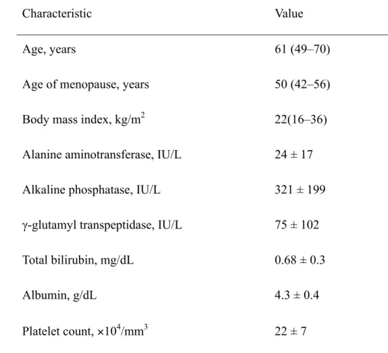
Table 1 provides a summary of the baseline characteristics of the 128 patients who fulfilled the inclusion criteria at the time of PBC diagnosis. The mean age of the patients was 61 years, and 70% of the patients were positive for anti-mitochondrial antibody. All patients had abnormal levels of γ-glutamyl transpeptidase (GGT), but did not show signs of impaired liver function, as the total bilirubin level, albumin level, and platelet count were all within normal limits.
Evaluation of clinical and histological characteristics as potential risk factors for osteoporosis in PBC patients
Following logistic regression analysis (Table 2), we found that the age at diagnosis and body mass index were significantly associated with low BMD (p: 0.020 and 0.015, respectively), while the levels of biliary enzymes such as alanine aminotransferase, alkaline phosphatase (ALP), and GGT were not. Findings related to liver function (total bilirubin, albumin, and platelet count) showed no significant correlations with low BMD. A similar lack of statistical significance was noted for the correlations between low BMD and PBC-specific histological findings.
9
Comparison of BMD between PBC patients and propensity-score matched healthy volunteers
Because our logistic regression analysis showed that the age at diagnosis and body mass index were significantly associated with osteoporosis in PBC patients, the propensity scores were calculated for age at diagnosis and body mass index, and 100 PBC patients were matched with 50 healthy controls.
After propensity-score matching, the matched subgroups of PBC patients and healthy controls showed similar age, age of menopause, body mass index, and BMD (Table 3).
Nevertheless, the diagnosis of low BMD and osteoporosis was significantly more frequent in the PBC group than in the control group (p = 0.050, Kruskal-Wallis test; Table 3). Similarly, the prevalence of patients with osteoporosis was significantly higher in the PBC group (26%) than in the control group (10%, p = 0.015, the Fisher exact probability test).
Markers of bone turnover in PBC patients
The mechanisms underlying reduced BMD in PBC patients were assessed by examining the levels of blood and urine markers of bone turnover (Figure 1). No decrease in the levels of vitamin D was observed among these patients, confirming the lack of liver dysfunction.
There were several patients with above-normal levels of intact parathormone or vitamin D (18% and 34% of patients, respectively). Nevertheless, the serum levels of calcium were within normal ranges in all patients. No patient showed decreased levels of bone formation markers such as bone-specific alkaline phosphatase (BAP), but many patients (95%) showed above-normal values. The urine marker of bone resorption type I collagen-cross-linked N-telopeptide showed variable levels among our sample of PBC patients.
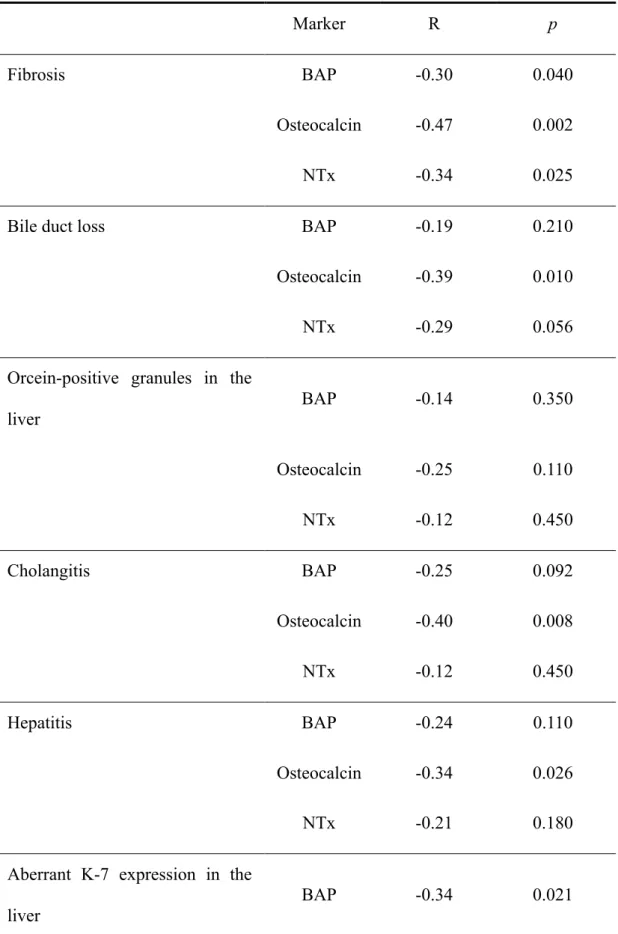
Associations between markers of bone turnover and PBC-specific histological findings
10
To further clarify the mechanisms underlying reduced BMD in non-cirrhotic PBC patients, we examined the associations between PBC-specific histological findings and markers of bone turnover (Table 4). Advanced fibrosis was significantly correlated with low levels of serum BAP, serum osteocalcin, and urine type I collagen-cross-linked N-telopeptide (p: 0.040, 0.0018, and 0.025, respectively; Spearman’s rank correlation coefficient), suggesting that advanced fibrosis is associated with low bone turnover with decreased bone formation and resorption. Cholangitis and hepatitis were associated with decreased bone formation, but showed no relationship with bone resorption. In terms of lobular cholestasis, the finding of orcein-positive granules in the liver showed no significant associations with bone turnover markers, while bile duct loss tended to indicate low bone turnover, showing weak associations with serum osteocalcin and urine type I collagen-cross-linked N-telopeptide (p: 0.01 and 0.056, respectively).
Associations between markers of bone turnover and aberrant K-7 expression
Lobular cholestasis, defined in terms of aberrant K-7 expression in hepatocytes, also indicated low bone turnover, showing significant negative correlation (the Spearman’s rank correlation coefficient) with both bone formation and resorption (Table 4 and Fig. 2).
Additionally, aberrant K-7 expression in hepatocytes was significantly correlated with GGT levels (R = 0.34, p = 0.0088), but not with ALP levels (R = 0.17, p = 0.21). The prevalence of osteoporosis was slightly higher in patients with K-7 expression of grade 3 or 4 than in those with K-7 expression of grade 1 or 2, but the difference was not statistically significant (36%
vs 31%, p = 0.22; the Fisher exact probability test).
11
DISCUSSION
We conducted the present case-control study to examine BMD in PBC patients without liver cirrhosis and to assess whether these patients have increased risks of osteoporosis. Our analysis involving propensity-score matched groups of middle aged postmenopausal women revealed that, compared to healthy controls, even non-cirrhotic PBC patients have higher risk of osteoporosis. Histological examinations suggested that PBC itself results in a further risk for low bone turnover associated with lobular cholestasis in non-cirrhotic PBC stages.
Osteoporosis is known to be associated with old age, female sex, reduced physical activity, and low body mass index.(6) Further factors such as a history of fracture, steroid treatment, smoking, alcohol abuse, and diabetes mellitus are thought to increase the risk of secondary osteoporosis. While hepatic osteodystrophy is a known complication in patients with liver cirrhosis,(10–12) it is occasionally observed in PBC patients without liver cirrhosis.(11) The present investigation represents the first case-control study to evaluate whether PBC itself may increase the risk of osteoporosis even in non-cirrhotic patients. We found that, compared with healthy controls, PBC patients in non-cirrhotic stages indeed have significantly higher risk of osteoporosis.
Our results indicated that low BMD occurred due to unbalanced bone resorption, despite increased bone formation. Post-menopausal women often have low levels of estrogen, which may trigger high bone turnover with increased bone resorption over bone formation.(6) On the other hand, low bone turnover with decreased bone formation and resorption was noted in patients with liver cirrhosis.(1) The present study enrolled only post-menopausal women in their 50s or 60s; all patients were non-cirrhotic and maintained good liver function with sufficient secretion of bile acids into the intestine. While it is possible that these patients had high bone turnover because of the low levels of estrogen, some of the patients with lobular cholestasis showed low bone turnover. It was previously noted that PBC patients occasionally
12
have lobular cholestasis with bile duct loss even in non-cirrhotic stages.(9) Taken together, these results suggest that this specific feature of PBC (i.e., lobular cholestasis) may cause secondary osteoporosis in PBC patients.
Patients with cholestatic liver diseases exhibit high serum levels of ALP and GGT. In recent reports, GGT has been identified as a novel bone-resorbing factor that stimulates osteoclast formation.(13–15) In a mouse model of cholestasis, elevated GGT levels were found to stimulate the expression of receptor activator of NF-κB ligand in osteoblasts, and to activate osteoclast formation.(14) On the other hand, it has been reported that higher ALP levels in PBC patients indicate severe cholangitis, but not severe cholestasis.(16) Indeed, the present results indicated that GGT level, but not ALP level, correlated significantly with cholestasis, suggesting that lobular cholestasis in PBC patients triggers bone resorption due to increased GGT levels.
Aberrant K-7 expression in hepatocytes is a more sensitive marker of lobular cholestasis than bile duct loss or orcein-positive granules in the liver, as previously reported.(8, 9) The results of the present study revealed that, in cholestatic patients, aberrant K-7 expression in hepatocytes is clearly associated with bone turnover, which reflects the rate of bone formation and resorption. Therefore, in patients with lobular cholestasis and aberrant K-7 expression in hepatocytes, early initiation of preventive care should be considered, in order to reduce the risk for future osteoporosis or fragility fractures. Based on our results, we believe that medical therapies focused on improving bone turnover may be effective in reducing the risk of secondary osteoporosis in PBC patients; such therapies may include vitamin K2 supplements, to support osteocalcin synthesis and avoid the expression of receptor activator of NF-κB ligand following osteoclast activation.(17)
Overall, the results of our study showed that, compared to healthy controls, even PBC patients in non-cirrhotic stages have significantly higher risk of osteoporosis. Furthermore,
13
lobular cholestasis was associated with low turnover, suggesting that lobular cholestasis itself may cause secondary osteoporosis in PBC patients.
DISCLOSURES
All authors state that they have no conflicts of interest.
ACKNOWLEDGEMENTS
This work was supported in part by the research program of intractable disease provided by the ministry of health, labor, and welfare of Japan. We thank Taiko Kameyama, and Toshie Ishii for technical assistance. Each author was involved in preparation of the manuscript as follows: Anna Seki,1 and Fusao Ikeda,1: acquisition, analysis and interpretation of data, and drafting of the manuscript; Hirokazu Miyatake,2 Koichi Takaguchi,3 Shosaku Hayashi,4 Toshiya Osawa,5 Shin-ichi Fujioka,5 Ryoji Tanaka,5 and Masaharu Ando,6: acquisition of data; Hiroyuki Seki,1 Yoshiaki Iwasaki,1 Kazuhide Yamamoto,5 and Hiroyuki Okada1: study concept and design.
14
REFERENCES
1. Parés A, Guañabens N. Treatment of bone disorders in liver disease. J Hepatol. 2006 Sep;45(3):445–53.
2. Zein CO, Jorgensen RA, Clarke B, et al. Alendronate improves bone mineral density in primary biliary cirrhosis: a randomized placebo-controlled trial. Hepatology. 2005 Oct;42(4):762–71.
3. Guañabens N, Parés A, Mariñoso L, et al. Factors influencing the development of metabolic bone disease in primary biliary cirrhosis. Am J Gastroenterol. 1990 Oct;85(10):1356–62.
4. Guañabens N, Parés A, Ros I, et al. Severity of cholestasis and advanced histological stage but not menopausal status are the major risk factors for osteoporosis in primary biliary cirrhosis. J Hepatol. 2005 Apr;42(4):573–7.
5. Kubota J, Ikeda F, Terada R, et al. Mortality rate of patients with asymptomatic primary biliary cirrhosis diagnosed at age 55 years or older is similar to that of the general population. J Gastroenterol. 2009 Sep;44(9):1000–6.
6. Orimo H, Nakamura T, Hosoi T, et al. Japanese 2011 guidelines for prevention and treatment of osteoporosis--executive summary. Arch Osteoporos. 2012 Dec;7(1-2):3–20.
7. Harada K, Hsu M, Ikeda H, et al. Application and validation of a new histologic staging and grading system for primary biliary cirrhosis. J Clin Gastroenterol. 2013
Feb;47(2):174–81.
8. Yabushita K, Yamamoto K, Ibuki N, et al. Aberrant expression of cytokeratin 7 as a histological marker of progression in primary biliary cirrhosis. Liver. 2001 Feb;21(1):50–
5.
9. Seki H, Ikeda F, Nanba S, et al. Aberrant expression of keratin 7 in hepatocytes as a predictive marker of rapid progression to hepatic failure in asymptomatic primary biliary
15
cirrhosis. Acta Med Okayama. 2015 Jun;69(3):137–44.
10. Parés A, Guañabens N. Osteoporosis in primary biliary cirrhosis: pathogenesis and treatment. Clin Liver Dis. 2008 May;12(2):407–24.
11. Newton J, Francis R, Prince M, et al. Osteoporosis in primary biliary cirrhosis revisited.
Gut. 2001 Aug;49(2):282–7.
12. Menon KV, Angulo P, Weston S, et al. Bone disease in primary biliary cirrhosis:
independent indicators and rate of progression. J Hepatol. 2001 Sep;35(3):316–23.
13. Hiramatsu K, Asaba Y, Takeshita S, et al. Overexpression of gamma- glutamyltransferase in transgenic mice accelerates bone resorption and causes osteoporosis. Endocrinology.
2007 Jun;148(6):2708–15.
14. Niida S, Kawahara M, Ishizuka Y, et al. Gamma-glutamyl transpeptidase stimulates receptor activator of nuclear factor-kappaB ligand expression independent of its
enzymatic activity and serves as a pathological bone-resorbing factor. J Biol Chem. 2004 Feb;279(7):5752–6.
15. Kawazoe Y, Miyauchi M, Nagasaki A, et al. Osteodystrophy in cholestatic liver diseases is attenuated by anti-γ-glutamyl transpeptidase antibody. PLoS One. 2015
Sep;10(9):e0139620.
16. Ikeda F, Okamoto R, Baba N, et al. Prevalence and associated factors with esophageal varices in early primary biliary cirrhosis. J Gastroenterol Hepatol. 2012 Aug;27(8):1320–
8.
17. Parés A. Old and novel therapies for primary biliary cirrhosis. Semin Liver Dis. 2014 Aug;34(3):341–51.
16
TABLES
Table 1. Clinical characteristics of patients with primary biliary cholangitis (n = 128)
Characteristic Value
Age, years 61 (49–70)
Age of menopause, years 50 (42–56)
Body mass index, kg/m2 22(16–36)
Alanine aminotransferase, IU/L 24 ± 17 Alkaline phosphatase, IU/L 321 ± 199 γ-glutamyl transpeptidase, IU/L 75 ± 102
Total bilirubin, mg/dL 0.68 ± 0.3
Albumin, g/dL 4.3 ± 0.4
Platelet count, ×104/mm3 22 ± 7
Data given as median (range) or mean ± standard deviation.
17
Table 2. Clinical and histological characteristics potentially associated with osteoporosis in patients with primary biliary cholangitis
Characteristic Odds ratio (95% CI) p
Age, years 1.1 (1.01–1.2) 0.020
Age of menopause, years 1.0 (0.88–1.1) 0.970 Body mass index, kg/m2 0.81 (0.68–0.95) 0.015 Blood test findings
Alanine aminotransferase, IU/L 1.0 (0.99–1.0) 0.170 Alkaline phosphatase, IU/L 1.0 (0.99–1.0) 0.290 γ-glutamyl transpeptidase, IU/L 1.0 (0.99–1.0) 0.490
Total bilirubin, mg/dL 0.61 (0.11–2.6) 0.520
Albumin, g/dL 0.61 (0.23–1.6) 0.310 Platelet count, ×104/mm3 1.0 (0.96–1.1) 0.660 Liver histology findings
Fibrosis 2.0 (0.93–4.3) 0.080
Bile duct loss 1.5 (0.90–2.5) 0.130
Orcein-positive granules in the liver 1.0 (0.51–1.9) 0.999
Cholangitis 1.2 (0.56–2.6) 0.710
Hepatitis 1.4 (0.32–5.8) 0.620
CI, confidence interval.
18
Table 3. Overview of risk factors for osteoporosis in patients with primary biliary cholangitis
Factor All patients
(n = 128)
Matched (n = 100)
Healthy control (n = 50) Age, years 61 ± 6 59 ± 5 60 ± 3 Age of menopause, years 50 ± 4 50 ± 3 50 ± 4 Body mass index, kg/m2 22 ± 3 23 ± 3 22 ± 2 Bone mineral density, mg/cm2 0.84 ± 0.2 0.86 ± 0.2 0.85 ± 0.1
Bone mineral density,a
normal/osteopenia/osteoporosis
71/24/33 58/19/23 33/12/5
Data given as mean ± standard deviation, unless otherwise specified.
aBone mineral density according to the classification proposed in the Japanese 2011 guidelines for prevention and treatment of osteoporosis.
19
Table 4. Correlation between markers of bone turnover and histological findings specific to primary biliary cholangitis
Marker R p
Fibrosis BAP -0.30 0.040
Osteocalcin -0.47 0.002
NTx -0.34 0.025
Bile duct loss BAP -0.19 0.210
Osteocalcin -0.39 0.010
NTx -0.29 0.056
Orcein-positive granules in the liver
BAP -0.14 0.350
Osteocalcin -0.25 0.110
NTx -0.12 0.450
Cholangitis BAP -0.25 0.092
Osteocalcin -0.40 0.008
NTx -0.12 0.450
Hepatitis BAP -0.24 0.110
Osteocalcin -0.34 0.026
NTx -0.21 0.180
Aberrant K-7 expression in the liver
BAP -0.34 0.021
20
Osteocalcin -0.48 0.001
NTx -0.39 0.009
BAP, bone-specific alkaline phosphatase; K-7, keratin 7; NTx, type I collagen-cross-linked N-telopeptide.
21
FIGURE LEGENDS
Fig. 1. Blood and urine markers of bone turnover: bone-specific alkaline phosphatase (A), osteocalcin (B), urine type I collagen-cross-linked N-telopeptide (NTx) (C), parathormone (D), vitamin D (E), alkaline phosphatase (F), -glutamyl transpeptidase (G). Upper and lower limits of the normal range are indicated in dashed lines. BCE, bone collagen equivalent; Cr, creatinine.
Fig. 2. Association between keratin 7 (K-7) expression in hepatocytes and markers of bone turnover: bone-specific alkaline phosphatase (A), osteocalcin (B), urine type I collagen-cross-linked N-telopeptide (NTx) (C), parathormone (D), vitamin D (E), alkaline phosphatase (F), -glutamyl transpeptidase (G). K-7 expression patterns in hepatocytes were classified as follows: grade 1, expression in bile duct epithelial cells and proliferated bile ductules; grade 2, expression in periportal hepatocytes; grade 3, expression also in intralobular hepatocytes; grade 4, K-7 diffusely stained throughout the lobules. BCE, bone collagen equivalent; Cr, creatinine.