VOL. 2 8 NO. 1 MAR C H 2 0 0 0
CONTENTS
Review
Kimura, M., Ohtomo, H., Kumagai, M. and Hiroshige, Y.
The Use of Malaria Diagnostic Test Kits by Travelers(in Japanese) 1‑7
Original article
Kobayashi, J., Sato, Y., Toma, H., Shimabukuro, I., Tasaki, T., Takara, M. and Shiroma, Y.
Epidemiological Features of Strongyloides Infection in Okinawa, Japan:
Comparative Study with Other Endemic Areas 9‑14
Iwanaga, M., Insisiengmay, S., Higa, N. and Sisavath, L.
Tetracycline Resistant and Polymyxin B Sensitive Vibrio cholerae O1 E1 Tor Isolated
from the Recent Epidemics 15‑18
Takaoka, H. and Saito, K.
Description of a New Species of Simulium (Nevermannia)from Japan(Diptera:Simuliidae) 19‑24
Takaoka, H. and Mulla, M.S.
Taxonomic Notes on Simulium siripoomense from Thailand(Diptera: Simuliidae): Descriptions of Female and Pupa, and Change of its Subgeneric Status
from Byssodon to Simulium s. str. 25‑28
Proceedings of Joint Conference:
40th Annual Meeting of Japanese Society of Tropical Medicine and
14th Annual Meeting of Japanese Association for International Health 29‑97
………
………
………
………
………
………
Japanese Journal of Tropical Medicine and Hygiene Vol.28 No.1
第28巻 第1号 平成12年3月
内 容
総 説
旅行者によるマラリア診断キット使用の問題
木村 幹男,大友 弘士,熊谷 正広,廣重 由可 1‑7
原 著
沖縄における糞線虫症の疫学的特徴:他の流行地域と比較して(英文)
小林 潤,佐藤 良也,當眞 弘,島袋 勲,田崎 力,
高良 政弘,城間 祥行 9‑14
テトラサイクリン耐性ポリミキシンB感受性エルトールコレラ菌によるコレラの流行(英文)
岩永 正明,Sithat Insisiengmay,比嘉 直美,Lay Sisavath 15‑18 日本産ブユ属ホソスネブユ亜属に属する1新種(双翅目:ブユ科)の記載(英文)
高岡 宏行,斎藤 一三 19‑24
タイ国産ブユSimulium siripoomense(双翅目:ブユ科)の分類学的検討:
雌成虫と蛹の記載およびByssodon亜属からSimulium亜属への帰属変更(英文)
高岡 宏行,Mir S. Mulla 25‑28
第40回日本熱帯医学会・第14回日本国際保健医療学会合同大会英文抄録
(日本熱帯医学会発表講演のみ) 29‑97
会報・記録
日本熱帯医学会会則(理事選挙細則・評議員選出細則・名誉会員選 細則・「日本熱帯医学会
研究奨励賞」選 規定) 99‑
日本熱帯医学会特別会計内規
2000(平成12)年度日本熱帯医学会役員名簿(2000年1月1日現在)
日本熱帯医学会雑誌編集委員名簿 ‑
投稿規定 ‑
著作権複写に関する注意
………
………
………
………
………
………
………
………
………
………
………
………
March 2000
総 説
旅行者によるマラリア診断キット使用の問題
木村 幹男 ,大友 弘士 ,熊谷 正広 ,廣重 由可
平成11年12月16日受付/平成11年12月20日受理
はじめに
世界におけるマラリアの患者数は年間3‑5億人,それに よる死亡者は150‑270万人と推定されており,他の感染症の 撲滅や制御が行われている現代においても,全般的なマラ リアの流行状況は少しも改善を見ていない。なかでも,熱 帯アフリカのマラリアの殆どを占める他,アジア,オセア ニア,南米などにも広く分布する熱帯熱マラリアは脳症,
急性腎不全,肺水腫/
ARDS,重症貧血, DIC
様出血傾向な どを生じて重症化したり死亡する危険のある疾患である。国際交流の活発化,それを可能にする大量航空機輸送の発 達により,日本人もマラリア流行地へ出かけることが頻繁 となり,帰国後の発症例として年間110‑120例程度が知られ るが,徐々に熱帯熱マラリアの占める割合が増加しつつあ ることが危惧される(大友,1998
;Suzaki et al.,
1999)。また日本人の問題として言えば,統計に上がって来ない国 外での発症例も多数あり,そこでの邦人の死亡例も散見さ れる。このような状況下で,マラリアから身を守るために 旅行者自身でも使えると期待されて,ヨーロッパの数か国 で市販もされているマラリア診断キットにつき,検討を加 えてみた。
マラリア予防,その中での緊急治療およびその問題点 マラリア予防で全ての場合に行うべき事は蚊に刺されな いように工夫することであるが,目的地での熱帯熱マラリ アの流行が高度で,旅行者の予想される行動から判断して も罹患の可能性が高い場合,オプションとしての薬物予防 が選択される。しかし,副作用が皆無であるはずもなく,
たとえば,予防効果に優れるメフロキンを選択した場合に は精神神経系の副作用が危惧されている(木村,藤井,1997
; Schlagenhauf,1999)。ただし,このような自覚的副作用に
ついては服用者の先入観にも大きく作用されるので,無作 為割付,二重盲験による判定が望ましいが,現在までに出 されているそれらのデータは少数例での検討であり,メフ ロキンの真の副作用がどの程度であるのかについてのコン センサスは得られていないのが現状である(木村,橋本,1998)。
別のオプションとして,発熱してマラリアが疑われる状 況であれば,旅行者自身の判断で治療薬を服用する緊急治 療(自己治療)が挙げられる。世界保健機関
WHOの定義
では,発熱して24時間以内に医療機関を受診できない場合 に緊急避難的に行うものとするが,あくまで急場しのぎで あることの理解が必要であり,緊急治療を行った後もでき るだけ医療機関を受診するよう努めるべきとされている(WHO,1999)。熱帯熱マラリアの流行が高度な地域に一定 期間滞在する場合には薬物予防を主体に行うべきとされて おり,緊急治療が適応となるのは流行度が低い地域へ行く 場合,短期間の滞在,短期間の滞在を頻回に繰り返す場合
(飛行機の乗務員など)などとされる(Schlagenhauf and
Steffen,1994)。しかし,薬物予防を行っている場合でも薬
剤耐性の原虫により罹患することもあるので,緊急治療の オプションを 慮すべきであるとも言える。緊急治療の問題点として,用法・用量の間違いが挙げら れる。内在的な問題として,実際にはマラリアでない場合 の使用が多くなる。これは初めから承知のことではあるが,
ドイツの調査からすると,マラリアの緊急治療を行った旅 行者の中で,後日抗体陽性によりマラリアであったと確認 されたのは10.4%のみであった(Nothdurft
et al.,1995)。
この数値は地域,旅行者自身の知識や経験などの因子によ り様々であろうが,熱帯地域での発熱原因としてはマラリ ア以外の方が圧倒的に多いことは事実である。マラリアで ない場合に緊急治療を行い,高度の副作用が出るなどは非 常に不幸なことである。そこで,正確な自己診断および緊 急治療を行うために,一般旅行者でも簡便に検査でき,し かも信頼性の高いマラリア診断キットの開発が望まれてき たが(Schlagenhauf
et al.,1995),ここ数年その期待がか
けられるimmunochromatography法を用いたキットが市販
されるようになった。ただし,これらは海外で市場に出回 るようになったが,日本国内での入手は現段階では不可能 である。1 東京大学医科学研究所感染免疫内科 2 東京慈恵会医科大学熱帯医学教室 3 海外勤務健康管理センター研修交流部
* 著者連絡先:〒108‑8639 東京都港区白金台4‑6‑1 東京大学医科学研究所感染免疫内科 TEL:03‑5449‑5338,FAX:03‑5449‑5427,E‑mail: mkimura@ims. u‑tokyo. ac. jp
マラリア診断キットの種類,感度,特異度 1)Histidine‑
rich protein 2
(HRP
‑2
)検出系 a.ParaSight TMF
Dipstick
形式のParaSight
TMF
が最初に市販されたが,これは熱帯熱マラリア原虫のみを検出する。モノクローナ ル抗体を吸着させたスティックの他に試薬として3種類含 まれ,それらは初めに血液を溶血させる試薬,スルフォ ローダミンBを吸着させた当該抗原に対するポリクローナ ル抗体を含む試薬,最後にスティックを洗浄する試薬など から構成される。必要な血液量は50μ
l,全体の反応時間は
15‑20分間,キットは2‑30℃での保存が可能である。感度・特異度に関しては様々なデータが報告されており,
低い場合は90%以下であるが,概して90%以上の感度が報 告されており,一部では100%の数値も挙げられている(表 1)。野外調査で多数検体を扱う場合などには,1例ごとの 検討が十分でない可能性があるので,それらのデータの解 釈には注意が必要である。例えば,三日熱マラリアの輪状 体しか見られない時期での顕微鏡検査で,熱帯熱マラリア と判定される可能性もある。これは,特に厚層塗抹標本の 場合に起こりやすい。そして抗原検出では陰性であるので,
感度の数値を低下させることとなる。我々も熱帯熱マラリ アに関して東大医科研と慈恵医大での症例57例につき検討 し,感度100%の成績を得ている(表1)。しかも,その半 数以上の症例では 18
S rRNA
遺伝子をターゲットするPCR
法(Kimura et al.,1995)を併用して顕微鏡法でのマラリ ア診断,原虫種の同定の裏付けを得ている。筆者らの印象 として,特にnon
‑immune患者を対象にする場合には通常
一定数以上の原虫数になるので,表1の数値を全体的に見 るよりも感度はより優れており,より100%に近いと えられる。しかし稀ではあるが,原虫数15,000/μ
l
で抗原陰性で あった例も報告されている(Humar et al.,1997)。また,抗原検出での感度が原虫感染赤血球数に依存する のは当然であり,1‑100/μ
l
の場合71.4%,101/μl
以上で あ れ ば95.5‑97.1%の 感 度 と す る データ(Kilian et al., 1997),21/μl
以上であれば100%とするデータ(Caraballoand Ache,1996)などが報告されている。
特異度に関しても様々な数値であり,マラリア流行地の 住民を対象とする野外調査ではやや低く86‑90%程度であ り,
non
‑immuneを対象とする場合には97‑99%と高くなる
(WHO,1996)。これは流行地の場合,例えば 1‑2週間前に 熱帯熱マラリアに罹患し,顕微鏡検査で原虫は検出できな くても抗原が陽性に出る例などがあるためである。また,
リウマチ因子陽性者の偽陽性反応が66.7‑68.4%に見られて いる(Laferi
et al.,
1997; Bartoloni et al.,
1998)。さら に,三日熱マラリアでの交差反応も知られており,我々も 52例の三日熱マラリアの中で5例(9.4%)の陽性例を見い だした。しかし,この場合には反応は弱いことが殆どであ る。このように,特異度は対象の種類に大きく影響される ことを 慮する必要がある。b.ICT Mamaria P.f.(
MalaQuick
TM)ParaSight
TMF
に3年程遅れて市場に出たキットであり,検出する抗原は同一である。しかし,カード型でしかも試 薬は1種類のみであり,操作が簡便であることが特徴で,
旅行者が携行するには便利である。溶血のための試薬,金 コロイドを吸着させた
HRP
‑2に対する抗体などはパッドの 中に固相化されている。キットの保存条件は2‑8℃となっ ているが,熱帯地域での高温にも十分耐えられるとのこと である(Schlagenhauf,私信)。血液量も10μlと少なく,検査
所要時間は10分程度で済む。特異度に関してはParaSight TM
Table 1 Summary of the reports on ParaSight TM F
Author Study country Performed by Reference standard No.HRP‑2(+) /No.P.f.(+)
Sensitivity (%)
No.HRP‑2(−) /No.P.f.(−)
Specificity (%)
Thick smear 226/242 93.4 659/671 98.2
Banchongaksorn et al.,
1996 Thailand Not described PCR 153/167 91.6 351/353 99.4
Caraballo and Ache,
1996 Venezuela Unclear Thick smear 268/309 86.7 1,081/1,089 99.3
Humar et al., 1997 Canada Hospital staff PCR 66/75 88.0 74/76 97.4
Singh et al., 1997a India Medical labora-
tory staff Thick smear 354/382 92.7 785/849 92.5
Kodisinghe et al.,
1997 Sri Lanka Not described Thick and thin
smears 202/224 90.2 1,069/1,079 99.1
Kilian et al., 1997 Uganda Health worker (after single day training)
Thick smear 68/78 87.2 98/102 96.1
Bustos et al., 1999 Philippines,
Japan Not described Thick and thin
smears 103/103 100.0 313/335 93.4
Current authors, un
published Japan Medical doctor Thick and thin smears, partially confirmed by PCR
57/57 100.0 239/256 93.4
-
Only those published since 1996.
2
F
と同程度と報告されることが多いが,感度についてはより 優れた数値が出されることが多く,100%あるいは殆ど100%に近い数値も報告されている(表2)。筆者らも少数例の検 討で感度96.2%の結果を得ているが(表2),偽陰性の1例 は原虫数330/μ
l
のlow parasitemia
であった。原虫数と感 度の関係については,1‑100/μl
で71.4%,101/μl
以上で 100%とする報告(Kilian et al.,1997)がある。しかし,稀ではあるが,原虫数が20,000/μ
l
で抗原陰性であった症例 も示されている(Jelinek et al.,1999a
)。リウマチ因子陽性者での偽陽性反応は問題にならず
(Mishra et al.,1999),筆者らの印象では三日熱マラリア での交差反応も殆どないようである。
あるメーカーはこの検査キットにアルコール綿,ラン セット,スポイト,バンドエイド,体温計を組み合わせた ものを発売しているが,これを用いると他には何も必要と しない。
c.ICT Malaria P.f/
P.v
最近,前述のカード型抗原検出キットに三日熱マラリア も対象に組み込んだものが発売されるようになり,今後は これが主に使われることになると推測される。熱帯熱マラ リア原虫検出に関しての感度・特異度などは従来の
ICT Malaria P.f.
の場合と同じであると予想されるが,インドネシアから感度95.5%,特異度89.8%と報告されている(表 2,Tjitra et al.,1999)。しかし,三日熱マラリア原虫検 出の感度は75%と低値であった。
2)原虫由来
pLDH
検出系(OptiMAL )これは熱帯熱マラリア原虫,あるいは三日熱マラリア原 虫に特異的な酵素
pLDH
を識別して検出するキットであ る。必要血液量は10μl,検査所要時間は15‑20分程度,保存
は 2‑4℃である。感度や特異度に関してのデータは
HRP
‑2検出系の場合ほ どには得られていないが,熱帯熱マラリアに関しては今の ところ感度89‑94%,特異度88‑98%である(Palmer et al., 1998;Jelinek et al.,1999 a;Cooke et al.,
1999)。HRP‑2 検出系では治癒例でも長期間陽性反応が続く傾向があるが,この
pLDH
検出系では治癒の場合早期に陰性化するので,治療経過の判定に有用であると期待される(Chiodini,私 信)。
一般旅行者を対象にしたマラリア診断キットの検討 以上のデータは殆どの場合,この種の手技にある程度習 熟した者が行って得られたものであるが,一般旅行者にこ れらのキットを携行させるアイディアが検討課題となる。
発熱を生じてマラリアを疑う状況のときに自己診断を行い,
その結果が陽性であれば緊急治療を行い,マラリアによる 重症化や死亡の発生を抑えることができるであろうとの期 待がもたれる。もちろん,感度が100%でなく熱帯熱マラリ アの見逃しが皆無とは言えない。Low parasitemiaの場合 だけでなく,稀ではあるが一定数以上の原虫数があっても 抗原陰性であった例が報告されている。それらの原因とし て,1) 血液サンプル処理の問題,2) 反応を抑制する抗体 の存在,3)
HRP‑2の抗原性の変異,などが えられるが
(Humar et al.,1997),これらの問題に関しては今後解明 されることを期待したい。このようにキットは完璧なもの でないにしても,マラリアを疑うときに医療機関へのアク セスがない場合もあり,また現地の医療機関での診断に問 題がありうることも えると,適切に使用すれば相当のメ リットをもたらすことができると えられる。
そこで,一般旅行者が
ParaSight
TMFと MalaQuick
TMを 使って正しく検査し,正しく判定できるかの検討がチュー Table 2 Summary of the reports on ICT Malaria P.f. or ICT Malaria P.f/P.v
Author Study country Performed by Reference standard No.HRP‑2(+) /No.P.f.(+)
Sensitivity (%)
No.HRP‑2(−) /No.P.f.(−)
Specificity (%) Garcia et al., 1996 Solomon Is. Not described Thick and thin smears 39/39 100.0 204/212 96.2 Singh et al., 1997b India Field worker Thick smear 201/201 100.0 120/142 84.5
Kilian et al., 1997 Uganda Health worker(after
single day training) Thick smear 72/78 92.3 94/102 92.2
Durrheim et al.,1998 South Africa Nurse Thick smear 68/69 98.6 191/195 97.9
Tjitra et al., 1999 Indonesia Health worker Thick and thin smears 236/247 95.5 281/313 89.8
Bustos et al., 1999 Philippines,
Japan Not described Thick and thin smears 102/103 99.0 332/335 99.1
Jelinek et al., 1999a Germany Not described Thick and thin smears, partially confirmed by PCR
49/53 92.5 175/178 98.3
Current authors, un
published Japan Medical doctor Thick and thin smears, partially confirmed by PCR
25/26 96.2 35/35 100.0
-
ICT Malaria P.f/P.v
Mixed data with ICT Malaria P.f. and ICT Malaria P.f/P.v
リヒで行われたが,予想に反した惨澹たる結果であった(Funk
et al.,1999)。当人が説明書を読んで理解するだけで,それ
以外の説明を与えない状況で自己診断を行わせたが,説明 書に述べられた試薬類などを理解できなかった人が40‑50%
以上に達したり,
ParaSight
TMF
の場合には血液量が多いの で,採血に困難を感じた人が多く見られた。実際の検査手 技についてもParaSight
TMF
の場合には16.9%に,Mala-Quick
TMの場合には8.6%に誤りが見られた。さらに,あらかじめ反応を終わっているスティックが用意され,説明書 の内容に沿って旅行者が判定したところ,0.1%以下の
low parasitemiaの血液で反応を行ったスティックの場合に
「陽 性」と 正 し く 判 定 し た 者 は,ParaSight TMF
で52.1%,MalaQuick
TMで10.8%に過ぎなかった。逆に2%以上のhigh parasitemiaの血液で反応を行ったスティックの場合に
「陽 性」であると正しく判定した者は,MalaQuick
TMでは96.8%であったが,ParaSight TM
F
では33.8%に過ぎなかった。また,ロンドンのトラベルクリニックでは帰国後発熱な どの症状を呈して受診した患者を対象とし,ICT Malaria
P.f.
による自己診断を行わせ,流行地で一般旅行者がマラリアの自己診断をするのと同じ状況を再現した。実際に熱帯 熱マラリアであった者は21人であったが,そのうち7人(33%)
はキットを用いて「陰性」との自己診断であった(Behrens
et al.,1999)。発熱を有する旅行者を対象にして ICT Malaria P.f.
による自己診断の検討をケニアで行った時にも,様々な段階での誤りが見られ,結果を判定できない者が31.7%に も達し,熱帯熱マラリア11人の中で正しく診断ができたの は1人に過ぎなかった(Jelinek et al.,1999
b
)。これらの結果から,一般人がキットを使うに際しての問 題は無視できないものであることが明らかになった。誤っ た使い方や判定により,旅行者が害悪を被ってはならない ことは言うまでもない。特に,熱帯熱マラリアの陽性反応 が出ているのに「陽性」の判定ができなかった例では,実 地ではマラリアの緊急治療を行わずに,重症化・死亡に至 る危険がある。逆に,本当は陰性なのに「陽性」と誤って 判定する間違いでは,マラリアでないのに抗マラリア薬を 服用して副作用の危険にさらすこと,他の診断が 慮され ることなく重症化する可能性,などの問題点が挙げられる。
マラリア診断キットの今後について
一般旅行者に無差別に診断キットを渡し,説明書通りに 行えばマラリアの自己診断が的確に行えるとアドバイスす るのは,危険であることが明らかになった。したがって,
これだけ多くの問題を有する診断キットなので,一般旅行 者での使用はあきらめ,医療機関においてのみ使用すべき なのであろうか? いや,上述のごとく,感度や特異度に おいて完璧でないにしろ,特に
non‑ immuneを対象にする
場合には100%に極めて近い優れたものと えられる。アフ リカの奥地などで,医療機関を受診するのに数日間を要す る所に滞在する者もある。また,現地でのマラリアの診断 は残念ながら信頼できないことも少なくない。前項に述べた旅行者が,自己診断を行う場合の悲観的なデータについ ては,その後の指導や訓練によって改善する余地が残され ている。この様な実際的な問題に取り組むのは旅行医学の 役割であり,今後も問題点およびそれらの解決策などが真 剣に検討され,旅行者がマラリア診断キットを正しく使え るようになることが望まれる。
具体的には,メ ー カ ー 側 が 成 す べ き 事 と し て,特 に
ParaSight
TMFの場合には検査の過程をより単純化すること
が望まれる。判定の仕方については図を用いて陽性と陰性 の場合を示してあるが,実際のスティックあるいはカード 上で起こった反応を正しく判定するのが困難なこともあり うる。陽性のバンドが余りにも強すぎると,逆に陽性と判 断されなくなる誤りも見られている(Schlagenhauf,私 信)。また,簡便であると評判の高いカード型キットの場 合,血液が濾紙上を一様に上って行かずに端の部分を上 がったり,時には全く上がらないこともある。このような 時にも,正しい判断をすることが必要となる。スティック あるいはカード上での反応として,実際にありうる様々な 状況をカラー写真で示すことなどが不可欠であろう。また,トラベルクリニックの側では旅行者に説明書を読 ませるだけでなく,口頭での説明を加えるべきである。説 明書を読むのみで正しく検査を行えた者は75%であったが,
口頭での説明を加えたら90%に上昇したとのデータもある
(Trachsler et al.,1999)。あるいは,ビデオでの学習を取 り入れるのも効果的であろう。いずれにしろ前もって最低 数回の実習を行わせ,正しく実施し正しく判定できるよう になったことを確認することが不可欠である。
さらに,ある程度生物・医学関係の実験の経験のある人 の場合には,適切に検査・判定できる可能性が高いので,
それらの人達に限って使用を勧めていくのも一案であろう。
そして彼らが他の人にも正しく教えれば,マラリア診断 キットでメリットを受ける人数が増加していくことが期待 できる。同時にやはり,キットを正しく使うことが絶対的 に必要であり,しかもそれは一般には簡単でなく,適切な 訓練を要することも周知させていく必要がある。
おわりに
マラリア診断キットは完璧ではないにしても,簡便性と 高い感度は貴重であり,旅行者自身が利用できる可能性を 追求すべきであると える。ただし,そのためには適切に 使うことが必須であり,しかもそれが簡単ではないことも 認識しなければならない。実際には,メーカーとトラベル クリニックの両者での様々な具体的な工夫が必要となる。
この問題に関する,旅行医学的視点からの進歩が望まれる。
文 献
1)Banchongaksorn,T.,Yomokgul,P.,Panyim,S.,Rooney, W.and Vickers,P.(1996): A field trial of the ParaSight F test for the diagnosis of Plasmodium falciparum infec- tion. Trans. R. Soc. Trop. Med. Hyg., 90, 244‑245 4
2)Bartoloni, A., Strohmeyer, M., Sabatinelli, G., Benucci, M., Serni, U. and Paradisi, F.(1998): False positive ParaSight F test for malaria in patients with rheumatoid factor. Trans.R.Soc.Trop.Med.Hyg.,92, 33‑34
3)Behrens, R.H., Whitty, C. and Armstrong, M.(1999): Comparison of laboratory diagnosis of malaria with immunochromatographic kits when self ‑administered by symptomatic patients. In: Abstracts of the 6th Conference of the International Society of Travel Medi- cine. p. 60.
4)Bustos, D.G., Olveda, R.M., Negishi, M. and Kurimura, T.(1999): Evaluation of a new rapid diagnostic test
“Determine Malaria Pf”against standard blood film, ICT Malaria P.f and ParaSight F. Jpn. J. Trop.
Med. Hyg., 27, 417‑425
5)Caraballo,A.and Ache,A.(1996): The evaluation of a dipstick test for Plasmodium falciparum in mining areas of Venezuela. Am. J. Trop. Med. Hyg., 55, 482 ‑484 6)Cooke, A.H., Chiodini, P.L., Doherty, T., Moody, A.H.,
Ries,J.and Pinder,M.(1999): Comparison of a parasite lactate dehydrogenase‑based immunochromatographic antigen detection assay(OptiMAL )with microscopy for the detection of malaria parasites in human blood samples. Am. J. Trop. Med. Hyg., 60, 173 ‑176
7)Durrheim, D.N., la Grange, J.J.P., Govere, J. and Mngomezulu, N.M.(1998) : Accuracy of a rapid im- munochromatographic card test for Plasmodium fal- ciparum in a malaria control programme in South Africa. Trans. R. Soc. Trop. Med. Hyg., 92, 32 ‑33 8)Funk,M.,Schlagenhauf,P.,Tschopp,A.and Steffen,R.
(1999): MalaQuick versus ParaSight F as a diagnos- tic aid in travellersʼmalaria. Trans.R.Soc.Trop.Med.
Hyg., 93, 268‑272
9)Garcia,M.,Kirimoama,S.,Marlborough,D.,Leafasia,J.
and Rieckmann,K.H.(1996): Immunochromatographic test for malaria diagnosis. Lancet, 347, 1549
10)Humar, A., Ohrt, C., Harrington, M.A., Pillai, D. and Kain, K.C.(1997): ParaSight F test compared with the polymerase chain reaction and microscopy for the diagnosis of Plasmodium falciparum malaria in trav- elers. Am. J. Trop. Med. Hyg., 56, 44‑48
11)Jelinek,T.,Grobusch,M.P.,Schwenke,S.,Steidl,S.,von Sonnenburg, F., Nothdurft, H.D., Klein, E.and Loscher, T.(1999a): Sensitivity and specificity of dipstick tests for rapid diagnosis of malaria in nonimmune travelers.
J. Clin. Microbiol., 37, 721‑723
12)Jelinek, T., Amsler, L., Grobusch, M.P. and Nothdurft, H.D.(1999b): Self‑use of rapid tests for malaria diagno- sis by tourists. Lancet, 354, 1609
13)Kilian, A.H.D., Mughusu, E.B., Kabagambe, G. and von Sonnenburg, F. (1997): Comparison of two rapid, HRP2‑based diagnostic tests for Plasmodium falcipar- um. Trans. R. Soc. Trop. Med. Hyg., 91, 666‑667 14)Kimura, M., Miyake, H., Kim, H.‑S., Tanabe, M.,Arai,
M., Kawai, S., Yamane, A. and Wataya, Y.(1995): Species‑specific PCR detection of malaria parasites by
microtiter plate hybridization: clinical study with malaria patients. J. Clin. Microbiol., 33, 2342 ‑2346 15) 木村幹男,藤井達也(1997):マラリア予防薬としてのメフ
ロキンの副作用の問題.日本醫事新報 № 3838,37‑41 16) 木村幹男,橋本麻希(1998):メフロキン薬物予防における
精神神経系副作用の問題.Clin. Parasitol.(日本臨床寄生 虫学会誌),9,82‑85
17)Kodisinghe, H.M., Perera, K.L.R.L., Premawansa, S., Naotunne, T. de S., Wickramasinghe, A.R.and Mendis, K.N.(1997): The ParaSight F dipstick test as a rou- tine diagnostic tool for malaria in Sri Lanka. Trans.R.
Soc. Trop. Med. Hyg., 91, 398‑402
18)Laferi, H., Kandel, K. and Pichler, H.(1997): False positive dipstick test for malaria. N.Engl.J.Med.,337, 1635‑1636
19)Mishra,B.,Samantaray,J.C.,Kumar,A.and Mirdha,B.
R.(1999): Study of false positivity of two rapid antigen detection tests for diagnosis of Plasmodium falciparum malaria. J. Clin. Microbiol., 37, 1233
20)Nothdurft, H.D., Jelinek, T., Pechel, S.M., Hess, F., Maiwald,H.,Marschang,A.,Sonnenburg,F.V.,Weinke, T. and Loscher, T.(1995): Stand‑by treatment of suspected malaria in travellers. Trop. Med. Parasitol., 46, 161‑163
21) 大友弘士(1998):特集 マラリア再び.日本におけるマラ リア.化学療法の領域,14,789‑794
22)Palmer, C.J., Lindo, J.F., Klaskala, W.I., Quesada, J.A., Kaminsky, R., Baum, M.K. and Ager, A.L. (1998): Evaluation of the OptiMAL test for rapid diagnosis of Plasmodium vivax and Plasmodium falciparum malaria.
J. Clin. Microbiol., 36, 203‑206
23)Schlagenhauf,P.(1999): Mefloquine for malaria chemo- prophylaxis 1992‑1998:a review. J.Travel Med.,6,122‑ 133
24)Schlagenhauf,P.and Steffen,R.(1994): Stand‑by treat- ment of malaria in travellers:a review. J. Trop. Med.
Hyg., 97, 151‑160
25)Schlagenhauf,P.,Steffen,R.,Tschopp,A.,Van Damme, P., Mittelholzer, M.‑L., Leuenberger, H. and Reinke, C.
(1995): Behavioural aspects of travellers in their use of malaria presumptive treatment. Bull. World Health Organ., 73, 215‑221
26)Singh, N., Singh, M.P. and Sharma, V.P.(1997a): The use of dipstick antigen‑capture assay for the diagnosis of Plasmodium falciparum infection in a remote forested area of central India. Am.J.Trop.Med.Hyg.,56,188 ‑ 191
27)Singh, N., Valecha, N. and Sharma, V.P. (1997b): Malaria diagnosis by field workers using an immuno- chromatographic test. Trans.R.Soc.Trop.Med.Hyg., 91, 396‑397
28)Suzaki, A., Kimura, M. and Ohtomo, H.(1999): The current situation of imported malaria in Japan. In:
Abstracts of the 6th Conference of the International Society of Travel Medicine. p. 84.
29)Tjitra, E., Suprianto, S., Dyer, M., Currie, B.J. and Anstey, N.M.(1999): Field evaluation of the ICT
Malaria P.f/P.v immunochromatographic test for detec- tion of Plasmodium falciparum and Plasmodium vivax in patients with a presumptive clinical diagnosis of malaria in eastern Indonesia. J. Clin. Microbiol., 37, 2412 ‑2417 30)Trachsler, M., Schlagenhauf, P. and Steffen, R.(1999):
Feasibility of a rapid dipstick antigen‑capture assay for self‑testing of travellersʼmalaria. Trop. Med. Int.
Health, 4, 442‑447
31)WHO(1996): A rapid dipstick antigen capture assay for the diagnosis of falciparum malaria. Bull.World Health Organ., 74, 47‑54
32)WHO(1999): 5.Health Risks and Their Avoidance.In:
International Travel and Health. Vaccination Require- ments and Health Advice.p.55‑89. World Health Orga- nization, Geneva
6
THE USE OF MALARIA DIAGNOSTIC TEST KITS BY TRAVELERS
M
IKIOK
IMURA, H
IROSHIO
HTOMO, M
ASAHIROK
UMAGAIand Y
UKAH
IROSHIGEReceived December 16, 1999/ Accepted December 20, 1999
Malaria prophylaxis measures of travelers include,in addition to chemoprophylaxis,stand‑by treatment (self treatment)which is indicated in areas of low transmission,for short‑term travelers and for those with brief,repeated exposure to malaria. The indication of stand ‑by treatment has now been expanded in some areas, partly due to an increasing concern about the possible adverse effects of malaria chemoprophylaxis.
However, the inappropriate use of stand‑by treatment in non‑malaria cases may expose persons to a significant drug risk, thus developing simple and reliable self diagnostic tests especially for Plasmodium falciparum malaria has been expected. Recently marketed kits for detecting P. falciparum histidine‑rich protein 2(HRP‑2)or pLDH could be good candidates for malaria self diagnosis by travelers. Especially a card‑type kit detecting HRP‑2 is characterized by its simplicity and reliability,often showing a sensitivity of>95%or even sometimes 100%. Although the sensitivity of those tests could not always be 100%,they could be very useful when performed properly,especially in cases travelers develop malaria ‑like symptoms in areas remote from reliable medical facilities. However,reports of self diagnosis with those kits performed by travelers showed discouraging results. Recent studies highlighted errors of performing these tests correctly and of interpreting previously reacted strips or cards. Travelers should not become a victim of potentially useful malaria diagnostic tests due to their incorrect use. Nonetheless,we still believe that the use of those kits by travelers should not be abandoned, rather they must have potential for application to travelersʼmalaria. This goal could be accomplished by improving the contents of the kitsʼinstructions and also by giving full oral explanation to travelers at travel clinics.
EPIDEMIOLOGICAL FEATURES OF STRONGYLOIDES INFECTION IN OKINAWA, JAPAN: COMPARATIVE STUDY WITH OTHER ENDEMIC AREAS
J
UNK
OBAYASHI, Y
OSHIYAS
ATO, H
IROMUT
OMA, I
SAOS
HIMABUKURO, T
SUTOMUT
ASAKI, M
ASAHIROT
AKARA ANDY
OSHIYUKIS
HIROMAReceived April 21, 1999/Accepted October 4, 1999
Abstract: The epidemiological features of Strongyloides stercoralis infection in Okinawa, Japan, were studied by comparing with those in Thailand, Laos and Brazil. The prevalence rates of Strongyloides infection in the present study were 9.6%(133/1,380)in Okinawa,47.6% (99/208)in Chiang Mai,Thailand, 23.8% (106/445)in Khammouane, Laos, and 12.0% (32/267)in Maceio, Brazil, respectively. The age inclination in aged subjects and sex dominance in males were significant features of Strongyloides infection in Okinawa, suggesting that new infection from the environment does not occur in present ‑day Okinawa.
The epidemiological feature was considered to provide a favorable field to investigate therapeutic efficacy unaffected by reinfection from environment after treatment. Absence of helminth infection other than Strongyloides was an additional feature in Okinawa. The features may also be convenient to study host response and pathogenicity in the Strongyloides infection unaffected by concurrent infection with other helminths.
Key words:Strongyloides stercoralis,strongyloidiasis, epidemiology, Okinawa, Thailand, Laos, Brazil
I
NTRODUCTIONStrongyloides stercoralis is a nematode parasite of man with a high prevalence in many tropical and sub- tropical countries. A warm and moist climate is essen- tial for its natural transmission,and man becomes typi- cally infected through skin contact, by penetration of infective larvae developing in the soil. It has also been well known that the parasite can multiply within host by internal autoinfection. Due to the autoinfection, the parasite can survive for many years in its host and it is frequently imported in temperate regions without out- side source of reinfection. The biological nature of the parasite influences not only the clinical manifestation of strongyloidiasis but also epidemiological feature in endemic and non
‑endemic areas.
Okinawa prefecture lies in the southernmost part of Japan and is the only prefecture located in subtropical
zone. Under the circumstance, the inhabitants in Okin- awa have been suffering in the past from many impor- tant parasitic diseases, such as malaria and filariasis.
However, these parasitic diseases have already been almost completely eradicated in recent years and only strongyloidiasis is currently highly prevalent in the prefecture. In the recent surveys, the mean prevalence rate was found to be as high as 10% among the inhabit- ants over 40 years old
(Asato et al.,1992
). On the other hand,the prevalence in the young generation is extreme- ly low, suggesting that new infection from the environ- ment rarely occurs among inhabitants in present
‑day Okinawa and that presumably the majority( more than 90%) of cases are long‑ standing cases acquired the infection in childhood and presisted until adulthood
(Sato et al.,1986
). Under the condition,epidemiological feature of strongyloidiasis in Okinawa is considered to be different from those of other endemic areas where S.
1 Department of Parasitology, Faculty of Medicine, University of the Ryukyus, 207 Uehara, Nishihara, Okinawa 903‑0125, Japan 2 Kumejima Family Clinic, 1134 Nakadomari, Gushikawa, Okinawa 901‑3124, Japan
3 Izumizaki Hospital, 1‑11‑2 Izumizaki, Naha, Okinawa 900‑0021, Japan Correspondence to:Jun Kobayashi,
Department of Parasitology, Faculty of Medicine, University of the Ryukyus, 207 Uehara, Nishihara, Okinawa 903‑0125, Japan E‑mail:jun@med.u‑ryukyu.ac.jp Fax:+81‑98‑895‑4914
Jpn. J. Trop. Med. Hyg., Vol. 28, No. 1, 2000, pp. 9‑14 9
stercoralis infection currently manifested among the inhabitants.
The purpose of the present study was to compare the epidemiological features of strongyloidiasis in Okin- awa with those of endemic areas in Thailand,Laos and Brazil, where many parasitic infections are currently prevalent among the inhabitants.
M
ATERIALS ANDM
ETHODSSurvey Areas and Populations Examined:
Okinawa prefecture,also referred as to the Ryukyu Islands, consisted of about 60 small islands. One of the islands, Kume Island, located about 100 km east of Okinawa Island was selected as survey area in the present study. The similar surveys were also conducted in two countries of Southeast Asia and in a country of South America. In Southeast Asia,a village( Sanpatong village
)in Chiang Mai province,northern Thailand,andtwo villages
(Sisamsung and Phavang villages
)in Khammouane province, southeast Laos, were selected as survey areas. These areas were located in similar tropical environment, however, socio
‑economic situa- tion was much different among the areas. Sanpatong village was located in rural area in Chiang Mai province, however, infrastructure of the village was well devel- oped. Although the two rural villages in Laos were self
‑
sufficient in food, incomes of the villagers was very low. The two slums
(Vila Brejal and Vila Aratu
)in Maceio City, Alagoas State, northern Brazil, were selected as survey areas in South America. The slums were located in the city, health facility in the slums being poorly developed as compared with other areas in the city.
The surveys were carried out on November 1991 in
Chiang Mai
(Thailand) ,on November 1993 in Okinawa
(Japan
), on October 1994 in Maceio
(Brazil
)and on September 1996 in Khammouane
(Laos
).
In Table 1,sex and age distributions of the subjects in the present study are shown. In Okinawa,all subjects were adults over 20 years old and more than 90% of them were occupied by the aged subjects over 40 years old. The age distribution pattern in Chiang Mai was similar to that in Okinawa although the aged subjects over 70 years old examined in the area were consider- ably few in the present survey. On the other hand, in Maceio and Khammouance, more than 50% of the sub- jects belonged to children and the young under 20 years.
The average age
(±SD)was 59.6(±13.4)in Okinawa,47.9
(±15.1)in Chiang Mai,22.6(±16.7)in Maceio,and25.5(±20.9)in Khammouane,respectively. Sex ratio of the subjects was almost the same in Chiang Mai and Khammouane but the number of female subjects was greater than that of males in Okinawa and Meceio.
Stool Examination:
The stool examinations were performed by fecal concentration
(formalin‑ ether concentration
),Harada
‑Mori fecal culture and an agar plate fecal culture methods. The last method was recently developed in Okinawa, in which fecal samples
(about 3 g) were placed on the primary agar plate for becterial culture and incubated at 28
℃for 3 days
(Arakaki et al.,1988
). After the incubation, the surface of the agar plate was examined carefully under a stereoscopic microscope to find out motile larvae that crawled out of the fecal mass on the agar plate. When found, the larvae were differ- entiated morphologically from those of hookworm and free
‑living Rhabditis . If the tracks left by the larvae were observed but no larvae were found on the agar
Table 1 Number of subjects examined in 4 survey areas
No. subjects examined (by age group)
Area Sex
‑9 10‑ 20‑ 30‑ 40‑ 50‑ 60‑ 70‑ 80‑ Total
Okinawa Male 0 0 7 44 78 97 166 116 25 533
(Japan) Female 0 0 11 78 110 163 293 162 30 847
Total 0 0 18 122 188 260 459 278 55 1,380
Chiang Mai Male 0 2 13 24 13 15 28 8 0 103
(Thailand) Female 0 0 11 31 13 19 26 5 0 105
Total 0 2 24 55 26 34 54 13 0 208
Khammouane Male 67 55 18 25 17 23 16 6 2 229
(Laos) Female 73 31 33 22 25 19 12 1 0 216
Total 140 86 51 47 42 42 28 7 2 445
Maceio Male 24 47 10 6 4 4 4 2 0 101
(Brazil) Female 34 40 39 20 19 8 5 1 0 166
Total 58 87 49 26 23 12 9 3 0 267
surface,the presence of larvae was assumed and further appropriate examinations
(i.e.fecal concentration and
/or Harada
‑Mori fecal culture
)were carried out for a correct diagnosis. For the above stool examination,two stool samples were collected from each subject on 2 different days.
Statistics:
The data were analyzed by the
χ (chi
‑suare
)testto determine significance level among the subject groups.A P value for smaller probability than 0.05 and 0.01 was considered to be significant and highly signifi- cant, respectively.
R
ESULTSIn Table 2, the prevalence rates of helminth para- sites among the subjects in the present surveys are shown. About 70% or more of the subjects were found to be positive for one or more helminth parasites in the
survey areas except in Okinawa. Ascaris lumbricoides and Trichuris trichiura were dominant infection in Maceio,Brazil. While in Chiang Mai and Khammouane, Southeast Asia, liver fluke, Opisthorchis viverrini, showed as the highest prevalence rate as 77.9% in Chiang Mai and 54.6% in Khammouane. A. lum- bricoides, T. trichiura and hookworm, although the species did not identified in the present study,were also common parasites among the subjects in Khammouane, but A. lumbricoides and T. trichiura infections were rare in Chiang Mai. A total of 133 subjects
(9.6%) were confirmed harboring parasite infection in Okinawa,but Strongyloides was the only helminth species detected among the subjects. The highest infection rate of S.
stercoralis was obtained in Chiang Mai, showing 47.6%, which was followed by 23.8% in Khammouane, 12.0 in Maceio and 9.6% in Okinawa,respectively. Hookworm infection was higher in rate than that of S. stercoralis in Maceio and Khammouane,but was only about a half of that of S. stercoralis in Chiang Mai.
Table 2 Prevalence of parasitic helminths among the subjects examined in 4 survey areas Okinawa
(n=1,380)
Chiang Mai (n=208)
Khammouane (n=445)
Maceio (n=267) No. positive for
helminth infection 133(9.6) 193(92.8) 384(86.3) 186(69.7)
Type of infection:
Strongyloides stercoralis 133(9.6) 99(47.6) 106(23.8) 32(12.0)
Ascaris lumbricoides 0(0) 0(0) 193(43.4) 111(41.6)
Trichuris trichiula 0(0) 7(3.4) 112(25.2) 118(44.2)
Hookworm 0(0) 50(24.0) 164(36.9) 77(28.8)
Enterobius vernicuralis 0(0) 6(2.9) 10(2.2) 2(0.7)
Opisthorchis viverrini 0(0) 162(77.9) 243(54.6) 0(0)
Schistosoma mansoni 0(0) 0(0) 0(0) 71(26.6)
Fasciola hepatica 0(0) 0(0) 2(0.4) 0(0)
Taenia sp. 0(0) 4(1.9) 17(3.8) 0(0)
Hymenolepis nana 0(0) 0(0) 0(0) 10(3.7)
The species did not identified in the present study.
Table 3 Prevalence of Strongyloides infection by age group in 4 survey areas
Age group Okinawa Chiang Mai Khammouane Maceio
‑9 ― ― 21/140(15.0) 5/58(8.6)
10‑ ― 1/2(50.0) 21/86(24.4) 13/87(14.9)
20‑ 0/18(0) 9/24(37.5) 16/51(31.4) 5/49(10.2)
30‑ 1/122(0.8) 26/55(47.3) 15/47(31.9) 3/26(11.5)
40‑ 4/188(2.1) 12/26(46.2) 9/42(21.4) 1/23(4.3)
50‑ 18/260(6.9) 17/34(50.0) 13/42(31.0) 1/12(8.3)
60‑ 55/459(12.0) 25/54(46.3) 9/28(32.1) 4/9(44.4)
70‑ 46/278(16.5) 9/13(69.2) 2/7(28.6) 0/3(0)
80‑ 9/55(16.4) ― 0/2(0) ―
Total 133/1,380(9.6) 99/208(47.6) 106/445(23.8) 32/267(12.0)
11
The positive rates of S. stercoralis by age groups of the subjects are compared among the 4 survey areas in Table 3. In Okinawa, more than 95% of the positive persons were aged subjects over 50 years old, although the number of younger subjects were very few in the present survey. The positive rate increased steadily with the age of the subjects in Okinawa. On the other hand, Strongyloides infection was highly confirmed in younger subjects in the other areas;the mean positive rate in the subjects under 20 years old was more than 10% in Khammouane
(18.6%) and Maceio
(12.4%) . Increasing tendency of the Strongyloides positive rate with subjectʼ s age was not clear in the areas in South- east Asia and South America.
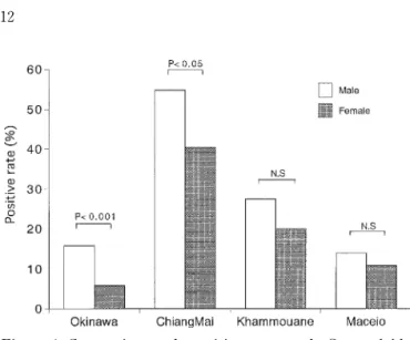
Fig. 1 shows sex difference of the positive rate in each area. The positive rate in males was consistently higher than that in female subjects. Especially in Okinawa, there was a great difference in positive rate between males and females; the rate in male subjects was three
‑times higher than that in females,this differ- ence being highly significant
(P
<0.001). On the other hand, sex difference of positive rate was not so signifi- cant in the other areas,although male positive rate was significantly higher in Chiang Mai
(P<0.05
).
D
ISCUSSIONThere have been several reports on the prevalence of Strongyloides infection in the countries surveyed here;
the positive rate reported ranged from 6% to 24% in Thailand
(Yamaguchi et al., 1982;Bayajian, 1992;Suk- havat,1994
),from 3% to 13% in Laos
(Sormmani et al., 1974;Chai et al., 1998
), and from 6% to 40% in Brazil
(Dias,1968;Asami,1970;Marzochi et al.,1978
),respec- tively. These results on prevalence of Strongyloides
infection, however, may extremely underestimate the true prevalence because of low efficacy of detection methods used in the past. In the previous study in which strongyloidiasis cases were reinfection
‑checked without treatment several months later, the conventional methods of fecal examination, such as direct smear, fecal concentration and Harada
‑Mori fecal culture methods,were only 15
‑24% effective in reconfirming the infection
(Sato et al.,1995
). On the other hand,an agar plate culture method, a newly developed fecal culture method, was confirmed to be much effective for detec- tion of chronic, low
‑level infection with Strongyloides.
In the present survey, therefore, the agar plate culture method was applied. The results indicate that Stron- gyloides infection was a major human public health problem in the survey areas. Especially in Chiang Mai, Thailand, positive rate as high as 47% was estimated among the subjects examined. The positive rate of 23%
in Khammouane, Laos, was also significantly higher than those reported in the past in this country. On the other hand, the positive rate in Okinawa, Japan, was greatly low as compared to those in the other countries.
One of the characteristic epidemiological features of strongyloidiasis in Okinawa, however, was observed in the age inclination of Strongyloides positive persons
(Sato, 1986;Sato et al., 1990
). Namely, most of Stron- gyloides positive people
(more than 95%)were included in middle to upper age groups over 40 years old in Okinawa. Only one positive case in 140 residents was found under the age of 40. Unfortunately, the subjects under 20 years old were not examined in this survey.
However, it was reported that no case of Strongyloides infection was found in 205 subjects under 20 years old in Okinawa
(Asato et al., 1983
). Whereas many young subjects were found infected with Strongyloides in the survey areas in Southeast Asia and South America. The extremely low incidence in the young generation in Okinawa suggests that the inhabitants have no opportu- nity to acquire new infection from environment in the island recently. From the epidemiological feature, Okinawa seemed to provide favorable field to investi- gate therapeutic efficacy of anthelmintics, because the follow
‑up examination can not be affected by reinfec- tion
‑infection after treatment. Although there have been many reports on therapeutic effect on stron- gyloidiasis, the results obtained were considerably dif- ferent from each other even in the case in which same drug was used in the similar regimen. The inconsistency may be attributed to the several factors. The most effective factor may be difference of applied methods in the follow
‑up fecal examination. Another factor affec-
Figure 1 Comparison of positive rate of Strongyloidesbetween male and female subjects in 4 survey areas.
ting therapeutic efficacy may be the duration of follow
‑up examination after the treatment. The Strongyloides infection has been known to relapse frequently in sev- eral months after seemingly successful treatment. The relapses due to unsuccessful treatment can not be distin- guished from reinfection from environment during the follow
‑up examination. On the basis of the above epidemiological background,the authors could examine the exact efficacy of several anthelmintics for as long as a year after treatment without consideration of reinfec- tion during the follow
‑up period
(Sato et al., 1992;
Takara et al., 1992;Toma et al., 1993
).
In Okinawa, the infections with soil
‑transmitted nematodes were highly prevalent until 1950ʼ s,but many of them have already been controlled. Ascariasis and trichuriasis were very rare in 1960ʼ s. Hookworm infec- tion was still observed in more than 30% of Okinawan people in 1960ʼ s, but the prevalence rate also decreased extremely to less than 0.1% in the following two decades
(Asato et al.,1990
). These changes are considered to be caused by the appropriate control measures, socio
‑eco- nomical and agricultural improvements. Apparently, most of Strongyloides carriers are also considered to acquire the infection during and
/or immediately after the World War II when the standard of living was very low and human excrements were used as fertilizer.
Long term of Strongyloides infection among aged people without new infection from environment may be due to internal autoinfection of the parasite. Therefore, the other epidemiological feature of strongyloidiasis in Okinawa seems to be that there is no concurrent infec- tion with other helminths occurred among the people with Strongyloides infection. Actually, no intestinal helminth infection other than Strongyloides was obser- ved in Okinawa in the present study, while helminth infections such as ascariasis and opisthorchiasis were concurrently detected in many subjects positive for Strongyloides infection in the other survey areas. This epidemiological feature may also be convenient for the investigation of immune response and clinical features of strongyloidiasis unaffected by concurrent infection with other helminth infection.
Finally, significant dominance of male subjects in prevalence of Strongyloides infection in Okinawa was also an epidemiological feature different from those in the other areas. In general,it has been recognized that males were more commonly infected with S. stercoralis than females
(Soroczan, 1976; Scagalia et al., 1984;
Walzer et al.,1982
). The infection rate in males was 3 times higher than that in females in Okinawa in the present study. One of the reasons for the high preva-
lence in males may be that males have had frequent opportunities for the infection in their life style. It is also possible to consider that males were more suscep- tible to the infection than females, as known in experi- mental infection model with S. ratti and animal hosts
(Katz, 1961; Dawkins et al., 1980
). In the previous study,the authors also demonstrated that the efficacy of treatment with various anthelmintics was significantly low in male subjects as compared to that in females
(Kobayashi et al., 1996
). Due to the significant resis- tance to treatment,male subjects harbor the parasite for many years, and the obstinacy in males might result in a significant accumulation of male subject positive for Strongyloides under the improved sanitary condition in which persons do not acquire new infection from envi- ronment.
In conclusion, Strongyloides infection in Okinawa showed a significant age inclination and sex dominance.
These epidemiological features may be derived from an environmental situation in which new infection from outside did not occur for many years,and also from the unique property of the parasite to maintain the infection for several decade years beyond the life span of the parasite. These epidemiological features in Okinawa are considered to provide many favorable conditions to investigate the parasite and its disease.
A
CKNOWLEDGEMENTSWe are especially grateful to Prof. C. Kham- boonruang, Chiang Mai University, and Prof. A.R.C.
Dacal, Escola de Ciencias Medicas de Alagoas for their generous help and arrangement. We thank the Institute of Malariology, Parasitology and Entomology( IMPE
), Lao PRD, and Japan International Cooperation Agency
(JICA) Laos Office for giving the opportunity of the study,and all stuff of malaria net work in Khammouane province, Lao PDR, for their cooperation in this study.
R
EFERENCES1)Arakaki, T., Hasegawa, H., Asato, R., Ikeshiro, T., Kinjo, F., Saito, A. and Iwanaga, M.(1988): A new method to detect Strongyloides stercoralis from human stool. Jpn. J. Trop. Med. Hyg., 16, 87 ‑90
2)Asami, K., Enomoto, Y. and Miura, S.(1970): Infesta- tions by ancylostomides and Strongyloides stercoralis in Pernambuco. Survey based on the identification of lar- vae. Rev. Inst. Med. trop. Sao Paulo, 12, 31‑35
3)Asato, R., Hasegawa, H., Takai, A. and Ikeshiro, T.
(1983): Epidemiological study of strongyloidiasis in Okinawa. Annual Report of Okinawa Prefectural Insti-
13
tute of Public Health, 17, 58‑63
4)Asato, R., Hasegawa, H. and Ikeshiro, T. (1990): Transition in the prevalence of intestinal parasitic infec- tions in Okinawa, Japan, after World War II.In: Col- lected Papers on the Control of Soil‑transmitted Hel- minthiasis, vol. IV, APCO Research Group(ed.), The Asian Parasite Control Organization (APCO), Tokyo, 39‑50
5)Asato, R., Nakasone, T., Yoshida, C., Arakaki, T., Ike- shiro, T., Murakami, H. and Sakiyama, M.(1992): Current status of Strongyloides infection in Okinawa, Japan. Jpn. J. Trop. Med. Hyg., 20, 169‑173
6)Bayajian,T.(1992): Strongyloidiasis on the Thai‑Cam- bodian border. Trans. Roy. Soc. Trop. Med. Hyg., 86, 661‑662
7)Chai,J.Y.and Bouasy,H.(1998): A small scale‑survey of intestinal helminthic infections among the residents near Pakse, Laos. Korean J. Parasitol., 36, 55 ‑58 8)Dawkins, H.J.S., Grove, D.I., Dunsmore, J.D. and Mit-
chell, G.F.(1980): Strongyloides ratti:susceptibility to infection and resistance to reinfection in inbred strains of mice as assessed by excretion of larvae. Int. J. Par- asitol., 10, 125‑129
9)Dias,J.C.P.(1968): Observacoes sobre a estrongiloidose no Oeste de Minas Gerais, Brasil. Rev. Inst. Med. trop.
Sao Paulo, 10, 305‑311
10)Katz, F.F.(1961): Differences in Strongyloides ratti worm burdens in male and female rats. J. Parasitol., 47 (Suppl.), 52
11)Kobayashi, J., Sato, Y., Toma, H., Tasaki, T., Takara, M. and Shiroma, Y.(1996): Sex‑related different effi- cacy of chemotherapy on human strongyloidiasis in Okinawa, Japan. Jpn. J. Parasitol., 45, 350 ‑354
12)Marzochi,M.C.and Carvalheiro,J.R.(1978): Studies on factors involved in the dissemination of intestinal para- sites:III.The distribution of some intestinal parasites in 2 social groups of Ribeirao Preto City, Sao Paulo, Brazil. Rev. Inst. Med. trop. Sao Paulo, 20, 31‑35 13)Sato, Y.(1986): Epidemiology of strongyloidiasis in
Okinawa.In: Collected Papers on the Control of Soil
‑transmitted Helminthiasis, vol. III, APCO Research Group(ed.), The Asian Parasite Control Organization (APCO), Tokyo, 20‑31
14)Sato, Y., Kobayashi, J., Toma, H. and Shiroma, Y.
(1995): Efficacy of stool examination for detection of Strongyloides infection.Am.J.Trop.Med.Hyg.,53,248 ‑ 250
15)Sato,Y.,Shiroma,Y.,Kiyuna,S.,Toma,H.and Kobaya- shi,J.(1992): Reduced effect of chemotherapy of stron- gyloidiasis in patients with concurrent HTLV‑1 infec- tion in Okinawa,Japan.Jpn.J.Trop.Med.Hyg.,20,183‑ 192
16)Sato, Y., Toma, H., Takara, M., Kiyuna, S. and Shir- oma, Y.(1990): Seroepidemiological studies on the concomitance of strongyloidiasis with T‑ cell leukemia viral infection in Okinawa.Jpn.J.Parasitol.,39,376 ‑383 17)Scaglia, M., Brustia, R., Gatti,S,Bernuzzi,A.M.,Stros-
selli,M.,Malfitano,A.and Capelli,D.(1984): Autochth- onous strongyloidiasis in Italy:An epidemiological and clinical review of 150 cases.Bull.Soc.Pathol.Exot.,77, 328‑332
18)Sornmani, S., Pathammavong, O. (1974): An epidemiological survey of human intestinal parasites in Vientiane, Laos. Southeast Asian J. Trop. Med. Public Health, 5, 541‑546
19)Soroczan,W.(1976):Strongyloides stercoralis in eastern and southeastern Poland. Wiad. Parazytol., 22, 51 ‑516 20)Sukhavat, K.(1994): Comparative efficacy of four
methods for the detection of Strongyloides stercoralis in human stool specimens.Ann.Trop.Med.Par.,88,95 ‑96 21)Takara, M., Toma, H., Kobayashi, J. and Sato, Y.
(1992): Effect of concurrent HTLV‑1 infection on the efficacy of pyrvinium pamoate treatment of stron- gyloidiasis. Jpn. J. Parasitol., 41, 202‑212
22)Toma,H.,Sato,Y.,Kobayashi,J.,Shiroma,Y.,Kiyuna, S., Takara, M. and Ohtomo, H.(1993): Treatment of strongyloidiasis with albendazole in Okinawa, Japan.
Jpn. J. Parasitol., 42, 300‑307
23)Walzer, P.D., Milder, J.E., Banwell, J.G., Kilgore, G., Klein,M.and Parker,R.(1982): Epidemiologic features of Strongyloides stercoralis infection in an endemic area of the United States.Am.J.Trop.Med.Hyg.,31,313 ‑319 24)Yamaguchi, T., Khamboonruang, C., Inaba, T., Huang,
W.H., Ishida, K., Fujimaki, Y., Asano, H., Thitasut, P.
and Vajrasthira, S.(1982): Studies on intestinal para- sitic infections in Chiang Mai Province,north Thailand.
Jpn. J. Parasitol., 31, 447‑459