β- tricalcium Phosphate/Collagen Composites Improve Bone Regeneration in Rat Calvarial Bone Defects
Yuichi TAKIGUCHI*, Yu KATAOKA and Takashi MIYAZAKI
Abstract : Autogenous bone grafting is the most widely accepted approach for repairing bone defects, yet limitations in the donor site and subsequent morbidity associated with harvesting bone from other sites remain as major concerns. Con- sequently, synthetic bone-like biocomposites have drawn much attention as a novel regenerative strategy. This study evaluated the regenerative properties of our pro- totype β-TCP/collagen composite in an animal model. We prepared the original β-TCP/collagen composite by mixing an acidic atelocollagen gel and alkaline col- loidal β-TCP, and Raman microspectroscopy of the composite revealed the typical spectral features attributable to bone after manual mixing. We then transplanted either the composite or collagen alone into a full-thickness trephine defect made in the calvarial bone of rats. At 8 weeks after implantation, the elastic modulus of regenerated bone that developed alongside the composite was comparable to that of native cortical bone. Decalcification and processing of the calvarial bones for histological observation revealed that the β-TCP/collagen enabled better bone regenerative properties compared to collagen alone. Our newly developed β-TCP/
collagen composite imitates the structural properties of bone, and thus provides a potentially useful scaffold material to support the mechanical integrity of regener- ated bone.
Key words : β- TCP/collagen composite, bone regeneration, biocompatibility, biode- gradability, osteoconductivity
Introduction
Bone regeneration is attracting increasing attention in the craniofacial, periodontal, and orthopedic fields. Bone grafts are used to replace or reconstruct critical-sized skeletal defects resulting from trauma, infection, cysts, or neoplasms in oral, maxillofacial, and reconstructive surgery, with allografts and autografts the most widely accepted strategies. Further, bone grafts involve osteogenic progenitor cells1, 2) and nonantigenic properties3), yet the inherent donor- site limitation and the potential risk of carrying infection4-6) necessitates the development of a synthetic bone-like biomaterial.
Bone is a natural biocomposite which primarily comprises inorganic calcium phosphates and a collagen matrix, while β- tricalcium (β-TCP) has excellent properties of biocompatibility, Original
Department of Conservative Dentistry, Division of Biomaterials and Engineering, Showa University School of Dentistry, 1-5-8 Hatanodai, Shinagawa-ku, Tokyo 142-8555, Japan.
* To whom corresponding should be addressed.
biodegradability, and osteoconductivity7-11). Accordingly, we have developed a β-TCP/collagen composite that is naturally synthesized and requires no additional treatment12, 13).
The bone-like β-TCP/collagen composite we developed might be useful for an effective bone substitute in bone defects. This study investigates bone regenerative properties of the β-TCP/
collagen composite in a critical defect of rat calvarial bone2, 14-17). Subsequent nanomechanical characterization enabled the qualification of regenerated bone associated with the composite.
Materials and methods Electrolytes
The composition of our electrolyte (mSBF)-generating β-TCP was documented previously12, 13). Preparation of colloidal β-TCP
A 10 10 0.1-mm platinum foil was connected to the cathode of a device developed in our laboratory, and a 50 100 0.1-mm platinum foil was used as the counter electrode. Each foil was immersed in 100 ml of mSBF. Discharge was generated between the electrolyte and the working electrode through a gas layer on the surface of the electrode, and then maintained at 2.5 A and 100 V for 270 s12, 13). β-TCP particles suspended in the electrolyte were precipitated and isolated, with the colloidal deposits prepared by removal of the supernatant. The pH values and particle diameters of the colloidal β-TCP were measured by a pH meter and scanning electron microscopy (SEM), respectively.
Preparation of β-TCP/collagen composite disc
Colloidal β-TCP (ml) was dispersed in the same volume of collagen solution (8 mg/ml, pH3.0; Nitta Gelatin, Osaka, Japan) and stirred for 30 s, immediately resulting in a shift to the gel phase. The β-TCP/collagen composite gel was then lyophilized at −80℃ for 24 h in a lyophilizer (FD-1000RE; Tokyo Rikakikai Co., Ltd., Tokyo, Japan), molded into 15 discs (9 mm diameter, 1 mm thickness, 30 mg weight), and then sterilized using γ-irradiation.
Scanning Electron Microscopy
The prepared discs were gold coated in a vacuum evaporation device (IB-2; Eiko Engineering, Ibaraki, Japan), and then observed by SEM (S-2360; Hitachi, Tokyo, Japan).
Raman spectroscopy
A Raman spectroscopy analyzer (RXN1, Kaiser Optical Systems Inc., Ann Arbor, MI, USA)
was used to measure the Raman spectra of the bone graft material (β-TCP/collagen composite). Electron probe microanalyzer (EPMA)
A sample of the β-TCP/collagen composite was also sputter coated with platinum-palladium and analyzed with an EPMA (EPMA1610, SHIMADZU, Kyoto, Japan) at 5,000 magnification.
Animal experiments
We used one 12-week-old male Wister rat, weighing approximately 250 g and kept in a metal cage at a constant temperature of 22℃ on a 12 h/12 h light/dark cycle. All procedures were approved by the Animal Research Committee of Showa University (2007; Approval Number:
2048983) and conducted according to the principles of laboratory animal care and the national laws.
Implantation procedure
The rat was anesthetized with intraperitoneal sodium pentobarbital at a dose of 50 mg/
kg body weight supplemented by ether inhalation. After shaving, an elliptical skin incision of approximately 20 mm in length was made in the scalp along the sagittal suture and continued down to the calvarium. The periosteum of the calvarium was ablated, and a critical-sized calvarial bone defect was surgically created using a trephine (h0891011; Micro Tech Co., Tokyo, Japan)14). A defect of 9 mm in diameter was carefully made in the calvarium including the mid- sagittal suture under continuous saline buffer irrigation to avoid injury to the dura mater, and a composite disc was implanted into the incision (Fig. 1). The ablated periosteum and skin were then sutured with nylon thread, and a postoperative antibiotic (Ampicillin; Gibco, Tokyo, Japan)
was administered. The rat was kept under observation until anesthesia recovery, and then returned to the cage.
Tissue preparations
The rat was sacrificed at 8 weeks after the operation and prepared for histology. The defect sites were removed along with the surrounding bone, fixed in 70% ethanol, and decalcified in 10% EDTA in 0.01 M PBS (pH7.4) for 4 weeks. The samples were dehydrated in ascending concentrations of ethanol and embedded in paraffin. Serial sections of 5-µm thickness was cut coronally (SM2000R; Leica, Jena, Germany) and stained with hematoxylin and eosin.
Photographs were taken with a photomicroscope (BX51; Olympus, Tokyo, Japan).
Fig. 1. Schematic illustration of animal surgery in the present study
Nanoindentation test
Nanoindentation experiments were performed on the regenerated bone with a diamond Berkovich tip attached to a quantitative nanomechanical test instrument (TI 950 TriboIndenter; Hysitron Inc., Minneapolis, MN, USA). To minimize errors arising from surface roughness, sufficiently smooth mineralized regions were chosen with a scanning range of 50 50µm and then 1 1µm. Fused quartz was used as the standard calibration material to determine the indenter tip area function and compliance of the instrument. The distance between indents was maintained at greater than 10µm to avoid any influence of residual stress from adjacent indentations. The thermal drift rate at a contact load of 2µN was monitored for 40 s prior to indentation to correct for thermal drift based on the time–mean drift rate over the previous 20 s.
The measured indentation data for each sample were assessed based on the effective tip area function in the contact depth range of 20–250 nm. The load applied to the sample was controlled using a closed-loop load-control algorithm, which involved initial withdrawal of the indenter tip from the surface to a set distance (lift height), and then determination of the re-captured sample surface based on a pre-load of 2µN. The reduced elastic modulus, Er, was calculated from the force-displacement curve using the standard unloading analysis procedure according to Equation (1), where S is the contact stiffness calculated as the slope of the unloading curve at the onset of unloading, and A is the projected area of the indenter tip as a function of contact depth.
Er=(S π)/(2 A) (1)
The elastic moduli of all samples were measured as a function of depth using the partial unloading technique, in which the load function involved 33 partial unloading cycles, each comprising a 1-s loading segment, a 1-s hold segment, and a 1-s unloading segment.
Results SEM
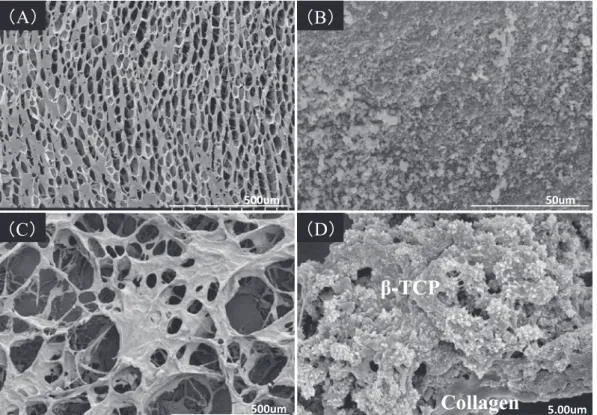
The apparent porosity of the β-TCP/collagen composite discs was tens of microns (Fig. 2A), while the length scale of the colloidal β-TCP particles was nanoscale-submicron (Fig. 2B), and the observed porosities of the composite at lower magnification were tens to hundreds of microns (Fig. 2C). At a high magnification, nanoscale β-TCP particles were clearly deposited on the micron-scale collagen fibers (Fig. 2D).
Raman microspectroscopy
Regarding the β-TCP/collagen composite, there seemed to be average spectral features attributable to bone. The composite showed two amide bands at around 1,665 cm-1 and 1,250cm-1 for amide I and amide Ⅲ, respectively. The peak at 960 cm-1 attributable to υ1 phosphate vibration mode was correlated to a calcium phosphate (Fig. 3).
Fig. 2. SEM images of collagen(A), β-TCP colloid(B), and β-TCP/collagen composites at low(C)
and high magnifications(D).
Fig. 3. A representative Raman spectrum from theβ-TCP/collagen composite, showing peaks around 1665 cm-1 for amide I and 1250 cm-1 for amide Ⅲ, The peak at 960 cm-1 is attributable to theυ1 phosphate vibration mode.
Chemical composition by EPMA
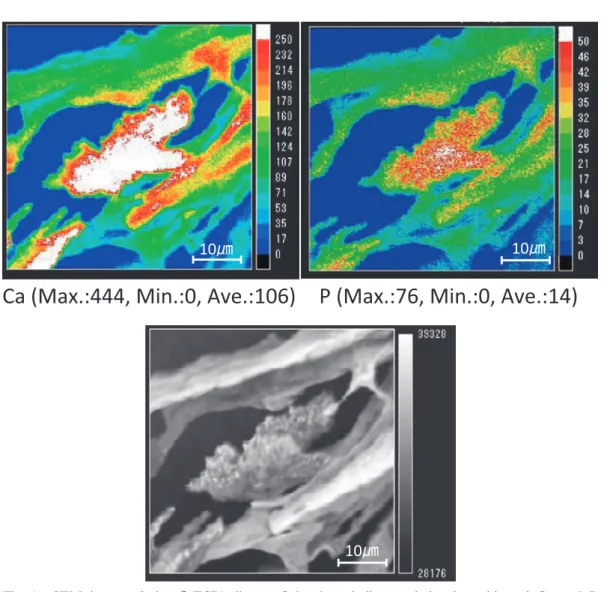
Both calcium and phosphate deposition were observed around a likely mineralized region on the β-TCP/collagen composite (Fig. 4).
Histology in tissue sections
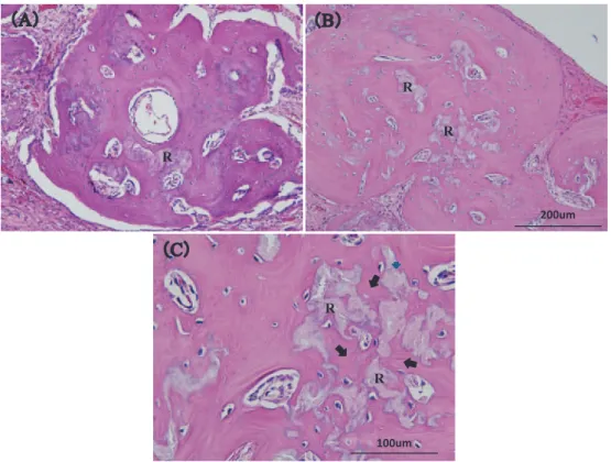
At 4 weeks after implantation, large amounts of composite were still observed (Fig. 5A), although these composite regions would likely be replaced by new bone over time (Fig. 5B and C). Elastic moduli of new bone evaluated by nanoindentation tests
The elastic modulus of the regenerated bone measured by nanoindentation tests was 8–10 GPa at 4 weeks (Fig. 6, red line), and 20–25 GP at 8 weeks (Fig. 6, blue line). The elastic moduli were almost constant between indenter contact depths of 100–200 nm.
Fig. 4. SEM image of the β-TCP/collagen. Color bars indicate relative intensities of Ca and P measured by EPMA. High intensities are expressed as red, whereas relatively lower intensities are blue.
Fig. 5. Tissue sections (20 magnification) of the bone defect implanted withβ-TCP/collagen composite at (A) 4 weeks and (B) 8 weeks. Tissue section (60 magnification) of the bone defect withβ-TCP/collagen composite (C) at 8 weeks. Growth of new bone
(arrow) into the remnantβ-TCP/collagen composite (R).
Fig. 6. The depth-dependent elastic moduli of the regenerated bone. red line: 4 weeks; blue line: 8 weeks. At least five indentation test data are merged and shown. Error bars show standard deviation of the all indentation data.
Discussion
Collagen is a widely used base scaffold material in tissue regeneration, although inorganic materials such as calcium phosphates are also needed in the mix to attain the mechanical properties sufficient to recover bone defects. Nevertheless, while calcium phosphate/collagen composites most closely mimic natural bone, they cannot be naturally synthesized without toxic chemical agents such as glutaraldehyde18, 19). Hence, naturally derived composites for bone repair and regeneration need to be developed.
We previously developed a naturally synthesized β-TCP/collagen composite12, 13, 20) that we predicted could provide improved bone regenerative potential while maintaining the required mechanical integrity based on fewer chemical cross-linking agents needed in preparing the composite. In this study, the elastic moduli of regenerated bone increased during implantation of our β-TCP/collagen composite into a rat bone defect. Assuming that the mineral density of regenerated bone increases as a function of time, these findings agree with theoretical predictions that the elastic modulus of mineralized tissue is entirely associated with bone mass or mineral density21). Indeed, at 8 weeks after implantation, the elastic modulus of regenerated bone was comparable to that of natural cortical bone. The primary requirement of bone as a supportive tissue is high stiffness due to mechanical integrity, and in this regard the quality of regenerated bone associated with our β-TCP/collagen composite was satisfactory.
The rate of bone regeneration depends partially on the biodegradability of the scaffold materials, so the newly formed bone can replace the space occupied by the scaffold22, 23). In this respect, reducing the diameter of β-TCP particles in the collagen matrix of the composite could allow a larger contact area resulting in a higher biodegradation rate and thus better regenerative properties in comparison with micron-scale granules or blocks of β-TCP.
During bone regeneration in a calvarial bone defect, the bone marrow, periosteum, or dura mater could provide osteoprogenitors able to differentiate into osteoblasts. In the present study, the source of osteoprogenitors might have been the periosteum, since newly formed bone was mainly observed beneath the periosteum of the calvarium away from the bone defect margin.
Conclusions
Our newly developed β-TCP/collagen composite demonstrated a high bone regenerative prop- erty at a critical defect in rat calvariae. The quality of regenerated bone seemed to be satisfac- tory, with an elastic modulus comparable to natural cortical bone.
Conflicts of interstt disclosure none.
References
1)Langer R, Vacanti JP. Tissue engineering. Science. 1993;260:920-926.
2)Kamakura S, Sasaki K, Honda Y, et al. Octacalcium phosphate combined with collagen orthotopically enhances bone regeneration. J Biomed Mater Res B Appl Biomater. 2006;79:210-217.
3)Sai S, Fujii K. beta-tricalcium phosphate as a bone graft substitute. Jikeikai Med J. 2005;52:47-54. (in Japanese). 4)Le Geros, RZ. Properties of osteoconductive biomaterials: calcium phosphates. Clin Orthop Relat Res. 2002;395:81-98.
5)Hirota M, Matsui Y, Mizuki N, et al. Combination with allogenic bone reduces early absorption of beta-tricalcium phosphate (beta-TCP) and enhances the role as a bone regeneration scaffold. Experimental animal study in rat mandibular bone defects. Dent Mater J. 2009;28:153-161.
6)Takamoto A, Tokikazu T, Dainobu K, et al. Questionnaire survey about uneasiness and satisfaction to bone aug- mentation surgery for oral implant. J Hiroshima Univ Dent Soc. 2012;44:29-35. (in Japanese).
7)Saito M, Shimizu H, Beppu M, et al. The role of beta-tricalcium phosphate in vascularized periosteum. J Orthop Sci. 2000;5:275-282.
8)Oyake Y, Beppu M, Ishii S, et al. Intramedullary anchoring strength of titanium rod with mixed beta-tricalcium phosphate and fibrin adhesive. J Orthop Sci. 2002;7:123-130.
9)Matsuno T, Nakamura T, Kuremoto K, et al. Development of beta-tricalcium phosphate/collagen sponge composite for bone regeneration. Dent Mater J. 2006;25:138-144.
10)Sugawara A. Kotsusaisei no tekunoroji -kotsusaisei no gainen to rinsho oyo-. rev ed. Tokyo: Zenith publication;
2011. (in Japanese).
11)Ogose A, Hotta T, Hatano H, et al. Histological examination of beta-tricalcium phosphate graft in human femur. J Biomed Mater Res. 2002;63:601-604.
12)Shibata Y, Yamamoto H, Miyazaki T. Colloidal beta-tricalcium phosphate prepared by discharge in a modified body fluid facilitates synthesis of collagen composites. J Dent Res. 2005;84:827-831.
13)Takashima H, Shibata Y, Kim TY, et al. Hydroxyapatite coating on a titanium metal substrate by a discharging method in modified artificial body fluid. Int J Oral Maxillofac Implants. 2004;19:66-72.
14)Bosch C, Melsen B, Vargervik K. Guided bone regeneration in calvarial bone defects using polytetrafluoroethylene membranes. Cleft Palate Craniofac J. 1995;32:311-317.
15)Sasano Y, Kamakura S, Homma H, et al. Implanted octacalcium phosphate (OCP) stimulates osteogenesis by osteoblastic cells and/or committed osteoprogenitors in rat calvarial periosteum. Anat Rec. 1999;256:1-6.
16)Dupoirieux L, Neves M, Pourquier D. Comparison of pericranium and eggshell as space fillers used in combina- tion with guided bone regeneration: an experimental study. J Oral Maxillofac Surg. 2000;58:40-46.
17)Rojbani H, Nyan M, Ohya K, et al. Evaluation of the osteoconductivity of alpha-tricalcium phosphate, beta-tricalcium phosphate, and hydroxyapatite combined with or without simvastatin in rat calvarial defect. J Biomed Mater Res A. 2011;98:488-498.
18)Chang MC, Tanaka J. FT-IR study for hydroxyapatite/collagen nanocomposite cross-linked by glutaraldehyde. Bio- materials. 2002;23:4811-4818.
19)Lickorish D, Ramshaw JA, Werkmeister JA, et al. Collagen-hydroxyapatite composite prepared by biomimetic process. J Biomed Mater Res A. 2004;68:19-27.
20)Ata F, Kataoka Y, Tamaki Y, et al. Micro-CT based quantitative evaluation of in vivo bone regeneration with collagen-based biomaterials. Dent Med Res. 2011;31:24-27.
21)Felsenberg D, Boonen S. The bone quality framework: determinants of bone strength and their interrelationships, and implications for osteoporosis management. Clin Ther. 2005;27:1-11.
22)Kamakura S, Sasaki K, Homma T, et al. The primacy of octacalcium phosphate collagen composites in bone regeneration. J Biomed Mater Res A. 2007;83:725-733.
23)Le Guehennec L, Goyenvalle E, Aguado E, et al. MBCP biphasic calcium phosphate granules and tissucol fibrin sealant in rabbit femoral defects: the effect of fibrin on bone ingrowth. J Mater Sci Mater Med. 2005;16:29-35.
[Received July 31, 2018 : Accepted September 20, 2018]