• 検索結果がありません。

Development of a high-intensity focused ultrasound exposure device for reducing skin burn risk

N/A
N/A
Protected

Academic year: 2021

シェア "Development of a high-intensity focused ultrasound exposure device for reducing skin burn risk "

Copied!
25
0
0

読み込み中.... (全文を見る)

全文

(1)

Development of a high-intensity focused ultrasound exposure device for reducing skin burn risk

Shogo Nishii

1

, Kohei Seo

1

, Aleksander Tatsuya Izdebski

1

, Miki Kushima

2

, Ryo Takagi

3

, Shin Yoshizawa

4

, Shin-ichiro Umemura

4

, Kiyotake Ichizuka

1

and Akihiko Sekizawa

1

1

Department of Obstetrics and Gynecology, School of Medicine, Showa University, Tokyo 142-8555, Japan

2

Department of Pathology, Showa University Koto Toyosu Hospital, Tokyo 135-8577, Japan

3

Theranostic Device Research Group, The National Institute of Advanced Industrial Science and Technology, Ibaraki 305-8564, Japan

4

Graduate School of Biomedical Engineering, Tohoku University, Miyagi 980-8579, Japan

Corresponding author Kohei Seo

Address: 1-5-8 Hatanodai, Shinagawa-ku, Tokyo, 142-8555, Japan Phone: +81-3-3784-8551

Fax: +81-3-3784-8355

E-mail: [email protected]

(2)

(Running title: HIFU system for reducing skin burn) Abstract

Purpose: High-intensity focused ultrasound (HIFU) can non-invasively irradiate inside the body. However, when used to treat fetuses, it can cause thermal burns in the mother’s abdominal wall at the skin interface. This study was carried out to determine whether a modified HIFU transducer enabling split-aperture irradiation can prevent thermal burns.

Methods: Two HIFU transducers were compared: a conventional transducer for full-aperture irradiation and a modified transducer enabling split-aperture irradiation. The modified transducer was divided into six sectors for split- aperture irradiation and had a larger surface area and a smaller F number (focal length/aperture diameter) than the unmodified one. HIFU was delivered to eight sites on the left and right legs of a three-month-old baby pig under general anesthesia, and the sites were assessed for thermal burning by two or more dermatologists. The same person performed all irradiations.

Results: Full-aperture irradiation with the conventional transducer caused deep dermal burns at all target sites, while split-aperture irradiation with the modified transducer caused only epidermal burns or superficial dermal burns.

Conclusion: Split-aperture irradiation with the modified HIFU transducer

having six sectors and a smaller F number reduced the severity of skin

(3)

Keywords: High-intensity focused ultrasound, Exposure protocol, Burn injuries

Introduction

Although improvements in prenatal diagnosis have increased the likelihood of identifying abnormalities in fetuses, there are a limited number of

effective strategies for treating the abnormalities, particularly in a non- invasive manner. To improve this situation, various new medical devices have been and are being developed, along with advanced control methods.

Before using these devices in clinical settings, the possibility of damage to the patient should be considered and assessed. Because many fetal

treatments involve the insertion of a device into the uterus from outside the body, treatment-related complications and infections can occur.

High-intensity focused ultrasound (HIFU) treatment uses a powerful

ultrasonic wave from a transmission source (transducer). Ultrasonic energy

is concentrated in the vicinity of a focal point in the target tissue, converted

to heat, and thermally coagulates the tissue (1, 2). HIFU has been used for

several categories of treatment, such as for the treatment of solid cancers,

including liver and prostate cancers (3–6). We used HIFU to treat twin

reversed arterial perfusion sequence for the first time in 2013 (7, 9). The

treatment had little influence on the intervening tissues outside the area of

focus although HIFU exposure duration had to be limited in order to avoid

possible skin burn of the mother. It was reported that skin burn injuries were

(4)

observed in 0.29% of the 27,053 patients who received HIFU treatment for benign uterine diseases (8).

HIFU can non-invasively irradiate the inside of the body, rendering it suitable for fetal treatment. The therapeutic actions of ultrasound are

attributable to its thermal and non-thermal effects. The thermal effect of ultrasound occurs when ultrasonic energy is absorbed and converted into thermal energy. Depending on the tissue absorption coefficient, it is possible to thermally induce tissue coagulation necrosis via HIFU irradiation for several seconds by raising the focal temperature to 60°C or higher (2).

The non-thermal effect of ultrasound typically occurs in the existence of microbubbles whether they have been generated in situ via acoustic

cavitation or transported from somewhere else. It has been reported that they can even enhance the thermal effect of ultrasound (15). Ultrasonically

activated microbubbles cause tissue and cellular damage, including the destruction of cell membranes and breakdown of capillary blood vessels. In addition to thermal coagulation necrosis and degeneration, HIFU can cause tissue destruction via the above mentioned mechanism, which can be an advantage of HIFU treatment as long as it is properly localized.

It has also been reported that acoustic cavitation can be induced more easily in a standing wave than in a progressive wave field because

microbubbles smaller than under the resonant size migrate toward the

(5)

through coalescence and rectified diffusion (14). During HIFU exposure, HIFU waves can be reflected by the interfaces of the coupling water bag as well as the biological tissue boundaries intervening between the transducer and the focus, resulting in standing wave components in the HIFU field. We hypothesized that such standing wave components had been one of the primary causes of the skin burn due to HIFU exposure. The hypothetical scenario is that cavitation occurs in the vicinity of the intervening skin due to standing wave components of the HIFU field and then the cavitated

microbubbles locally enhances the heating effect of HIFU, which will cause skin burn.

Based on this hypothesis, two types of HIFU transducers were prototyped: a conventional HIFU transducer and a modified HIFU transducer enabling split-aperture exposure. It takes time in the order of milliseconds for a microbubble to grow to the resonant size by the effect of standing waves, whose duration can be controlled by the exposure duration.

Therefore, the modified transducer was designed to enable intermittent exposure at the intervening tissues while maintaining continuous exposure at the HIFU focus. The biological effect of conventional continuous exposure using the conventional HIFU transducer and that of intermittent split-

aperture exposure using the modified transducer were compared in an

animal experiment in this paper.

(6)

Materials and Methods

HIFU transducer and system

Two HIFU transducers using PZT (C-213, Fuji Ceramic, Fujinomiya, Japan) were prototyped. Both air-backed transducers in aluminum housing had a central hole 34 mm in diameter reserved for an imaging probe. The

conventional HIFU transducer (Figure 1a) 78 mm in diameter had a

spherical curvature radius of 75 mm. The modified HIFU transducer (Figure 1b) 100 mm in diameter had a spherical curvature radius of 85 mm. Its aperture was divided into six sectors to allow split-aperture exposure as shown in Figure 2. In this exposure sequence, the most tissues intervening between the transducer and the focus are exposed intermittently while the tissue at the focus is exposed continuously.

The transducer with an imaging probe was sealed with a latex-free ultrasound probe cover (sterile, 17.8 × 147 cm, telescopically folded cover;

Civ-Flex; Civco, Kalona, IA, USA) with circulating water inside, degassed and cooled below 20°C by a Sonachill cooling system (Sonablate 500;

SonaCare Medical, LLC, Charlotte, NC, USA). The conventional HIFU

transducer was driven by a radiofrequency amplifier (RF Power Amplifier

Model 1040L; E&I, Rochester, NY, USA) amplifying a sine wave from a

function generator (WF1974, NF Corp., Yokohama, Japan). Each three of

(7)

the six sectors of the modified HIFU transducer was driven by such an amplifier.

Laboratory animal experiment

The experiments were conducted using a 3-month-old baby pig (Landrace × Large White × Duroc three-way cross-head, weight: 35 kg). General

anesthesia was administered with the animal in the supine position. The epidermis of the pig’s thigh was regarded as a model of the epidermis of the mother’s body, and the pig bristles were shaved. Blood pressure, pulse rate, respiration rate, and blood oxygen concentration were continuously

measured before and after irradiation. The pig was sacrificed by intravenous potassium chloride drip and necropsied after death; the muscle and skin were carefully examined for gross pathological lesions. The experimental

protocols were approved by the Institutional Animal Care and Use

Committee (Approval Number:IVT17-11), which operates in accordance with the Japanese Government for the care and use of laboratory animals.

HIFU irradiation method

Each HIFU transducer was driven at an instantaneous acoustic power of 200

W and a frequency of 1.1 MHz for 10 s. These parameters were chosen

based on the previous basic and clinical research studies (10). The ultrasonic

energy had been sufficient to denature tissue (7, 9, 11, 12). Each of the three

(8)

of the six sectors of the modified transducer was driven alternately one after the other with a switching period of 50 µs. This irradiation sequence is referred to as the split-aperture irradiation, while the other is the full- aperture irradiation. In both irradiation sequences, a high-intensity short pulse at an instantaneous acoustic power of 760 W for 0.1 ms was irradiated every 100 ms to generate cavitation around the focal point. Even in the split- aperture sequence, the full aperture was used for this short pulse irradiation.

The same operator performed all irradiations, which were separately delivered to the left and right legs (Figure 3).

Evaluation method

Gross thigh skin evaluation. We irradiated eight randomly chosen sites on the femoral skin: four on the right and four on the left side of the animal.

The irradiated femoral skin was excised, along with the muscle, and subsequently examined by two or more dermatologists.

Histological evaluation of the thigh. A total of eight sites in the skin and

muscle were histologically evaluated. The tissue specimens were stained

with hematoxylin and eosin.

(9)

Evaluation of complications. Blood pressure, pulse rate, respiratory rate, and blood oxygen concentration were analyzed for irradiation-related changes.

The experimental protocols were conducted in compliance with the Animal Management Act, under the approval of the Institutional Animal Care and Use Committee.

Results

Gross thigh skin evaluation

In the full-aperture protocol, white lesions near the hardened areas of all irradiation sites were observed, as was redness in the surrounding tissue. The white lesions were determined to be deep tissue burn injuries (Figure 4). In the split-aperture protocol, scattered white lesions were observed in some areas, and all sites had epidermal burns or superficial dermal burns (Table 1).

Histological evaluation of the thigh skin

After full-aperture irradiation, heat denaturation was observed in almost all

layers of the dermis, and the capillaries in the dermis exhibited vacuolar

degeneration (Figure 5). After split-aperture irradiation, heat denaturation

(10)

was observed in the upper half of the dermis only, and there was no vacuolar degeneration of the blood vessels.

Evaluation of complications. Blood pressure, pulse rate, respiratory rate, and arterial oxygen saturation in the pig before and after HIFU exposure were similar for full and split irradiation.

Discussion

The pathology of the tissue specimens was reviewed, and the burns were diagnosed by two or more dermatologists. As a result, the full-aperture exposure using the conventional HIFU transducer caused deep dermal burns in all irradiated areas, while the split-aperture exposure using the modified transducer did not, and at basically the same ultrasonic power at the same frequency delivered to the focal region, which could form similar focal lesions. This result is consistent with the proposed hypothesis that

microbubbles were generated in the vicinity of the intervening skin by the standing wave components of the conventional HIFU field and locally enhanced the heating effect, causing skin burn; therefore, the skin burn will be decreased by suppressing sanding wave formation.

In the split-aperture exposure, a period of 50 µs was chosen for

alternating irradiation using each three of the six sectors of the modified

(11)

between the transducer and the focus, standing waves due to the overlap of more than two components will occur if the ultrasonic pulse train is longer than the round-trip distance between the reflector and the transducer. The switching period of 50 µs, corresponding to a pulse train length of 75 mm, was chosen so that it could be shorter than the round-trip distance expected to be around the focal length, which was 85 mm for the modified transducer.

The F number of the modified transducer was 0.85, while it was 0.96 for the conventional transducer. The smallness of the F number of the modified transducer may have also contributed to suppressing standing waves. Assuming a semi-planar reflecting boundary such as skin, a standing wave field can be formed easily by plane waves. It is reasonable to

understand that a standing wave field can be formed by the focused field with a smaller F number less easily than that with a larger F number.

Regarding pathological evaluations, it is known that when HIFU irradiation is applied to blood vessels, the cells of the vessel wall become vacuolated (12). After the full-aperture irradiation, we observed vacuolar degeneration in the blood capillaries of the true skin, which may have been caused by cavitation in vessel walls. In contrast, with the split-aperture irradiation, there was no vacuolar degeneration in the true skin blood capillaries. Based on these findings, the extent of the burn injury was clearly reduced by the split-aperture irradiation. Under the same power and conditions, we

irradiated the kidney of the pig percutaneously. After both full-aperture and

(12)

split-aperture irradiation, similar reddish lesions corresponding to each HIFU focus were observed in the kidney as shown in Figure 6. This

proposed irradiation protocol using the modified HIFU transducer may have immense potential as the next generation HIFU treatment.

The present study has some limitations. The radiation targets were pig skin, fat, and muscles, which differ from human counterparts. In addition, human HIFU therapy is performed while the patient is conscious. Because our experiment was conducted under general anesthesia, the influence of muscle relaxants, body movements, and circulation must be considered.

Another limitation is the lack of an assessment of the efficacy of the

transducer in detail; although burn injuries were reduced, the definitive goal is the complete prevention of burns. Thus, further improvements in the transducer and irradiation protocol may be needed.

In conclusion, the proposed split-aperture irradiation protocol reduced the severity of skin thermal burns in this animal experiment. Using the modified HIFU transducer and adjusting the HIFU irradiation protocol, the risk of skin burns will be reduced also in human subjects. Thus, this

combination is proposed for the next- generation HIFU treatment in the

clinical setting.

(13)

Acknowledgment

We gratefully acknowledge the work of the past and present members of our laboratory.

Ethical statement

All institutional and national guidelines for the care and use of laboratory animals were followed.

Conflicts of interest

Shogo Nishii, Kohei Seo, Aleksander Tatsuya Izdebski, Ryo Takagi, Shin

Yoshizawa, Shin-ichiro Umemura, Akihiko Sekizawa, and Kiyotake

Ichizuka declare that they have no conflicts of interest.

(14)

References

1. Dubinsky TJ, Cuevas C, Dighe MK, et al. High-intensity focused

ultrasound: current potential and oncologic applications. Am J Roentgenol.

2008;190:191–199.

2. Sofuni A, Moriyasu F, Sano T, et al. The current potential of high-intensity focused ultrasound for pancreatic carcinoma. J Hepatobiliary Pancreatic Sci. 2011;18:295–303.

3. Chaussy CG, Thuroff SF. Robotic high-intensity focused ultrasound for prostate cancer: what have we learned in 15 years of clinical use. Curr Urol Rep. 2011;12:180–187.

4. Rebillard X, Gelet A, Davin JL, et al. Transrectal high-intensity focused ultrasound in the treatment of localized prostate cancer. J Endourol.

2005;19:693–701.

5. Stewart EA, Rabinovici J, Tempany CM, et al. Clinical outcomes of focused ultrasound surgery for the treatment of uterine fibroids. Fertil Steril.

2006;85:22–29.

6. Goldberg SH, Cohen MS, Young M, et al. Thermal tissue damage caused by ultrasonic cement removal from the humerus. J Bone Joint Surg.

2005;87:583–591.

7. Okai T, Ichizuka K, Hasegawa J, et al. First successful case of non-invasive

in-utero treatment of twin reversed arterial perfusion sequence by high-

(15)

8. Liu Y, Zhang WW, He M, et al. Adverse effect analysis of high-intensity focused ultrasound in the treatment of benign uterine diseases. Int J Hyperthermia. 2018;35:56–61.

9. Ichizuka K, Hasegawa J, Nakamura M, et al. High-intensity focused ultrasound treatment for twin reversed arterial perfusion sequence.

Ultrasound Obstet Gynecol. 2012;40:476–478.

10. Seo K, Ichizuka K, Okai T, et al. Evaluation of second-generation HIFU systems: less-invasive fetal therapy for TRAP sequence. Showa Univ J Med Sci. 2017;29:241–251.

11. Ichihara M, Sasaki K, Umemura SI, et al. Blood flow occlusion via

ultrasound image-guided high-intensity focused ultrasound and its effect on tissue perfusion. Ultrasound Med Biol. 2007;33:452–459.

12. Ishikawa T, Okai T, Sasaki K, et al. Functional and histological changes in rat femoral arteries by HIFU exposure. Ultrasound Med Biol. 2003;29:1471–

1477.

13. Azuma T, Kawabata K, Umemura S, et al. Bubble Generation by Standing Wave in Water Surrounded by Cranium with Transcranial Ultrasonic Beam.

Japanese Journal of Applied Physics. 2005;44:4625–4630.

14. Umemura S, Kawabata K, Sasaki K, et al. In vivo acceleration of ultrasonic

tissue heating by microbubble agent. IEEE Transactions on Ultrasonics,

Ferroelectrics, and Frequency Control. 2005;52:1690–1698.

(16)

15. Gnanaskandan A, Hsiao C-T, Chahine, G. Modeling of Microbubble- Enhanced High-Intensity Focused Ultrasound. Ultrasound in Medicine &

Biology. 2019;45:1743–1761.

(17)

Table 1. Dermal findings according to irradiation protocol Exposure site Irradiation

protocol ED SDB DDB

1 Full ○

2 Full ○

3 Full ○

4 Full ○

5 Split ○

6 Split ○

7 Split ○

8 Split ○

ED: epidermal burn, SDB: superficial dermal burn, DDB: deep

dermal burn

(18)

Figure Captions

Fig. 1. The unmodified and modified second-generation high-intensity focused ultrasound (HIFU) transducers. a: The unmodified unit. The

imaging probe was located in the coaxial radiation area, and the diameter of the transducer was 78 mm. b: The modified HIFU unit. The diameter of the modified HIFU transducer was 100 mm. The F number of the modified HIFU transducer was improved from 1.00 to 0.85, and the transducer was divided into six sectors to split the ultrasonic exposure pulse. c: Making the surface area in a skin interface large made the thermal burn in the skin reduce without changing a focal distance. F ; focal length , D ; aperture diameter, F number : focal length/aperture diameter

Fig. 2. Full and split irradiation. The total acoustic power of the high- intensity focused ultrasound (HIFU) was 200 W, the frequency was 1.1 MHz, and the irradiation time was 10 s. These settings were the same for both irradiation protocols.

a. Full irradiation.

b. Split irradiation.

The modified HIFU transducer was divided into six sectors to split the

ultrasonic irradiation pulse. The path of the ultrasonic exposure was changed

(19)

Fig. 3. High-intensity focused ultrasound (HIFU) irradiation. General anesthesia was administered in with the animal in the supine position. Full irradiation and split irradiation were delivered to four sites on the left and four sites on the right internal region of the posterior limbs (within the yellow squares). All exposures were performed by the same person.

Fig. 4. Injuries at the irradiated sites. Red circles: Sites that received full irradiation. Blue circles: Sites that received split irradiation.

Fig. 5. Histopathologic evaluation. a. Heat denaturationin all layers of the dermis after full irradiation. Hematoxylin and eosin, ×4. b. Heat denaturation in the upper half of the dermis after split irradiation. Hematoxylin and eosin,

×4. c. Vacuolar degeneration (yellow arrow) in the blood capillaries of the dermis after full irradiation. Hematoxylin and eosin, ×40. d. There was no vacuolar degeneration in the blood capillaries of the dermis after split irradiation. Hematoxylin and eosin, ×40.

Fig. 6. Kidney evaluation. Reddish lesions were observed for both full-

aperture irradiation (arrow ‘a’) and split-aperture irradiation (arrow ‘b’).

(20)

Fig. 1

78 mm

100 mm

a b

c

(21)

Fig. 2

a. Full irradiation.

b. Split irradiation.

a

b

50 μs

(22)

Fig. 3

(23)

Fig. 4

(24)

Fig. 5

a b

c d

(25)

Fig 6

Table 1.  Dermal findings according to irradiation protocol  Exposure site  Irradiation
Fig. 2  a. Full irradiation. b. Split irradiation.a  b  50 μs

参照

関連したドキュメント

It is suggested by our method that most of the quadratic algebras for all St¨ ackel equivalence classes of 3D second order quantum superintegrable systems on conformally flat

We show that a discrete fixed point theorem of Eilenberg is equivalent to the restriction of the contraction principle to the class of non-Archimedean bounded metric spaces.. We

[3] Chen Guowang and L¨ u Shengguan, Initial boundary value problem for three dimensional Ginzburg-Landau model equation in population problems, (Chi- nese) Acta Mathematicae

We present sufficient conditions for the existence of solutions to Neu- mann and periodic boundary-value problems for some class of quasilinear ordinary differential equations.. We

It turns out that the symbol which is defined in a probabilistic way coincides with the analytic (in the sense of pseudo-differential operators) symbol for the class of Feller

Then it follows immediately from a suitable version of “Hensel’s Lemma” [cf., e.g., the argument of [4], Lemma 2.1] that S may be obtained, as the notation suggests, as the m A

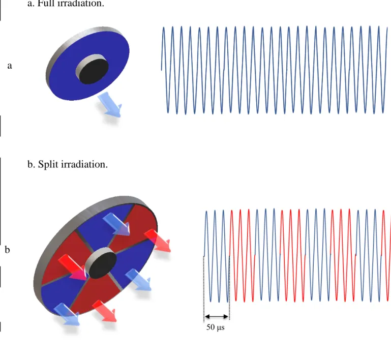
While conducting an experiment regarding fetal move- ments as a result of Pulsed Wave Doppler (PWD) ultrasound, [8] we encountered the severe artifacts in the acquired image2.

We now prove that the second cohomology groups of irreducible peculiar modules which are not mentioned in the formulation of theorem 1.1 are trivial.. The lists of highest weights