日呼吸誌 3(4),2014
緒 言
Epidermal growth factor receptor(EGFR)遺伝子変 異が発見され,EGFR 変異肺癌に対するゲフィチニブ
(gefitinib)・エルロチニブ(erlotinib)といった EGFR チロシンキナーゼ阻害薬(EGFR-TKI)の効果が注目さ れている
1)2).日本において EGFR 変異肺癌は肺腺癌の 約 40%に認められ,肺癌治療成績の向上において重要 である
3).しかしながら,EGFR-TKI による耐性の問題 も指摘されている
4)5).EGFR-TKI の耐性機序として T790M 変異・MET 遺伝子増幅の頻度が高いが,腺癌 からの小細胞癌化の報告もされている
6)〜8).今回,我々 は肺腺癌の小細胞癌化を認めた 1 例を経験したので報告 する.
症 例 患者:59 歳,女性.
主訴:腹部膨満.
既往歴:なし.
生活歴:喫煙歴 20 本/日×35 年.
現病歴:2011 年 10 月労作時呼吸困難を主訴に国立国 際医療研究センター病院を受診した.胸部 X 線写真にて 右胸水,胸部 CT にて右肺に腫瘤影を認めた.また頭部
MRI にて右大脳半球に浮腫を伴う転移がみられた(Fig.
1A〜C).血液検査にて CEA 2,694 ng/ml,NSE 58.4 ng/
ml と上昇を認めた.局所麻酔下胸腔鏡検査を施行し,
壁側胸膜に多数の腫瘤を認めた(Fig. 1D).生検を行い 病理結果から腺癌を認め CEA,Napsin A,TTF-1 が陽 性であり,肺腺癌 T3N2M1b(脳転移)Stage IV EGFR 遺伝子変異陽性(Exon19 E746-A750 欠失)と診断した.
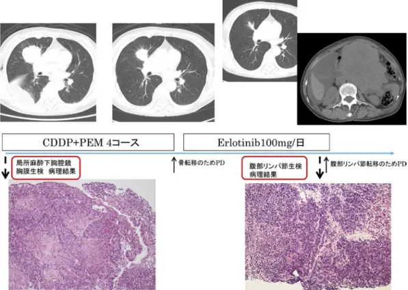
脳転移に対して全脳照射 30 Gy 施行,化学療法としてシ スプラチン(cisplatin,75 mg/m
2)+ペメトレキセド
(pemetrexed,500 mg/m
2)を 4 コース施行した.4 コー ス終了後の評価にて胸部病変は 48%の縮小を認めたが,
骨盤・右大腿骨転移が出現し progressive disease(PD)
と判断した.その後エルロチニブ(erlotinib)に変更,
国立国際医療研究センター病院における臨床研究への参 加了解を得て,100 mg/日で内服開始した.胸部病変な どは縮小傾向であり,内服開始から 3ヶ月の時点で標的 病変は−26%の stable disease(SD)であった.しかし erlotinib の内服を開始してから 4ヶ月後に腹部膨満感が 出現し,腹部 CT にて腹部リンパ節腫大を認めた.精査 目的に 2012 年 11 月入院となった.
入院時現症:performance status(PS)2(腹部膨満),
体温 37.1℃,脈拍数 86 回/min,血圧 121/72 mmHg,
経皮的動脈血酸素飽和度 98%(室内気),呼吸数 15 回/
min.呼吸音:異常なし,心音:異常なし,腹部:心窩 部に腫瘤を触知.
入院時検査所見:腫瘍マーカーは CEA 2.4 ng/dl と正 常であったが,NSE 178 ng/ml,Pro-GRP 92 pg/ml と 高値であった.生化学所見では総ビリルビン 3.5 mg/dl,
直接ビリルビン 2.3 mg/dl,AST 226 U/L,ALT 97 U/L
●症 例
肺腺癌の小細胞癌化を認めた 1 例
正木 晴奈 石井 聡 竹田雄一郎 渡辺 翔 放生 雅章 杉山 温人
要旨:症例は 59 歳,女性.主訴は腹部膨満.肺腺癌 T3N2M1b(脳転移,悪性胸水)Stage IV EGFR 遺伝 子変異陽性(Exon19 E746-A750 欠失)に対して,抗癌剤治療(cisplatin+pemetrexed → erlotinib)など を行った.胸部病変は制御できたが腹部リンパ節転移の出現を認めた.リンパ節生検にて TTF-1 陽性・
synaptophysin 陽性であり,肺腺癌小細胞癌化が疑われた.小細胞癌に対する抗癌剤治療を行うことにより,
リンパ節の縮小を認めた.肺腺癌の小細胞癌化に関して文献的考察を含め報告する.
キーワード:EGFR チロシンキナーゼ阻害薬,小細胞癌化,耐性化機序
EGFR-TKI, Transformation to SCLC, Resistance-gaining mechanism
連絡先:正木 晴奈
〒162‑8655 東京都新宿区戸山 1‑21‑1 国立国際医療研究センター病院呼吸器内科
(E-mail: [email protected])
(Received 18 Sep 2013/Accepted 18 Feb 2014)
541
と肝機能障害を認め,腹部リンパ節腫大の影響と考えら れた.また BUN 14 mg/dl,Cre 1.6 mg/dl と腎機能低下 を認めた.
画像所見:胸部 X 線写真:2011 年 10 月の胸部 X 線 では右胸水貯留を認めていたが,2012 年 11 月入院時は 結節影のみ認めた(Fig. 2A).胸腹部 CT:胸部 CT で は右中葉に結節影を認めた(Fig. 2B).腹部 CT では腹 腔内に長径12.7 cmの腫瘤を認め,腹水も伴っていた(Fig.
3A).
入院後経過:経皮的に腹部リンパ節生検を施行した.
病理組織診において腫瘍細胞は小さく,胞巣形成を伴い,
核は円形で細胞質は少なく,核小体は目立たなかった.
TTF-1 陽性・synaptophysin 陽性・chromogranin 陽性 であった.またリンパ節生検で得た検体の EGFR 遺伝 子変異を調べたところ,陽性(Exon19 E746-A750 欠失)
であった.T790M は陰性であった.以上の結果から,
肺腺癌の小細胞癌化の診断に至った.腹部リンパ節腫大 に伴う腹部膨満が増強したため化学療法を追加する方針 となり,erlotinib(100 mg/日)+カルボプラチン[car- boplatin,area under the curve(AUC)=5]+エトポ シド(etoposide,80 mg/m
2)による治療を開始した.2 コース施行後,腹部リンパ節は 12.7 cm から 10.4 cm と 縮小傾向を示し,また腹水も減少した(Fig. 3B).リン パ節の縮小により肝機能は改善傾向となったが,骨髄抑 制が遷延し次の治療を開始するまでに 6 週間を要し,そ の間に腹部リンパ節は再増大した.3 コース目の抗癌剤 治療を検討したが,PS が 4 となったため抗癌剤治療は 終了し,best supportive care となった.
考 察
EGFR 遺伝子変異をもつ非小細胞肺癌患者の 70〜80%
は EGFR-TKI に効果を示す.しかし,それも約 1 年で耐
Fig. 1 (A) Chest X-ray at the first medical examination. The right pleural effusion was ob-served. (B) Chest CT image at the first medical examination. The right pleural effusion and the lung tumor were observed. (C) Brain MRI. It showed a metastatic lesion in the right cere- bral hemisphere. (D) Finding of thoracoscopy under local anesthesia. The pleural dissemina- tion was observed.
腺癌の小細胞癌化を認めた 1 例
性を獲得することが知られている
9)10).EGFR-TKI への耐 性の機構は 2 つに大別され,EGFR 遺伝子の 2 次変異に よる耐性化と,MET 遺伝子増幅などによる側副経路の 活性化に分かれる
11).EGFR 遺伝子の 2 次変異獲得によ る耐性化は T790M 変異が知られており,EGFR-TKI に 耐性が生じた EGFR 変異症例の約 50%に認められる
4). 側副経路の活性化としては,約 20%に MET 遺伝子増 幅が認められる
12).また頻度は少ないが,近年 Sequist ら により腺癌からの小細胞癌化の報告がされており,小細 胞肺癌の化学療法に対して感受性があることが報告され ている
6).
当症例の小細胞癌化までの経過を Fig. 4 に示した.局 所麻酔下胸腔鏡による壁側胸膜生検の結果,病理組織診 では癌細胞が充実性胞巣を形成して増殖し,腺腔形成を
認めた.表面では低乳頭状のパターンを呈する部分もみ られた.免疫染色にて CEA,Napsin A,TTF-1 が陽性 であることから,腺癌と診断した(Fig. 5).Erlotinib を 開始し標的病変は縮小傾向であったが,経過中に出現し た腹部リンパ節転移を生検し,小細胞癌の診断に至った.
2 次癌の可能性も考慮したが,腹部リンパ節転移の組織 から小細胞癌が認められ,TTF-1 陽性・synaptophysin 陽性・chromogranin 陽性であり(Fig. 6),なおかつ EGFR 遺伝子変異を認めたため,肺腺癌の小細胞癌化の診断に 至った.
本例を進展型小細胞肺癌としての初回治療と考えた場 合,現在の肺癌診療ガイドラインにおいて年齢や PS か らはイリノテカン(irinotecan)が第一選択薬として推 奨されている.一方で,小細胞癌化に対してはこれまで
Fig. 2 (A) Chest X-ray on admission. A nodule was observed in the right middle lung field.The right pleural effusion detected in Fig. 1A had disappeared. (B) Chest CT image on ad- mission. A nodule was observed in the right middle lobe, and the size was smaller than in Fig.
1B.
Fig. 3 (A) Plain abdominal CT before treatment with chemotherapeutic agents (carboplatin+
etoposide). Maximum tumor diameter was 12.7 cm. (B) Plain abdominal CT after two cycles of carboplatin+etoposide. The maximum diameter of the abdominal tumor had decreased to 10.4 cm.
543
Fig. 4 Clinical course of this patient until transformation to small cell carcinoma.
Fig. 5 Microscopic findings of biopsy specimen from the metastatic pleural nodules. (A) He- matoxylin-eosin staining. (B) Immunohistochemical staining of TTF-1. The cancer cells were positive for TTF-1. (C) Synaptophysin was partially positive.
腺癌の小細胞癌化を認めた 1 例
に症例報告ではあるが irinotecan でも etoposide でも効 果が報告されている
6)7).本例では etoposide を選択し carboplatin+etoposide による治療を行い,効果を認め た.
過去の文献
7)8)においては,EGFR-TKI から別の抗癌 剤に切り替えてから小細胞癌化したとの報告が多く,EG- FR-TKIを使用していて小細胞癌化したとの報告はない.
また,EGFR 遺伝子変異陽性非小細胞肺癌で EGFR-TKI 既投与例に対し,PD 後に EGFR-TKI を継続する治療法
(beyond PD)を有効とする報告が,後方視的な研究では あるが散見される
13)14).当症例では carboplatin+etopo- side を 20%減量したうえで erlotinib を継続したが,この
治療が正しかったかは証明できない.
免疫染色の結果から腺癌から小細胞癌に移行している 症例は,当症例も含めて 8 例報告されている(Table 1)
6)〜8). 過去の症例を検討すると,平均 24.6ヶ月間は EGFR-TKI を使用できているが,当症例はEGFR-TKI開始から約4ヶ 月で耐性が生じた.当症例が早期に耐性を生じた理由は,
重喫煙者であったことや erlotinib の投与量が関与して いる可能性が考えられる.また,治療開始前の検体を再 度見直したところ,局所麻酔下胸腔鏡によって得た胸膜 生検の検体は TTF-1 陽性であるが部分的に synaptophy- sin・chromogranin も陽性であり,同部位に小型の細胞 が集簇していた(Fig. 5).胸膜生検の CD56 は陽性であっ
Fig. 6 Microscopic findings of biopsy specimen from abdominal lymph node. (A) Hematoxy-lin-eosin staining. The component of small cell carcinoma was observed. (B) Immunohisto- chemical staining of TTF-1. The cancer cells were still positive for TTF-1. (C) Synaptophysin was strongly positive at immunostaining.
Table 1 List of case reports of transformation from lung adenocarcinoma to small cell carcinoma
No Age Sex Baseline histology Period of EGFR-TKI Smoking mutation Reference
1 67 F Adenocartinoma 22 months Unknown L858R 6)
2 54 F Adenocartinoma 36 months Unknown Exon19 del 6)
3 56 F Adenocartinoma 14 months Unknown L858R 6)
4 40 F Adenocartinoma 24 months Unknown Exon19 del 6)
5 61 F Adenocartinoma 18 months Unknown L858R 6)
6 46 F Adenocartinoma 20 months Never Exon19 del 7)
7 73 F Adenocartinoma 38 months Never L858R 8)
8 59 F Adenocartinoma 4 months + Exon19 del This case
EGFR-TKI:EGFR-tyrosine kinase inhibitor
545
の組織をポリメラーゼ連鎖反応にて調べたところ同一の 塩基配列を認めた.以前より小細胞癌と腺癌が共通の幹 細胞から分化しているとの仮説が提唱されているが,証 明はされておらず,今後も検討していくことが必要であ る.
今回の症例報告の制限として,T790M 変異が陰性で あることは判明しているが,他の耐性機序(MET 遺伝 子増幅・PIK3CA など)に関しては調べられていないこ とがあげられる.
以上,肺腺癌の小細胞癌化を認めた症例を経験し,文 献的考察を加えて報告した.耐性機序において小細胞癌 化の頻度は少ないが,見落とさず適切な治療を行うこと が必要である.
著者の COI(conflicts of interest)開示:本論文発表内容 に関して特に申告なし.
引用文献
1)Paez JG, et al. EGFR mutations in lung cancer: cor- relation with clinical response to gefitinib therapy.
Science 2004; 304: 1497‑500.
2)Lynch TJ, et al. Activating mutations in the epider- mal growth factor receptor underlying responsive- ness of non-small-cell lung cancer to gefitinib. N Engl J Med 2004; 350: 2129‑39.
3)Tokumo M, et al. The relationship between epider- mal growth factor receptor mutations and clinico- pathologic features in non-small cell lung cancers.
Clin Cancer Res 2005; 11: 1167‑73.
4)Kobayashi S, et al. EGFR mutation and resistance of non-small-cell lung cancer to gefitinib. N Engl J Med 2005; 352: 786‑92.
5)Pao W, et al. Acquired resistance of lung adenocar- cinomas to gefitinib or erlotinib is associated with a second mutation in the EGFR kinase domain. PLoS
non-small-cell lung cancer. J Clin Oncol 2007; 25:
587‑95.
10)Rosell R, et al. Screening for epidermal growth fac- tor receptor mutations in lung cancer. N Engl J Med 2009; 361: 958‑67.
11)Nguyen KS, et al. Acquired resistance to epidermal growth factor receptor tyrosine kinase inhibitors in non-small-cell lung cancers dependent on the epi- dermal growth factor receptor pathway. Clin Lung Cancer 2009; 10: 281‑9.
12)Engelman JA, et al. MET amplication leads to gefi- tinib resistance in lung cancer by activating ERBB3 signaling. Science 2007; 316: 1039‑43.
13)Nishie K, et al. Epidermal growth factor receptor tyrosine kinase inhibitors beyond progressive dis- ease: a retrospective analysis for Japanese patients with activating EGFR mutations. J Thorac Oncol 2012; 7: 1722‑27.
14)Faehling M, et al. EGFR-tyrosine kinase inhibitor treatment beyond progression in long-term Cauca- sian responders to erlotinib in advanced non-small cell lung cancer: A case-control study of overall sur- vival. Lung Cancer 2013; 80: 306‑12.
15)Tatematsu A, et al. Epidermal growth factor recep- tor mutations in small cell lung cancer. Clin Cancer Res 2008; 14: 6092‑6.
16)Zakowski MF, et al. EGFR mutations in small-cell lung cancers in patients who have never smoked. N Engl J Med 2006; 355: 213‑5.
17)Okamoto I, et al. EGFR mutation in gefitinib-respon- sive small-cell lung cancer. Ann Oncol 2006; 17:
1028‑9.
18)Fukui T, et al. Epidermal growth factor receptor mutation status and clinicopathological features of combined small cell carcinoma with adenocarcino- ma of the lung. Cancer Sci 2007; 98: 1714‑9.
腺癌の小細胞癌化を認めた 1 例