日本整形外科スポーツ医学会
ORTHOPAEDIC SPORTS
MEDICINE
Japanese Journal of
目 次
1.思春期のスポーツ選手に発症した足根骨癒合症の手術経験 SurgicalTreatmentofTarsalCoalitioninAdolescentAthletes
金沢大学附属病院整形外科 高橋 竜平ほか 109
2.第 5 中足骨疲労骨折に対する髄内スクリュー固定術─再手術例の検討─
IntramedullaryScrewFixationinProximalFifth─metatarsalStressFracture
─ AnalysisofFailure ─
金沢大学大学院医学系研究科整形外科学 中瀬 順介ほか 114
3.TheIncitingEventsoftheAnteriorCruciateLigamentInjuriesinFemale BasketballPlayers
女子バスケットボール選手における膝前十字靱帯損傷の受傷時状況に関する検討 日本福祉大学健康科学部 小林 寛和ほか 119
4.中学生ハンドボール選手および指導者の膝前十字靱帯損傷に関するアンケート調査 AnAttitudeSurveyofACLInjuriesforJuniorHighSchoolHandballPlayersand Coaches
金沢大学附属病院卒後臨床研修センター 阿部 健作ほか 126
5.男子大学剣道選手の腰痛と脊柱 Alignment および脊柱筋の特徴との関係
CharacteristicsoftheSpinalAlignmentsandSpinalMusclesinMaleUniversity KendoPlayers
角谷整形外科病院リハビリテーション科 貴志 真也ほか 130
6.脛骨顆間隆起骨折に対する関節鏡視下骨接合術
─ Zhao 変法によるプルアウト法の 2 症例
ArthroscopicSutureFixationforTibialEminenceFracture
─ ReportofTwoCaseswithModifiedZhaoMethod
岡山大学病院整形外科 横山 裕介ほか 137
7.野球選手の胸郭出口症候群の特徴と術後成績の検討
ClinicalAspectofThoracicOutletSyndromeinBaseballPlayersanditsSurgical Results
福島県立医科大学整形外科学講座 大歳 憲一ほか 142
8.足部内在屈筋の筋力トレーニングについて
─筋力・足アーチ・動的検査項目への効果検証─
StrengthTrainingfortheIntrinsicFlexorMusclesoftheFoot:
EffectsonMuscleStrength,theFootArch,andDynamicParameters
順天堂大学大学院スポーツ健康科学研究科 橋本 貴幸ほか 149
9.超音波検査を用いた踵腓靱帯の動態評価とストレス X 線検査の関連性 RelationshipofTheDynamicUltrasoundEvaluationofCalcaneofibular LigamentandStressRadiography
JFA メディカルセンター 中條 智志ほか 155
10.サッカー選手に生じた閉鎖筋損傷 3 例の検討 ObturatorMuscleStrainintheSoccerPlayers
山形徳洲会病院整形外科 大沼 寧ほか 160
11.腰椎疲労骨折(成長期腰椎分離症)治療期間の短縮
StressFractureofLumbarSpine(LumbarSpondylolysisinAdolescence)
ShorteningofaTreatmentPeriod
医療法人大場整形外科 大場 俊二 164
12.少年期野球肘内側障害に対する保存療法における理学療法の重要性
ConservativeTreatmentFeaturingPhysiotherapyforMedialTypeBaseball ElbowinAdolescences
船橋整形外科スポーツ医学センター 戸野塚久紘ほか 171
13.フットサルトップチームにおける傷害の管理
─トップとサテライトの比較─
MedicalManagementofFutsalProfessionalTeam
─ theComparisonofTopTeamandSatelliteTeam ─
名古屋市立大学整形外科 西森 康浩ほか 176
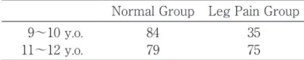
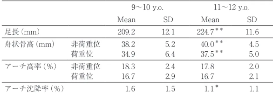
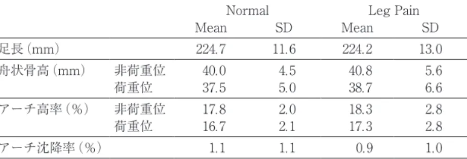
14.成長期サッカー選手の内側縦アーチと下肢疼痛の関係
RelationshipBetweenMedialLongitudinalArchandLegPainofSoccer PlayersinGrowthPeriod
国立病院機構徳島病院リハビリテーション科 川道 幸司ほか 180
15.高齢者転倒に関与する危険因子としての運動機能の検討
─第 7 回旧宮川村検診結果より─
EpidemiologyofRiskFactorsforFalling
三重大学大学院医学系研究科スポーツ整形外科 西村 明展ほか 185
16.スポーツ選手における ACL 再建術後 6 ヵ月でのスポーツ復帰の現状
ReturntoFullSportsActivitySixMonthsAfterAnteriorCruciateLigament ReconstructioninAthletes
横浜市スポーツ医科学センター 清水 邦明ほか 189
17.肩甲帯機能を考慮し開発された上半身ウエアが ゴルフパフォーマンスおよび身体機能に及ぼす効果
ANewDevelopedWearWouldInducePositiveEffectsforScapular, TrunkFunctionandGolfPerformance
船橋整形外科スポーツ医学センター理学診療部 小山 泰宏ほか 195
はじめに
足根骨癒合症は発生頻度 1%以下とまれな疾患で あり,また見逃されやすい疾患である 1).しかし近 年,CT や MRI などの画像技術の進歩により,早期 の診断率が飛躍的に向上し,無症候性も含めると,
10%を超えると報告されている 2, 3).また治療につ いては,保存的加療に抵抗する場合,関節固定術や 癒合部切除術が一般的に行なわれる.とくに思春期 のスポーツ選手に対しては,癒合部切除術をするこ
とで早期競技復帰が可能であると報告されてい
る 4, 5).思春期のスポーツ選手に発症した足根骨癒
合症について,癒合部切除術を施行した症例の治療 成績の評価を行なった.
対象と方法
対象は 2003 年 9 月から 2010 年 3 月までに当科を 受診し,スポーツを契機に発症した思春期の足根骨 癒合症のうち,癒合部切除術を施行した 6 例 7 足と した.手術時平均年齢は 14.6 歳(13〜16 歳),男性
思春期のスポーツ選手に発症した 足根骨癒合症の手術経験
Surgical Treatment of Tarsal Coalition in Adolescent Athletes
高橋 竜平1) Ryohei Takahashi 北岡 克彦2) Katsuhiko Kitaoka 中瀬 順介1) Junsuke Nakase 五嶋 謙一1) Kenichi Goshima 土屋 弘行1) Hiroyuki Tsuchiya
● Key words
Surgical:Tarsal coalition:Adolescent
●要旨
足根骨癒合症は比較的見逃されやすい足部痛や足関節痛の原因の 1 つである.思春期のス ポーツを契機に症状が出現し,手術を施行した足根骨癒合症の 6 例 7 足を経験したので報告 する.足根骨癒合症の内訳は,距・踵骨癒合症が 2 例 2 足,舟状・第 1 楔状骨癒合症が 2 例 2 足,舟状・立方骨癒合症が 1 例 1 足,第 1 楔状・中足骨癒合症が 1 例 2 足であった.手術時平 均年齢は 14.6 歳であった.治療に関しては,保存的加療に抵抗したため,全例癒合部切除 術を行なった.競技復帰の時期,術後再癒合と関節症性変化の有無,疼痛について評価した.
スポーツ復帰は平均 10.8 週と良好な結果となった.また再癒合や関節症性変化を認めた症 例はなかった.
1 1
高橋竜平
〒 920─8641 金沢市宝町 13─1 金沢大学附属病院整形外科
TEL 076─265─2374/FAX 076─234─4261
1)金沢大学附属病院整形外科
Department of Orthopaedic Surgery, Kanazawa University 2)木島病院整形外科
Department of Orthopaedic Surgery, Kijima Hospital
2 例 3 足,女性 4 例 4 足で,術後経過観察期間は平 均 6.1 ヵ月(5〜8 ヵ月)であった.症例の内訳は,距 踵骨癒合症 2 例 2 足,舟状第 1 楔状骨癒合症 2 例 2 足,舟状立方骨癒合症 1 例 1 足,楔状第 1 中足骨癒 合症 1 例 2 足であった.スポーツの種類はハンドボ ールが 2 例,陸上が 2 例,剣道とバスケットボール がそれぞれ 1 例ずつであった(表 1).術後再癒合の 有無,術後関節症性変化の有無,競技復帰時期,熊 井ら 6)の方法に準じた優・良・可・不可と 4 つの疼 痛評価(表 2)を評価項目とした.
結 果
経過観察期間中は再癒合や関節症性変化を全例認 めなかった.競技復帰は平均 10.8 週(7〜16 週)であ り,疼痛評価は優 5 例,良 1 例と良好な結果を認め た(表 3).また病理組織所見は全例線維軟骨性であ
った.
症例提示
症例①:13 歳男性,スポーツは陸上,長距離選 手.X─p と CT(図 1─a,b)で距踵骨癒合症と診断 し,癒合部切除術を施行した.手術はイメージ下で 距踵骨癒合部を確認した後,ノミを用い,正常関節 面が確認されるまで癒合部を切除した(図 2).切除 部の病理所見では,線維軟骨性組織が確認された
(図 3).術後再癒合や関節症性変化は認めず(図 4─a, b),術後 6 週よりランニング開始し,術後 12 週よ りスポーツ復帰となり,運動時や歩行時とも疼痛は 認めず,疼痛評価は優であった.
症例②:16 歳女性,スポーツはバスケットボー ル.X─p と CT(図 5─a, b)で舟状第 1 楔状骨癒合症 と診断し,癒合部切除術を施行した.術後再癒合や
1 11
表 1 症例の内訳
症例 部位 年齢 性別 患肢 スポーツ
症例 1 距・踵骨癒合症 15 歳 女性 左 ハンドボール 症例 2 距・踵骨癒合症 13 歳 男性 右 陸上(長距離)
症例 3 舟状・第 1 楔状骨癒合症 13 歳 女性 右 剣道
症例 4 舟状・第 1 楔状骨癒合症 16 歳 女性 右 バスケットボール 症例 5 舟状・立方骨癒合症 15 歳 女性 左 ハンドボール 症例 6 楔状・第 1 中足骨癒合症 16 歳 男性 両 陸上(長距離)
表 3 結果:競技復帰は平均 10.8 週であった
症例 部位 再癒合 関節症性
変化 競技復帰 疼痛評価
症例 1 距・踵骨癒合症 ─ ─ 10 週 優
症例 2 距・踵骨癒合症 ─ ─ 12 週 優
症例 3 舟状・第 1 楔状骨癒合症 ─ ─ 12 週 優 症例 4 舟状・第 1 楔状骨癒合症 ─ ─ 8 週 優
症例 5 舟状・立方骨癒合症 ─ ─ 7 週 優
症例 6 楔状・第1中足骨癒合症(右) ─ ─ 16 週 良
〃 楔状・第1中足骨癒合症(左) ─ ─ 16 週 良 表 2 疼痛評価
優……歩行時,運動時とも疼痛なし
良……歩行時または運動時にときどき疼痛あり 可……術前と疼痛程度が不変
不可…術前より疼痛が増悪
関節症性変化は認めず(図 6),術後 5 週よりランニ ング開始し,術後 8 週でスポーツ復帰となった.疼 痛評価は優であった.
考 察
足根骨癒合症は 2 つかそれ以上の先天的な足根骨 の癒合であり,組織学的には骨性と線維軟骨性に分 けられる.骨性は完全型癒合であり,症状は乏し く,臨床的に問題となりやすいのは,不完全型癒合 の線維軟骨性である.これは,不完全ながらも可動 性を認めるため,癒合部自身に過度の応力が加わる と考えられている 2).自験例でも全例線維軟骨性で
あった.
また足根骨癒合症の分類は,さまざまなタイプが ある.欧米では距踵骨癒合症と踵舟状骨癒合症が約 半数ずつ,合計で約 90%を占めている.その一方日 本では距踵骨癒合症が 60%で,その次に舟状第 1 楔 状骨癒合症が 30%を占めており,アジア人やヒスパ ニック系の人種に報告が多いことから,人種特異性 が示唆されている 6).
思春期のスポーツ選手に対する治療に関しては,
競技活動などの制限,消炎鎮痛剤内服,不完全癒合 部における動きを軽減させる目的でのアーチサポー トやギプス固定などの保存的加療がまず選択され
1 111
図 1 初診時画像所見(症例①)
a:単純 X 線側面像 b:CT axial 像 距踵骨の癒合を認める.
図 2 術中所見(症例①)
正常関節面が確認されるまで線維軟骨癒合部を 切除した.
図 4 術後画像所見(症例①)
a:単純 X 線側面像 b:CT axial 像 再癒合や関節症変化は認めなかった.
図 3 病理所見(症例①)(H─E 染色)
切除部の病理所見では,線維軟骨性組織が確認 された.
4
1 11
る.Elkus らは 2 週間のギプス固定によって約 1/3 の患者が症状の軽減を認めたと報告した 7).しかし 競技復帰に関しては,どのくらいで可能になるかは 明らかではない.
手術的治療では,以前は関節固定がされていた.
しかし早期診断に留意すれば,癒合部切除術で対処 可能 8)であり,癒合部切除術が失敗したときのサル ベージ手術と考えるべきである 9)と報告されてい る.また侵襲が大きく,競技復帰までに時間がかか るといったこともあるため,その適応は関節症性変 化の大きい場合や後足部の高度な外反変形を認める 場合のみと報告されている 4).
一方,癒合部切除術は競技復帰平均 10.3 週といっ た良好な成績 4)や,80〜100%の競技復帰率という良 好な結果が報告されている 8, 10).自験例でも競技復 帰平均 10.8 週と良好な結果となった.思春期のス ポーツ選手にとって,ベストパフォーマンスを行な うためには,可能な限り正常な癒合関節の可動性や 機能が必要となる 4).つまり癒合部切除術を施行す ることによって,正常関節により近くなり,これが 早期競技復帰につながるのではと考えられた.
結 語
スポーツを契機に発症した思春期の足根骨癒合症 6 例 7 足に対して,癒合部切除術を施行した.競技 復帰は平均 10.8 週と良好な結果となった.思春期の スポーツ選手の足根骨癒合症では,早期診断を行な い,癒合部切除術を施行することが早期競技復帰に つながると考えられた.
文 献
1) Stornont DM et al:The relative incidence of tarsal coalition. Clin Orthop Relat Res, 181:28─
36, 1983.
2) 熊井 司ほか:成長期の足部障害─足根骨癒合 症─.MB Orthop, 20:19─26, 2007.
3) Solomon LB et al:A dissection and computer tomography study of tarsal coalitions in 100 cadaver feet. J Orthop Res, 21:352─358, 2003.
4) Saxena A et al:Tarsal coalitions ─Activity levels with and without surgery─. J Am Podia- tr Assoc, 93:259─263, 2003.
5) O’ Neill DB et al:Tarsal coalition ─A followup of adolescent athletes. Am J Sports Med, 17:
図 5 初診時画像所見(症例②)
a:単純 X 線正面像 b:CT axial 像 舟状第 1 楔状骨の癒合症を認める.
図 6 術後画像所見(症例②)
a:単純 X 線斜位像
再癒合や関節症変化は認めなか った.
1 11
544─549, 1989.
6) 熊井 司ほか:舟状骨─第 1 楔状骨間癒合症の 臨床像.整形外科,45:679─686, 1994.
7) Elkus RA:Tarsal coalition in the young ath- lete. Am J Sports Med, 14:477─480, 1986.
8) Scranton PE:Treatment of symptomatic talo- calcaneal coalition. J Bone Joint Surg Am, 69:
533─539, 1987.
9) Lemley F et al:Current concepts review:
Tarsal coalition. Foot Ankle Int, 27:1163─
1169, 2006.
10) Dutoit M:Talocalcaneal bar resection. J Foot Ankle Surg, 37:199─203, 1998.
6
はじめに
第 5 中足骨疲労骨折は難治性で治療に長期間を要 するため,スポーツ選手では観血的治療が推奨され
ている 1, 2).その術式は髄内スクリュー固定術が一般
的に行なわれ,良好な治療成績が報告されている 3). しかし,まれではあるがスポーツ復帰後の再骨折や 骨癒合不全例の報告もある 4, 5).今回,われわれは第
5 中足骨疲労骨折に対し髄内スクリュー固定術を行 ない,再手術が必要となった 3 例を経験したため報 告する.
対象と方法
対象は,第 5 中足骨疲労骨折に対し,髄内スクリ ュー固定術を行ない,その後再手術が必要となった 3 例で,全例男性であった.平均年齢は 18.7 歳(16 〜
第 5 中足骨疲労骨折に対する髄内スクリュー固定術
─再手術例の検討─
Intramedullary Screw Fixation in Proximal Fifth─metatarsal Stress Fracture
─ Analysis of Failure ─
中瀬 順介 Junsuke Nakase 北岡 克彦 Katsuhiko Kitaoka 五嶋 謙一 Kenichi Goshima 高橋 竜平 Ryohei Takahashi 土屋 弘行 Hiroyuki Tsuchiya
● Key words
第 5 中足骨疲労骨折,髄内スクリュー固定術,再手術例
●要旨
第 5 中足骨疲労骨折は難治性でかつ保存療法後の再発率が高いことから,手術療法が選択 されることが多い.術式は髄内スクリュー固定術が一般的に行なわれ,良好な成績が報告さ れている.一方,まれに再骨折や癒合不全が生じるといわれている.今回,第 5 中足骨疲労 骨折の髄内スクリュー固定術後に再手術を必要とした3例を経験したので報告する.2例は,
固定力不足が,1 例は早すぎたスポーツ復帰が再手術の原因と考えた.髄内スクリュー固定 術を行なう際には,なるべく内側から刺入し,可能な限り長く太いスクリューを使用してい る.術後は,インソールを使用し,過度な足部外側荷重を改善させるような下肢・体幹の運 動指導を行なっている.
中瀬順介
〒 920─8641 金沢市宝町 13─1 金沢大学附属病院整形外科 TEL 076─265─2374
金沢大学大学院医学系研究科整形外科学
Department of Orthopaedic Surgery, Graduate School of Medical Science, Kanazawa University
22 歳)で,スポーツの種類はバスケットボールが 2 例,サッカーが 1 例であった.
再手術となった原因,第 5 中足骨骨軸の長さに対 するスクリュー長の割合(以下,スクリュー長の割 合),第 5 中足骨骨軸の長さに対する骨折部の割合
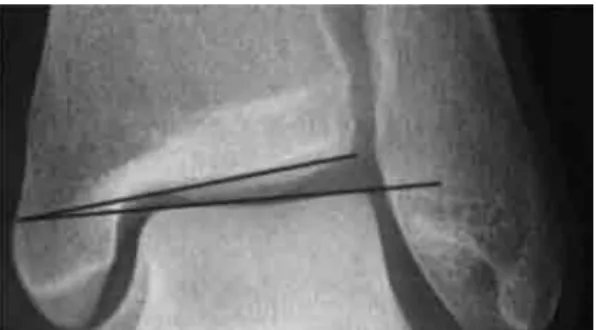
(最近位を 0 と定義),発症から手術までの期間と初 回手術後スポーツ復帰までの時期を調査した.スク リュー長の割合は高橋らの報告に準じ 6),足部斜位 レントゲン像で評価し(図 1),骨折部の割合も同様 に足部斜位レントゲン像で評価した.
結 果
症例 1 と 2 は骨癒合不全が,症例 3 は再骨折が再 手術の原因であった.スクリュー長の割合は症例 1 が 0.65,症例 2 が 0.6,症例 3 が 0.45 で,骨折部の 割合は症例 1 が 0.31,症例 2 が 0.36,症例 3 が 0.28 であった.また,発症から手術までの期間は症例 1 が 12 ヵ月,症例 2 が 10 ヵ月で症例 3 が 1 ヵ月であっ た.初回手術後のスポーツ復帰時期は症例 1 が術後
4 ヵ月,症例 2 が術後 2 ヵ月でいずれも単純レント ゲン像での骨癒合前であった.また,症例 3 は単純 レントゲンで骨癒合確認後の術後 2.5 ヵ月でスポー ツ復帰していた(表 1).
症例 1 16 歳,男性 サッカー選手
近医で左第 5 中足骨疲労骨折と診断され,保存的 に加療されたが,左足部痛が持続するため発症から 12 ヵ月後に髄内スクリュー固定術をうけた.術後 4 ヵ月で練習に復帰したころ左足部痛が再燃し,当院 に紹介となった.当院初診時レントゲン写真では骨 癒合は得られていなかった(図 2a).スクリュー長 の割合は 0.6 であるが,スクリューの方向が悪く固 定力が不足していたこととレントゲン像での骨癒合 前にスポーツに復帰したことが骨癒合不全の原因と 考え,再手術を施行した.再手術は 5mm 中空海面 骨スクリュー(ストライカー社)を用い,骨移植は 併用しなかった(図 2b).術後 8 週で単純レントゲ
図 1 第 5 中足骨骨軸の長さに対する スクリュー長の割合(スクリュー
長の割合) 図 2 a.当院初診時単純 X 線像
b.再手術直後単純 X 線像 c.再手術 8 週後単純 X 線像
a b c
表 1 結果
症例 1 症例 2 症例 3
再手術の原因 骨癒合不全 骨癒合不全 再骨折 スクリュー長の割合 0.65 0.6 0.45 発症から手術までの
期間 12 ヵ月 10 ヵ月 1 ヵ月 スポーツ復帰時期 4 ヵ月
(骨癒合前) 2 ヵ月
(骨癒合前) 2.5 ヵ月
(骨癒合後)
8
ン上骨癒合が得られたため,スポーツ復帰を許可し た(図 2c).術後 2 年の現在,自覚症状なくサッカー を継続している.
症例 2 18 歳,男性 バスケットボール選手 近医で右第 5 中足骨疲労骨折と診断され,保存的 に加療されたが,右足部痛が持続するため当院に紹 介となり(図 3a),発症から 10 ヵ月後にアキュトラ ックスクリュー(アキュメッド社)を用いて髄内ス クリュー固定術を施行した(図 3b).骨が硬く予定 したスクリューが挿入できずスクリュー長の割合は 0.6 であった.術後右足部痛がほとんどなく,単純 レントゲン像での骨癒合前の術後 2 ヵ月(図 3c)で スポーツに復帰したところ,一旦は骨癒合傾向にあ ったが骨折部周囲に骨吸収像が出現した(図 3d).
高校生最後の大会が近く,右足部痛がほとんどなか ったためプレーを継続し,高校卒業後の術後 1 年 4 ヵ月(図 3e)で再手術を施行した.5mm 中空海面骨 スクリュー(ストライカー社)で再固定し,さらに 骨折部には脛骨から採取した自家骨移植を追加した
(図 3f).術後経過は良好で,単純レントゲン上骨癒
合が完成した再手術後 3 ヵ月(図 3g)でスポーツ復 帰を許可し,大学に進学した現在もバスケットボー ルを継続している.
症例 3 21 歳,男性 バスケットボール選手 前医で右第 5 中足骨疲労骨折に対し,髄内スクリ ュー固定術を受けた(図 4a).術後 2.5 ヵ月で単純レ ントゲン写真上骨癒合したため,スポーツに復帰し た(図 4b).スポーツ復帰後 3 週間で右足部痛が再 燃し,右第 5 中足骨疲労骨折再骨折と診断され,当 院に紹介となった(図 4c).スクリューの径および 長さの不足が原因と考えアキュトラックスクリュー
(アキュメッド社)を用いて再手術を施行した.術 後経過は良好で,単純レントゲン上骨癒合が完成し た再手術後 1.5 ヵ月(図 4d)でスポーツ復帰を許可 した.術後 3 年の現在,自覚症状なくバスケットボ ールを継続している.
考 察
第 5 中足骨疲労骨折の発生メカニズムは足の外側
6
図 3 a.当院初診時単純 X 線像 b.手術直後単純 X 線像 c.手術 2 ヵ月後単純 X 線像 d.手術 1 年後単純 X 線像 e.手術 1 年 4 ヵ月後単純 X 線像 f .再手術直後単純 X 線像 g.再手術 3 ヵ月後単純 X 線像
a b c d e f g
7
荷重により解剖学的に弯曲している第 5 中足骨がし なり底側に張力が加わり,足が地面から離れた瞬間 にその力は背側の圧縮力となり,その結果靱帯で覆 われていない最も力学的に脆弱な部分で横骨折が生 じると報告されている(図 5) 7).
第 5 中足骨疲労骨折は観血的治療が推奨され,そ の術式は髄内スクリュー固定術が一般的に行なわれ ている.合併症には,スポーツ復帰後の再骨折や骨 癒合不全例が報告されている.その原因には,足部 外側に荷重が偏位すること,スクリューの固定力が 不足していることや骨癒合前のスポーツ復帰があげ られる.
外側荷重の原因には下肢内反による下肢アライメ ント異常 8)や股関節外転筋力低下 9)が報告されてお り,われわれは現在術後に足底挿板を使用し股関節 外転筋力と体幹筋力の強化指導を行なっている.
スクリューの長さに関しては,スクリュー長の割 合が 0.55 以下で再発例が多かったと報告されてい る 6).今回のわれわれの症例でもスクリュー長の割 合が 0.45 であった症例で再骨折が生じたため,ス クリューはなるべく長く太いスクリューを使用して いる.その際,スクリューをなるべく長く挿入する ために第 5 中足骨の形状を考慮し,刺入点は可能な 限り内側にしている.スクリューの種類は,以前は アキュトラックスクリューを使用していたが,現在
は中空海面骨スクリューを使用している.その理由 は,本手術の目的は第 5 中足骨の力学環境を変える ことにあり,そのためにはなるべく長いスクリュー を挿入する必要がある.アキュトラックスクリュー は固定力に優れているが,そのためトルクが強くか かりすぎ,硬い骨では長いスクリューが挿入できな いことを経験した.なるべく長いスクリューを挿入 し,第 5 中足骨の力学環境を変えることが再発にも 繋がると考えている.
骨癒合前のスポーツ復帰による骨癒合不全症例が 報告されており 4, 5),スポーツ復帰は局所の圧痛や 腫脹の消失などと単純レントゲン上の骨癒合終了後 に許可するようにしている.とくに発症から手術ま での経過が長い症例では骨癒合まで時間を要するこ とがあり,注意が必要と考えている.
また,再手術時の自家骨移植の是非に関しては一 定の見解が得られておらず,われわれは個々の症例 に応じて適応を判断している.
ま と め
第 5 中足骨疲労骨折に対し,髄内スクリュー固定 術を行なう際には,可能な限り長く太いスクリュー を使用し,術後は再骨折を予防するために足底板の 使用や下肢 ・ 体幹の運動指導を行ない,第 5 中足骨 の力学環境を変える工夫が必要と考える.
文 献
1) Portlamd G et al:Acute surgical management 図 5 第 5 中足骨疲労骨折発生メカニズム
(文献 7)から一部改変し引用)
図 4 a. 前医手術直後単純 X 線像,b.手術 2.5 ヵ月 後単純 X 線像,c.当院初診時時単純 X 線像,
d.再手術 1.5 ヵ月後単純 X 線像 d c
b a
0
8
of jones’ fractures. Foot Ankle Int, 24:829─
833, 2003.
2) Mologne T et al:Early screw fixation versus casting in the treatment of acute jones frac- tures. Am J Sports Med, 33:970─975, 2005.
3) Porter D et al:Fifth metatarsal jones fracture fixation with a 4.5─mm cannulated stainless steel screw in the competitive and recreational athlete. Am J Sports Med, 33:726─733, 2005.
4) Wright R et al:Refracture of proximal fifth metatarsal(jones)fracture after intramedul- lary screw fixation in athletes. Am J Sports Med, 28:732─736, 2000.
5) Larson C et al:Intramedullary screw fixation of jones fractures. Am J Sports Med, 30:55─
60, 2002.
6) 高橋良正ほか:プロサッカー選手の Jones 骨折 に対する手術成績の検討.中部整災誌,46:69─
70, 2003.
7) 戸祭正喜:中足骨疲労骨折.整形外科,58:
972─977, 2007.
8) 出家正隆ほか:サッカー選手に発生した Jones’
fracture の 3 例.臨スポーツ医,8:89─91, 1991.
9) 小粥智浩ほか:第五中足骨疲労骨折のリハビリ テーション.臨スポーツ医,23:175─182, 2006.
Introduction
It is widely accepted that anterior cruciate ligament
(ACL)injuries frequently occur especially in female basketball players. Arendt et al 1) reported that the incidence of the ACL injury in female basketball
players was 0.29 per 1,000 athlete exposures while that in male basketball players was 0.07 per 1,000 athlete exposures based on the National Collegiate Athletic Association(NCAA)database. The survey of Japan Basketball League 2)also showed that ap prox
i mate ly 20% of the female players had experienced at least more than one ACL injury during their
The Inciting Events of the Anterior Cruciate Ligament Injuries in Female Basketball Players
女子バスケットボール選手における 膝前十字靱帯損傷の受傷時状況に関する検討
Hirokazu Kobayashi 小林 寛和1) Sanae Kitaoka 北岡さなえ2)
Sentaro Koshida 越田専太郎3) Koji Miyashita 宮下 浩二4)
Takuya Shimizu 清水 卓也5) Kiyoshi Yokoe 横江 清司2)
● Key words
Female basketball players:Anterior cruciate ligament injury:Inciting events
● Abstract
The objective of this study was to demonstrate the detailed description of the inciting events of anterior cruciate ligament(ACL)injuries in female basketball players. We conducted a retrospective interviewing study with 235 female basketball players who had suffered from an ACL injury. Our study demonstrated that the ACL injury occurred more frequently on offense than on defense. We also found that the number of the ACL injuries which occurred at jump─landing and side─step cutting was larger than that at deceleration on offence, while the number of the injuries at the side─step cutting and deceleration was significantly larger than that at the jump─landing injury on defense. Finally, the result revealed that the ratio of the “jump─landing” ACL injury was relatively high in the contact situation compared to that in the noncontact situation. We may be able to utilize the detailed profiles of the inciting events for ACL injury prevention.
1 11
小林寛和
〒 475─0012 半田市東生見町 26─2 日本福祉大学健康科学部 TEL 0569─20─0118
1)Faculty of Health Science, Nihon Fukushi University 2)Institute of Sports Medicine and Science
3)Faculty of Health Sciences, Ryotokuji University 4)College of Life and Health Sciences, Chubu University
5)Graduate School of Health and Sport Science, Chukyo University
1
athletic career, which was approximately twice as high as male basketball players.Although surgical intervention and rehabilitation usually allow the injured athletes to return to the previous activity level, the ACL injury still deprives them of much of their time in sports participation. In addition, it has been reported that the risk of posttraumatic degenerative joint disease may increase regardless of the surgical reconstruction of the ligament. There
fore, the sports medicine practitioners and researchers have investigated the risk factors of the injury from various research aspects to develop the effective ACL injury prevention strategy.
Previous studies have suggested that most of the ACL injuries occurred with noncontact mechanism 1〜7)
and the risk factors of the noncontact ACL injury have been proposed from standpoints of the environ
ment 8, 9),anatomical structures 4, 10, 11),hormones 12, 13)
and biomechanics 1〜7, 10, 14).The findings of the previous studies have enhanced our understanding of the ACL injury and have helped us to develop the injury prevention strategy. However, if the main purpose is to prevent the ACL injury from occurr ing, it may also be necessary to develop the traditional ap proach
es to description of situations where the injury occurs.
Bahr et al 15)proposed the comprehensive model for injury causation, based on the epidemiological study of Meeuwisse 16).In the model, the inciting events of injuries(i.e. playing situation, player/opponent behavior, etc.)are regarded as a final link in chain that causes an injury and is usually directly as so ci
at ed with the onset of an acute injury. As we noted, many studies have focused on intrinsic and extrinsic factors of the ACL injury occurrence to establish a better strategy for the ACL prevention, whereas few studies have closely investigated the detailed description of the inciting events of the ACL injury.
Therefore, the aim of this study was to investigate the inciting events of the ACL injury and identify the high risk situation in female basketball players.
We believe this information will lead us to a future prevention strategy for the ACL injury in female
basketball players.
Materials and Methods
Subjects
The subjects were 235 female basketball players who suffered from the ACL injury during a practice or a game and visited the orthopedic clinic in the Institute of Sports Medicine and Science, Aichi, Japan, where they were diagnosed as having the ACL injury(Mean±SD age 18.6±3.9 years, height 164.6
±7.8cm, body weight 58.8±8.0kg).All the ACL injuries were confirmed by magnetic resonance imaging and/or arthroscopic procedure. The subjects received physical therapies periodically for the period of 20 years from June 1988 through June 2008. Approximately seventy percent of the subjects visited the clinic within one month and 46 percent of the subjects visited the clinic within one week after the ACL injury incident. We chose the subjects whose ages were fifteen or more so that they were included in this study. We also excluded the subjects from the study when the description of the injury mechanism was unclear or when the injury occurred by an accident not related to basketball activity. The group of the subjects consisted of 149 high school basketball players(63.4%),33 collegiate basketball players(14.0%),53 post─collegiate basketball players
(22.6%).One hundred and sixteen subjects(51.1%)
played at an amateur regional level, 68 subjects
(30.0%)played at an amateur national level, 19 subjects(8.4%)played at a semi─professional level and 8 subjects(3.5%)played at an international and/
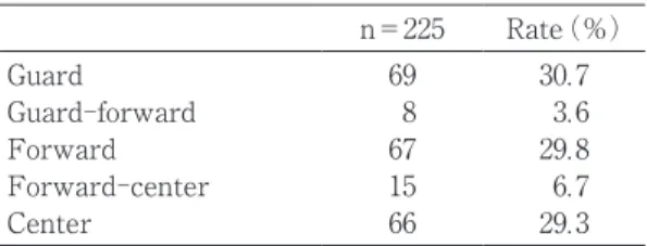
or a professional level. We were able to obtain the information on the playing position of 225 out of 235 subjects consisting of 69 “guard” players (30.7%),
8 “guard─forward” players(3.6%),67 “forward”
(29.8%),15 “forward─center”(6.7%)and 66 “center”
players(29.3%)(Table 1).One hundred and twenty─
nine(54.9%)subjects had their right side of the knee injured, and 106 subjects(45.1%)had the left side of the knee injured. They all suffered from the
1 1
unilateral ACL injury, so we investigated 235 ACL injury cases in total. The study protocol was approved by the Ethics Committee of the Institute of Sports Medicine and Science.
Data acquisition
We used the medical records of the orthopedic surgery and the physical therapy of the subjects as the database. When the subjects visited the institute, skilled physical therapists themselves interviewed the subjects and prepared and kept their medical records. They interviewed the subjects about the inciting events at the time of the injury in details.
In addition, the therapists confirmed the injury mechanism by having the subjects reproduce the scene of the injury occurrence with the use of the unaffected leg.
We investigated the following items:
1.Injury mechanisms
We classified the injury mechanism into the following three categories:
1) Noncontact:no contact with another player at the time of the injury
2) Contact:physical contact on body parts other than the lower extremity through being hit, pushed, or held by another player
3) Collision:direct physical contact with another person on the affected lower extremity
2.Playing situation at the time of the injury We classified the playing situation into the four categories:“offense,” “defense,” “other,” “unknown.”
We defined the category of “other,” as situations on which it was difficult to determine whether the situation was on offence or on defense such as
“rebound,” and “loose ball.”
3.Player’s behavior at the time of the injury We classified the player’s behavior at the time of the injury into the following six categories:“jump─
landing,” “jump─take off,” “deceleration,” “side─step cutting,” “other,” and “unknown.”
1) Jump─landing:landing on one(injured side)
or both legs after the player had jumped up to shoot or catch the ball
2) Jump─take off:taking off on one(injured side)
or both legs when the player had tried to jump up to shoot or catch the ball
3) Deceleration:decelerating or stopping running in any direction without jump stop
4) Side─step cutting:side─stepping and changing direction sideways to pass by or to mark the opponent
5) Other:player’s behavior that was not described clearly by the subjects
6) Unknown:the player’s data missing in medical records
Statistical analysis
We carried out a chi─square test to elucidate the interrelation between the playing position, the injury mechanisms, the playing situation and the player’s behavior at the time of the injury. We used the SPSS Ver.17J for Windows(SPSS Japan Inc., Shibuya, Tokyo, JAPAN)for the statistics. The significant level was set at 5% in this study.
Results
1.Injury mechanisms(Figure 1)
We were able to obtain the information on the injury mechanisms from all the subjects. The result showed that the number of “noncontact” cases(188 cases)was the greatest among the categories of the
1 1 1
Table 1 Playing position(n=225):we were not able to obtain the data from 10 out of 235 subjects.
n=225 Rate(%)
Guard 69 30.7
Guard─forward 8 3.6
Forward 67 29.8
Forward─center 15 6.7
Center 66 29.3
14
1 1
injury mechanisms, accounting for 80% of all the cases. The number of “contact” cases(40 cases)was also larger than that of “collision” cases(7 cases).
2.Playing situation(Figure 2)
We investigated the playing situation at the time of the injury in 187 out of 235 cases. The result showed that the number of ACL injury incidents which occurred in the “offense” situation (132 cases)
was the largest among the categories, accounting for about 70% of all the cases. Moreover, the numbers of ACL injuries occurred in the “defense”
and the “other” situations were 37 cases and 18 cases respectively.
3.Player’s behavior(Figure 3)
Finally, we were able to obtain the information on player’s behavior at the time of the injury from 232 out of 235 subjects. The result showed that the number of “jump─landing” case was the largest(77 cases),which accounted for about 33%, followed by
“side─step cutting(64 cases, 27.6%),” “deceleration
(40 cases, 17.2%),” “jump─take off(16 cases, 6.9
%),” and “other”(35 cases, 15.1%)respectively.
4. Relationship between injury mechanisms and player’s behavior(Table 2)
We were able to perform the cross tabulation analysis between injury mechanisms and player’s behavior at the time of the injury in 186 out of 235 cases. We performed the cross tabulation analysis between the injury mechanisms and player’s behav
ior after the categories of the direct and the other mechanisms were combined. Significant differences in the subjects behavior were found between noncon
tact and the other injury mechanisms [χ(df=8)2
=36.83, p<0.001]. The numbers of noncontact ACL injury which occurred during “side─step cutting,”
“jump─landing,” “deceleration,” and “jump─take off”
were 55 cases, 45 cases, 32 cases and 15 cases respectively, whereas “jump─landing” injury cases accounted for approximately 50% in the other Fig.1 Injury mechanism(n=235)
0 50 100 150 200
Number of subjects
Noncontact Contact Collision
Fig.2 Playing situation at the time of the in- jury(n=187):we were not able to obtain the data from 48 out of 235 subjects.
Fig.3 Player’s behavior at the time of the injury(n=232):we were not able to obtain the data from 3 out of 235 subjects.
Table 2 Injury mechanisms and player’s behavior at the time of the injury(n=186).
Noncontact Contact Collision n=152 Rate(%) n=34 Rate(%)
Jump─landing 45 29.6 16 47.1
Jump─take off 15 10.0 1 2.9
Deceleration 32 21.0 0 0.0
Side─step cutting 55 36.2 7 20.6
Other 5 3.3 10 29.4
χ2(df=4)=36.83 p<0.001
mechanism.
5. Relationship between playing situation and player’s behavior(Table 3)
We conducted the cross tabulation analysis to dem on strate the relationship between playing situation and player’s behavior at the time of the injury in 169 out of 235 cases. We excluded the
“other” category of the playing situation from the cross tabulation analysis. The result demonstrated that there was a significant difference in the subject’s behavior between the “offence” and the “defense”
situations [χ2(df=8)=15.50, p<0.001].The number of the ACL injury during “jump─landing”
(44 cases)and “side─step cutting”(42 cases)were relatively large in the offence situation compared to “deceleration”(20 cases)and “jump─take off”(15 cases).On the other hand, 20 cases of ACL injury occurred during “side─step cutting,” which accounts for about 50% in the defense situation, followed by “deceleration”(10 cases)and “jump─landing”(4 cases),respectively.
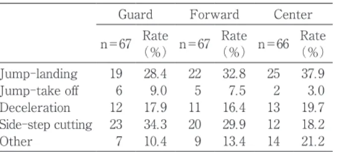
6. Relationship between playing position and player’s behavior(Table 4)
Finally, we conducted the cross tabulation analysis to demonstrate the relationship between playing position and player’s behavior at the time of the injury in the 200 cases out of 235. We excluded the subjects in the “guard─forward”(n=8),the “forward─
center”(n=15),and “unknown”(n=12)categories from the cross tabulation analysis. The result of
the cross tabulation analysis showed that there is no significant difference in player’s behavior among the playing positions [χ(df=8)=9.14, p=0.412].2
Discussion
In the same way as shown in the previous reports 1〜7),our result showed that the number of the noncontact injuries was larger than that of the other injuries in the female basketball players. Our study also demonstrated that the ACL injury occurred more frequently on offense than on defense. The defense situation may require reactive tasks with sharp acceleration/deceleration more frequently than the offence situation does and the moment─to─moment reactions might increase the risk of the ACL injury. However, such physical demands on the defense situation did not reflect the result of our study. In addition, Krosshaung 14) reported that the number of the ACL injury in basketball players which occurred in the offense situation was greater than that in the defense situation:29 cases of the injury occurred in the offense situation and 5 cases of the injury occurred in the defense situation. Both the previous and our results indicate that ACL injury risk may be higher when the players are in the offence situation than in the defense situation.
One of the notable findings of our study is that there may be differences between the offense and the defense situations in the high risk player’s
1 1
Table 3 Playing situation and player’s behavior at the time of the injury(n=169).
Offense Defense
n=132 Rate(%) n=37 Rate(%)
Jump─landing 44 33.3 4 10.8
Jump─take off 15 11.3 0 0
Deceleration 20 15.2 10 27.0
Side─step cutting 42 31.8 20 54.1
Other 11 8.3 3 8.1
χ2(df=4)=15.50 p<0.001
Table 4 Playing position and player’s behavior at the time of the injury(n=200).
Guard Forward Center
n=67 Rate
(%)n=67 Rate
(%) n=66 Rate
(%)
Jump─landing 19 28.4 22 32.8 25 37.9 Jump─take off 6 9.0 5 7.5 2 3.0 Deceleration 12 17.9 11 16.4 13 19.7 Side─step cutting 23 34.3 20 29.9 12 18.2
Other 7 10.4 9 13.4 14 21.2
χ2(df=8)=9.14 p=0.412
16
behaviors for the ACL injury. The cross tabulation analysis between player’s behavior and playing situation revealed that greater numbers of the ACL injury in offence situation occurred at jump─
landing and side─step cutting, whereas the number of side─step cutting and deceleration injury cases was larger than the jump─landing injury case in the defense situation. In the offense situation, players usually perform more jumping activities such as a lay─up shot, a jump shot and a catch of a ball passed high from another player than they do in the defense situation. As we described in the last paragraph, on the other hand, players are more involved with side─cutting with sharp acceleration/deceleration in order to react the opponent’s attack in the defense situation. Different movement tasks and neu ro mus
cu lar loads required during the play between on offence and on defense may be associated with the current result.
Our study also demonstrated the ratio of the ACL injury by “jump─landing” was relatively high in the “contact” situation compared to that in the
“noncontact” situation. Physical contact by the opponent while the player is jumping up in the air will give a large effect on the dynamic alignment of the lower extremity at a landing. It may be beneficial for players to perform the ACL injury prevention exercise in the contact situation at
“jump─landing” in order to be able to improve the body control skill during a jump, which may result in minimizing the effect of the physical contact on the alignment at the landing.
The result of the cross tabulation analysis between the playing position and player’s behavior at the time of the injury showed that player’s behavior at the time of injury was not significantly different among the playing position. Center players are usually more involved with such jumping tasks as rebounding, shooting and catching a ball passed high by another player in the area near the basket, whereas guard players are more involved with side─step cutting and sharp deceleration tasks while dribbling the ball.
Regardless of different physical demands between playing positions, we were not able to demonstrate a different trend on the player’s behavior at the time of the ACL injury.
In this study we analyzed the medical records of female basketball players who suffered from the ACL injury to identify the profile of the playing position and the inciting events(i.e. injury mechanisms, playing situation and player’s behavior)at the time of the ACL injury and their interrelationships.
These results may be able to add a new insight to the traditional ACL injury prevention program.
Previous ACL prevention studies stated that a prophylactic neuromuscular and proprioceptive training program might reduce the number of ACL injuries 17〜19). In addition to the traditional preventive strategies, it may be beneficial for the prevention of the ACL injury to have basketball players recognize the high risk situations of the ACL injury found in this study and have them improve the dynamic control of lower extremity in the simulated but controlled situations. We believe that this kind of prevention strategy will be able to decrease the risk of the injury in the real basketball situation.
References
1) Arendt E et al:Knee injury patterns among men and women in collegiate basketball and soccer NCAA data and review of literature.
Am J Sports Med, 23:694─701, 1995.
2) Narita T et al:Anterior cruciate injuries in Japan basketball league players. The Journal of Clinical Sports Medicine, 19:75─79, 2002.
(in Japanese)
3) Agel J et al:Anterior cruciate ligament injury in National Collegiate Athletic Association basketball and soccer:A 13 year Review. Am J Sports Med, 33:524─530, 2005.
4) Gray J et al:A survey of the anterior cruciate ligament of the knee in female basketball players.
1 1 4
Int J Sports Med, 6:314─316, 1985.
5) Kobayashi H:Kinematic analysis of the anterior cruciate ligament injuries in female basketball players. Journal of Japanese Physical Therapy Association, 21:537─540, 1994.(in Japanese)
6) Olsen OE et al:Injury mechanisms for anterior cruciate ligament injury in team handball:a systematic video analysis. Am J Sports Med, 32:1002─1012, 2004.
7) Boden BP et al:Mechanisms of anterior cruciate ligament injury. Orthopedics, 23:573─578, 2000.
8) Orchard J:Is there a relationship between ground and climatic conditions and injuries in football ? Sports Med, 32:419─432, 2002.
9) Dowling AV et al:Shoe─surface friction influ
ences movement strategies during a sidestep cutting task: implications for anterior cruciate ligament injury risk. Am J Sports Med, 38:478─
485, 2010.
10) Masujima A et al:Investigation of sports injuries in female athletes(second edition).Nihon Taiiku Kyokai Sports Kagaku Kenkyu Hokokushu, Tokyo:34─39, 1986.(in Japanese)
11) Anderson AF et al:Analysis of the intercon
dylar notch by computed tomography. Am J Sports Med, 15:547─552, 1987.
12) Hewett TE et al:Effects of the menstrual cy
cle on anterior cruciate ligament injury risk:a systematic review. Am J Sports Med, 35:659─
668, 2007.
13) Deie M et al:The influence of female hormone to anterior cruciate ligament injuries. Journal of clinical sports medicine, 19:991─994, 2002.
14) Krosshaung T et al:Mechanism of anterior cruciate ligament injury in basketball:video analysis of 39 cases. Am J Sports Med, 35:359─
367, 2006.
15) Bahr R et al:Understanding injury mecha
nisms:a key component of preventing injuries in sport. Br J Sports Med, 39:324─329, 2005.
16) Meeuwisse W:Assessing causation in sport Injury: a multifactorial model. Clin J Sport Med, 4:166─170, 1994.
17) Hewett TE et al:Anterior cruciate ligament injuries in female athlete. Am J Sports Med, 34:490─498, 2006.
18) Sell TC et al:The effect of direction and reac
tion on the neuromuscular and biomechanical characteristics of the knee during tasks that stimulate the noncontact anterior cruciate liga
ment injury mechanism. Am J Sports Med, 34:
43─54, 2006.
19) Noyes FR et al:The drop─jump screening test:difference in lower limb control by gen
der and effect of neuromuscular training in fe
male athletes. Am J Sports Med, 33:197─207, 2005.
1 1 5
18
中学生ハンドボール選手および
指導者の膝前十字靱帯損傷に関するアンケート調査
An Attitude Survey of ACL Injuries
for Junior High School Handball Players and Coaches
阿部 健作1) Kensaku Abe 北岡 克彦3) Katsuhiko Kitaoka 中瀬 順介2) Junsuke Nakase 五嶋 謙一2) Kenichi Goshima 高橋 竜平2) Ryohei Takahashi 土屋 弘行2) Hiroyuki Tsuchiya
● Key words
前十字靱帯損傷,意識調査,中学生
ACL injury:Attitude survey:Junior high school students
●要旨
近年,膝前十字靱帯(以下 ACL)損傷の予防に注目が集まり,予防トレーニングとその有 効性が報告されている.この予防トレーニングを普及させるには,まずスポーツ現場におけ る ACL 損傷に対する知識を明確にさせる必要がある.本研究の目的は,全国中学生ハンド ボール選手権大会に出場した選手および指導者に対し,ACL 損傷に関するアンケート調査 を行ない,その現状を把握することである.
選手権大会に出場した選手および指導者を対象としてアンケート調査を行ない,回答が得 られた 55 チーム,選手 704 名およびその指導者 96 名の結果を解析した.
ACL 損傷を知っている選手は 21.4%しかおらず,受傷機転については 4.4%,予防トレー ニングについては 2.2%しか知らなかった.また指導者の 88.5 %が ACL 損傷を知っていた が,受傷機転については 50%しか知らず,予防トレーニングについては 13.5 %しか知らな かった.そして指導者の 27.1%の指導者が ACL 損傷を受傷した選手を受け持ったことがあ り,92.7 %の指導者が予防トレーニングを実践したいと考えていた.
この結果から大きく次の三点,「中学生選手の ACL 損傷に対する低い認知度」「選手と指導 者の認識の乖離」「指導者の予防トレーニングを実践したいという強い要望」が汲み取れた.
1 1
阿部健作
〒 920─8641 金沢市宝町 13─1
金沢大学附属病院卒後臨床研修センター TEL 076─265─2058/FAX 076─234─4320
1)金沢大学附属病院卒後臨床研修センター
Post Graduation Resident Center, Kanazawa University Hospital 2)金沢大学附属病院整形外科
Department of Orthopaedic Surgery, Kanazawa University Hospital 3)木島病院整形外科
Department of Orthopaedic Surgery, Kijima Hospital
はじめに
ACL 損傷はハンドボールやバスケットボールな どを行なうスポーツ選手に多く発生し,スイスで行 なわれた前向き研究では,14 歳から 20 歳の若手ス ポーツ選手において,ACL 損傷の発生率が 1,000 人 に 0.18 人(1 年間あたり)と報告されている 1).また,
本邦においても年間 2 万から 3 万人の ACL 損傷が 発生するといわれている 2).ACL 損傷後のスポーツ 復帰には長期間を要し,保存療法後のスポーツ復帰 では半月板損傷や軟骨損傷のリスクを有する 3)う え,満足のいくパフォーマンスを得ることが難し い 4).また治療の有無に関わらず,ACL 損傷受傷者 は非受傷者に比べ,10 倍変形性膝関節症に進行し やすいと報告されている 5).以上より,ACL 損傷は スポーツ選手にとって最も避けたいスポーツ外傷の 1 つである.
近年,ACL 損傷の予防に注目が集まり,ACL 損 傷予防トレーニング方法とその有効性が報告されて いる 6).欧米では ACL 損傷予防に関する複数の大規 模な介入試験が行なわれているのに対し 7),本邦で はそのような研究が少なく,立ち遅れているのが現 状である.予防トレーニングを普及させるために は,まず選手および指導者に ACL 損傷について認 識させることが重要であると考えるが,これまでに 中学生スポーツ選手およびその指導者を対象とした ACL 損傷に関するアンケート調査の報告は渉猟し 得た範囲ではない.
本研究の目的は,全国中学生ハンドボール選手権 大会に出場した選手および指導者に対し,ACL 損 傷に関する意識調査を行ない,その現状を把握する ことである.
対象と方法
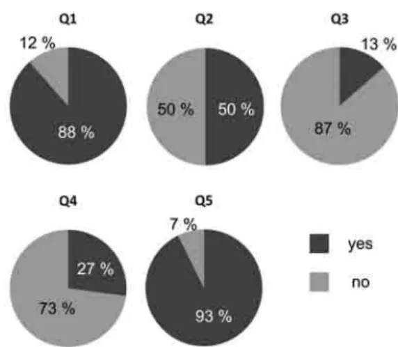
全国中学生ハンドボール選手権大会に出場した選 手および指導者にアンケート調査を依頼し,回答が 得られた 55 チーム,選手 704 名(男子 343 名,女子 361 名)およびその指導者 96 名を対象としてアンケ ート調査を行なった.アンケート内容は表 1 の通り で,選手には Q1)〜Q3)を,指導者には Q1)〜Q5)
を調査した(表 1).また,Q1)〜Q3)に関しては,
選手と指導者の回答を 2 群間で比較検討した.統計 学的検討にはχ2独立性の検定を用いて有意水準は 5%未満とした.
結 果
アンケート回収率は 58.5%(55 チーム/94 チーム)
で,回答率は男子が 52.8%(343 名/650 名),女子が 60.9%(361 名/593 名),指導者が 58.2%(96 名/165 名)であった.
その結果,選手に関しては 21.4%しか ACL 損傷 を知らず,受傷機転については 4.4%,予防トレー ニングについては 2.2%しか知らなかった.指導者 に関しては 88.5%が ACL 損傷を知っていたが,受 傷機転については 50%しか知らず,予防トレーニ ングについては 13.5%しか知らなかった.また 27.1
%の指導者が ACL 損傷を受傷した選手を受け持っ たことがあり,92.7%の指導者が予防トレーニング を実践したいと考えていた.Q1)〜Q3)のすべての 項目で選手・指導者間に統計学的有意差を認めた
(p<0.01)(図 1, 2).
この結果から,「選手の ACL 損傷に対する低い認 知度」と「選手と指導者との認識の乖離」,「指導者の 予防トレーニングを実践したいという強い要望」が 汲み取れた.
考 察
ACL 損傷はスポーツ選手にとって最も避けたい スポーツ外傷の 1 つであり,その予防に注目が集ま っている.現在の中学生選手および指導者の ACL に関する知識および ACL 損傷予防トレーニングに
1 1
表 1 アンケート内容 Q1)ACL 損傷を知っていますか.
Q2)ACL 損傷の受傷機転を知っていますか.
Q3) ACL 損傷の予防トレーニングを知っています か.
Q4) ACL 損傷を受傷した選手を受け持ったことが ありますか.
Q5) ACL 損傷の予防トレーニングを実践したいで すか.
0
関する知識の現状を把握することが今後,予防トレ ーニングの普及を行なうために有用であると考え,
本アンケート調査を行なった.その結果,「選手の ACL 損傷に対する低い認知度」と「選手と指導者と の認識の乖離」,「指導者の予防トレーニングを実践 したいという強い要望」が浮かび上がった.
1990 年代より ACL 損傷予防の取り組みが本格的 にスタートした.多くの報告では,予防トレーニン グを行なった群で ACL 損傷の発生頻度が減少し,
その有効性が述べられている 8〜12).また予防トレー ニングを対象となる種目や世代とうまく適合させる ことができれば,ACL 損傷予防の効果が期待でき ると考えられている 7).
次に ACL 損傷予防プログラムの介入時期につい て考察する.ACL 損傷は 10 歳代から 30 歳代に多 く,とくに 10 歳代で急激に増加する 11).一方,成 長期に神経筋協調訓練を行なうことで望ましい神経 筋機能の発達が引き起こされ,それによって膝関節 の理想的な動きが実現し,競技パフォーマンスが向 上すると報告されている 12).また本邦でのスポーツ は学校でのクラブ活動を中心に行なわれており,中 学校から本格的なクラブ活動が始まることが多い.
以上より,予防プログラムの介入時期は中学生がよ いのではないかと考えている.
本研究において,中学生選手とその指導者との ACL 損傷に関する知識の認知度を比較すると,選手 の認知度が有意に低いという結果が得られた.ACL 損傷予防プログラムの効果を高めるためには疾患に 対する正確な知識とコンプライアンスが重要であ
り 13, 14),中学生選手に対しても ACL 損傷の知識の
普及が必要であると考える.しかし,現実的にはす
べての中学生選手に対して,われわれが ACL 損傷 に関しての知識を直接伝達することは困難である.
今回の研究の結果をフィードバックし,ACL 損傷 に関するわかりやすいパンフレットや DVD を作成 し各大会で配布したり,また地域ごとの講演会など でわれわれのもつ知識の伝達を積極的に行なったり することが重要ではないかと考える.また「選手と 指導者との認識の乖離」があることから,指導者が 選手に対して自らもつ知識を伝達するように指導す ることも重要であると考えられる.そして「指導者 の予防トレーニングを実践したいという強い要望」
に対しては理学療法士やトレーナーと協力し,講演 会やセミナーなどを開催することで指導者に伝達し ていきたいと考えている.
結 語
全国中学生ハンドボール選手および指導者に対 し,ACL損傷に関するアンケート調査を行なった.
ACL 損傷の知識に関しては,選手と指導者との認 知度の乖離を埋めることが重要であり,また ACL 損傷予防トレーニングに関しては,具体的な方法を 指導者に提示することが効率よく予防トレーニング を普及させるために必要であると考えた.
文 献
1) de Loes M et al:A 7─year study on risks and
1 1 8
図 1 選手へのアンケート結果
図 2 指導者へのアンケート結果