Abbreviations: ATLL, adult T-cells leukemia-lymphoma; BAL, bronchoalveolar lavage; bp, base pair; DLco, pulmonary diffusing capacity for carbon monoxide; HAB, HTLV-I associated bronchopneumopathy; HABA, HTLV-I associated bronchiolo-alveolar disorder; HAM, HTLV-I associated myelopathy; HTLV-I, human T-cell lymphotropic virus type I; LDH, lactic acid dehydrogenase; PBMC, peripheral blood mononuclear T-cell; PCR, polymerase chain reaction; sIL-2R, soluble interleukin-2 receptor; TBLB, transbronchial lung biopsy; UIP, usual interstitial pneumonia; VC, vital capacity
Interstitial Pneumonia Developed in HTLV-I Carriers: Report of Two
Cases
Naoto Burioka, Hisashi Suyama, Yuji Sugimoto, Hiroki Chikumi, Hiroki Yajima, Katsuyuki Tomita, Eiji Hoshino, Yukio Matsumoto, Takao Sasaki and Makoto Motoi*
Third Department of Internal Medicine and *Second Department of Pathology, Faculty of Medicine, Tottori University, Yonago 683, Japan
Two carriers of human T-cell lymphotropic virus type I (HTLV-I) with interstitial pneumonia are described. The first case, a 60-year-old man, was admitted with cough and dyspnea on exertion. Light microscopy of a lung specimen obtained by a trans-bronchial lung biopsy (TBLB ) showed thickening of the alveolar walls with infiltration of lymphocytes and fibrosis of the pulmonary parenchyma. Immunohistochemical analy-sis of the TBLB specimen showed positive staining in the lymphocytes for UCHL-1. This case was suspected as HTLV-I associated bronchiolo-alveolar disorder. The second case, a 74-year-old man, visited our hospital because of a persistent productive cough and dyspnea on exertion. Light microscopy of the TBLB showed a slight thickening of the alveolar walls and fibrosis of the pulmonary parenchyma with minimal infiltration of lymphocytes. Only 2.2% of the bronchoalveolar lavage fluid consisted of lymphocytes. The findings of the second case suggest that some factors other than T-lymphocytes may be related with the development of interstitial pneumonia in HTLV-I carriers. Interstitial pneumonia in HTLV-I carriers may be caused by as yet undiscovered mecha-nisms. A cohort study involving residents of an area where HTLV-I is endemic should be conducted to clarify the mechanism of pulmonary involvement in HTLV-I carriers.
Key words: human T-cell lymphotropic virus type I; HTLV-I associated bronchiolo-alveolar
disorder; HTLV-I associated bronchopneumopathy; interstitial pneumonia; pulmonary fibrosis
Human T-cell lymphotropic virus type I (HTLV-I) is an oncogenic retrovirus. HTLV-I is endem-ic in the southwestern region of Japan. Most HTLV-I infected subjects are non-symptomatic carriers. HTLV-I has been implicated in the development of such diseases as HTLV-I asso-ciated myelopathy (HAM) (Roman and Osame, 1988), HTLV-I associated bronchiolo-alveolar disorder (HABA) (Kimura, 1992) [also called
as HTLV-I associated bronchopneumopathy (HAB) (Maruyama et al., 1988)], autoimmune thyroiditis (Akamine et al., 1996) and HTLV-I associated uveitis (Mochizuki et al., 1992). Inflammatory disorders can occur in the carrier state in HTLV-I infected individuals without adult T-cell leukemia-lymphoma (ATLL). We report two carriers of HTLV-I who exhibited interstitial pneumonia.
Patient Report Case 1
A 60-year-old Japanese man, a resident of Oki Island in Shimane Prefecture, was admitted to our hospital in March 1991 because of cough and dyspnea on exertion. He smoked 60 ciga-rettes daily for 30 years but quit smoking 2 years prior to admission. Auscultation of the lungs revealed fine crackles in both lower lung fields. Laboratory data showed an elevated white blood cell count (11,300/mm3; segmental neutrophils 44%, band neutrophils 4%, lympho-cytes 35%, monolympho-cytes 7%, eosinophils 10%) and lactic acid dehydrogenase (LDH; 257 IU/ L). Atypical lymphocytes were not detected in peripheral blood. Arterial blood gas analysis showed hypoxemia (PaO2; 67 mmHg). He tested negative for hepatitis C
virus antibody, rheumatoid factor and anti-nuclear antibody. HTLV-I antibody was positive (2560-fold). Bronchoalveolar lavage (BAL) was performed in the right B4 (Table 1). Although the total cell count in the BAL fluid was
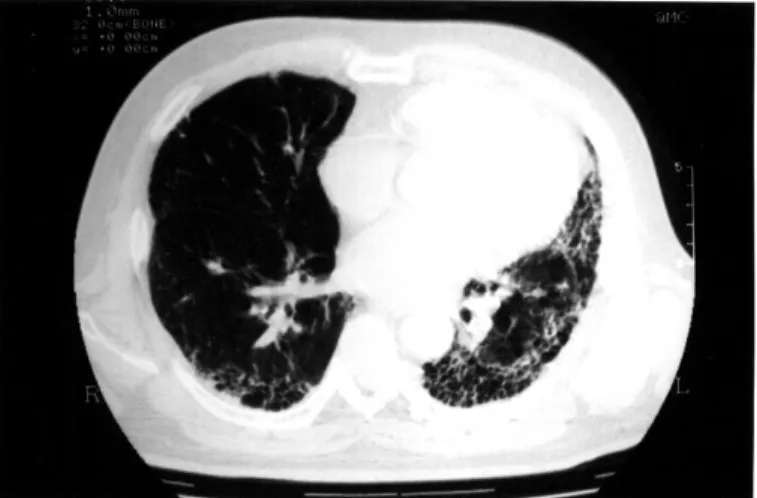
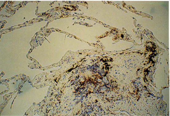
elevated, the ratio of lymphocytes was within the normal range (The BAL Cooperative Group Steering Committee, 1990). Spirography revealed a decreased vital capacity (VC) and a restrictive pattern (%VC, 68.4%). The pulmonary diffusing ca-pacity for carbon monoxide (DLco) was decreased (%DLco, 59.3%). Computed tomography of the chest demonstrated bilateral reticulo-nodular and honey-combed shadows in the lung fields (Fig.1). Light mi-croscopy of the transbronchial lung biopsy (TBLB) specimen showed thickening of t h e a l v e o l a r w a l l s w i t h i n f i l t r a t i o n o f lymphocytes and fibrosis of the pulmonary parenchyma. No atypical lymphocytes were detected in the infiltrating cells. Immunohisto-chemical analysis of the TBLB specimen show-ed positive staining of lymphocytes for UCHL-1 (Nichirei Co., Tokyo, Japan) by the strepto-avidin-biotin method [HISTOFINE SAB-PO(MULTI) KIT, Nichirei Co.] (Fig. 2).
This patient developed interstitial pneumo-nia and died in December 1992. The autopsy revealed the following: the lung—diffuse honey-combed lungs, usual interstitial pneumo-nia (UIP) and acute bronchopneumopneumo-nia; the heart—hypertrophy of the heart; the pancreas— chronic pancreatitis; the thyroid—adenomatous goiter; the bone marrow—no atypical findings in the bone marrow; the lymph nodes—no atypical lymphocytes in the lymph nodes.
Table 1. Results of bronchoalveolar lavage
Case 1 Case 2 Total cell count (106/dL) 23.0 66.0 Cell differentiation Macrophages (%) 86.0 97.6 Lymphocytes (%) 10.0 2.2 Neutrophils (%) 1.2 0.2 Eosinophils (%) 2.6 0 Basophils (%) 0.2 0 Lymphocytes subset CD3 (%) 39.6 83.4 CD4 (%) 24.7 60.8 CD8 (%) 19.3 16.4 CD4/CD8 1.3 3.7
Fig. 1. Computed tomography of the
Case 2
A 74-year-old Japanese man who lived in Yonago city in Tottori Prefecture visited our hospital in October 1995 because of a persistent productive cough, and dyspnea on exertion. He had stopped smoking 20 years previously, but until then, he had smoked 40 cigarettes daily for about 35 years. Abnormal findings on a chest radiograph were detected when he was about 60 years old. Chest auscultation revealed fine crack-les in both lower lung fields. Laboratory data revealed an increased erythrocyte sedimenta-tion rate (70 mm/h), but normal hematological findings (white blood cell count; 8900/mm3; segmental neutrophils 64%, band neutrophils 2%, lymphocytes 25%, monocytes 9%). No atypical lymphocytes were found in the peri-pheral blood. Serum LDH was within the nor-mal range (190 IU/L). Analysis of arterial blood gas showed hypoxemia (PaO2;71 mmHg). The patient tested negative for hepatitis C virus antibody, rheumatoid factor and antinuclear antibody. HTLV-I antibody was positive
(32,000-fold). Serum soluble interleukin-2 receptors (sIL-2R) were elevated (784 U/mL), but those in the BAL fluid were not (< 85 U/ mL). BAL was performed in the right B4 (Table 1). Although the total cell count in the BAL fluid was markedly elevated, only a low percent (2.2%) was lymphocytes. Pulmonary function testing demonstrated a restrictive pattern of disturbance (%VC, 54%) with a decreased DLco (%DLco, 61%). Computed tomography of the chest demonstrated bilateral reticulo-nodular and honey-combed shadows in the lung fields (Fig. 3). Light microscopy of the TBLB speci-men showed a slight thickening of the alveolar walls and fibrosis of the pulmonary paren-chyma with minimal infiltration by lympho-cytes. No atypical lymphocytes were detected in the infiltrating cells. Immunohistochemical analysis of the TBLB specimen showed partly positive staining of lymphocytes for UCHL-1 (Fig. 4).
To detect the HTLV-I provirus in the pe-ripheral blood lymphocytes of this patient, we examined the separated peripheral blood
mono-Fig. 2. Immunohistochemical staining of the specimen obtained from TBLB. Positive reactions of
nuclear cells (PBMCs). The PBMCs were incubated for 15 min at 70˚C with 1 mL of cell lysis buffer that consisted of 10 mM Tris-HCl, pH 7.5/10 mM EDTA, 1% sodium dodecyl sul-fate and protease K. Protease K was added once more and the solu-tion was incubated overnight at 37˚C. Total cellular DNA was ex-tracted according to the phenol-chloroform method. The poly-merase chain reaction (PCR) was performed by using the primers designed to amplify a 159-base pair (bp) segment in the pX re-gion of HTLV-I (Seiki et al.,
1983) (5'-CGGATACCCAGTCTACGTGT and 5'-GAGCCGATAACGCGTCCATCG, respectively) (Hatanaka and Kawanishi, 1990). Sample DNA (1 µg) was denatured at 95˚C, after which PCR was performed by step cycle at 94˚C for 20 s, at 50˚C for 30 s, and at 72˚C for 30 s for a total of 35 cycles. The amplified product was electrophoresed in 3% agarose gel and
Fig. 4. Immunohistochemical staining of the specimen obtained from TBLB. Positive reactions of a few
infiltrating lymphocytes for UCHL-1 are shown. × 100.
Fig. 3. Computed tomography of the chest in case 2.
transferred to Hybond N+ (Amersham Japan Co., Ltd., Tokyo, Japan). A 25-base oligo-nucleotide probe (5'-TACTGGCCACCTGTC CAGAGCATCA) that hybridized to an inter-vening sequence between two primers of HTLV-I was labeled with horseradish peroxy-dase by using ECL direct nucleic acid labeling and a detection system (Amersham Japan).
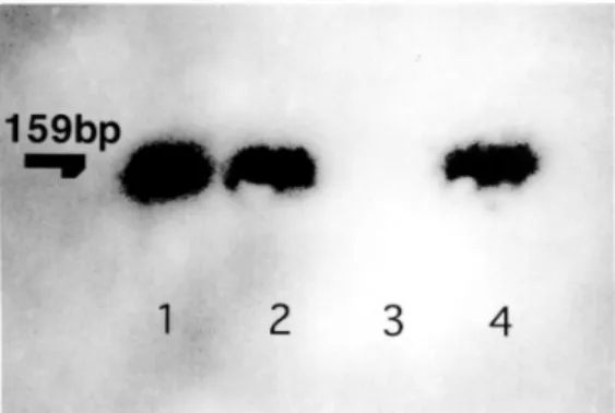
Results of PCR-Southern analysis of this pa-tient are shown in Fig. 5. A markedly amplified and hybridized product was detected.
Discussion
The incidence of pulmonary complications is markedly higher in ATLL than in other hemato-logical malignancies (Yoshioka et al., 1985). However, most of the HTLV-I infected subjects are non-symptomatic carriers. Even so, HTLV-I is implicated as a cause of such inflammatory disorders as HABA (Kimura, 1992) [or HAB (Maruyama et al., 1988)]. The pulmonary in-volvement seen in HABA (HAB) is mainly man-ifested as interstitial pneumonia (Maruyama et al., 1988; Setoguchi et al., 1991; Kimura, 1992) or a diffuse panbronchiolitis-like lesion (Kimura, 1992; Mukae et al., 1994; Kikuchi et al., 1996). The mechanism of pulmonary involvement with HTLV-I remains unclear. Salahuddin and coworkers (1984) reported that T-cells trans-formed in vitro by HTLV-I acquired the ability to release a considerable number of biologic response modifiers. Recent reports show that T-lymphocyte alveolitis is common in the chronic spastic myelopathy that is associated with HTLV-I infection (Sugimoto et al., 1987; Vernant et al., 1988). Sugimoto and coworkers (1989) reported that sIL-2R was increased in the BAL fluid of HAM patients, and suggested that the increased T-lymphocytes in the lung were activated locally to produce sIL-2R. Therefore, immune mechanisms may be im-portant in the development of pulmonary le-sions. The activation of T-lymphocytes by the HTLV-I tax gene may trigger an effect in the immune system (Tanaka et al., 1990). Maruyama and colleagues (1988, 1989, 1992) reported that HAB seemed to occur in healthy carriers as well as in patients with HAM. CD4+ lymphocytes and the ratio of CD4/CD8 were increased in the BAL fluid of these patients. These investi-gators also reported a high rate of T cells infil-tration in the lung specimens.
We encountered two HTLV-I carriers with pulmonary involvement. Case 1 died from
Fig. 5. PCR-Southern analysis of HTLV-I provirus
in peripheral blood lymphocytes of the patient in case 2. Lane 1, case 2; Lane 2, a HAM patient who is an HTLV-I carrier; Lane 3, an idiopathic inter-stitial pneumonia patient without HTLV-I; Lane 4, an HTLV-I carrier.
developed interstitial pneumonia in December 1992. The autopsy revealed that this patient was an HTLV-I carrier. In case 2, no atypical lymphocytes were detected in the peripheral blood. Since the HTLV-I provirus was detected in his peripheral blood by PCR (Fig. 5), we be-lieve that case 2 was also a carrier of HTLV-I.
The chest radiograph findings and histo-logical analysis in case 1 suggested a diagnosis of UIP. The total cell count in his BAL fluid was elevated compared with that of healthy ex-smokers (The BAL Cooperative Group Steer-ing Committee, 1990). The infiltrative lympho-cytes in the lung showed positive staining for UCHL-1 on immunohistochemical analysis (Fig. 2). UCHL-1 recognizes specifically the 180 kD low molecular weight isoform of CD45RO or leukocyte common antigen family (Terry et al., 1988). UCHL-1 labels most of the thymocytes, a subpopulation of resting T cells within both the CD4 and CD8 subsets, and the mature activated T cells. Although cells of the myelomonocytic series are also labeled by UCHL-1, normal B cells or NK cells are consis-tently negative (Smith et al., 1986). The UCHL-1-positive infiltrating lymphocytes were thought to be mainly T-cells in this case. We suspected that case 1 was HABA in broad sense, which has been reported (Kimura, 1992). The histological findings in case 2 revealed fibrosis of pulmonary parenchyma and minimal infiltration of lymphocytes that stained partly
positive for UCHL-1 (Fig.4). The total cell count of BAL fluid was markedly elevated, but the percentage of lymphocytes was low (2.2%) compared with that of healthy ex-smokers (The BAL Cooperative Group Steering Committee, 1990). The value of sIL-2 in the BAL fluid was not elevated. These findings differ from those of previous reports (Maruyama et al., 1988, 1989, 1992), and suggest that undetermined mechanisms may cause interstitial pneumonia in this HTLV-I carrier. Although the mecha-nism is unclear, activated alveolar macrophages may have played an important role in the development of pulmonary fibrosis (Martinet et al., 1987) in case 2, as suggested by the marked increase in the total count of alveolar macro-phages in his BAL fluid. Abnormalities shown on the chest radiograph in this case had been pointed out at least 14 years previously, demon-strating that long-term survival of HTLV-I carriers with interstitial pneumonia is possible. A cohort study involving residents of a region where HTLV-I is endemic will be needed to clarify the mechanism of pulmonary involve-ment in carriers of HTLV-I.
References
1 Akamine H, Takasu N, Komiya I, Ishikawa K, Shinjyo T, Nakachi K, et al. Association of HTLV-I with autoimmune thyroiditis in patients with adult T-cell leukaemia (ATL) and in HTLV-I carriers. Clin Endocrinol 1996;45:461–466. 2 The BAL Cooperative Group Steering
Commit-tee. Bronchoalveolar lavage constituents in healthy individuals, idiopathic pulmonary fibro-sis, and selected comparison groups. Am Rev Respir Dis 1990;141:s169–s202.
3 Hatanaka S, Kawanishi K. Understanding of HTLV-I through PCR work. Tanpakusitsu Kakusan Koso 1990;36:3055–3060 (in Japa-nese).
4 Kikuchi T, Saijo Y, Sakai T, Abe T, Ohnuma K, Tezuka F, et al. Human T-cell lymphotropic virus type I (HTLV-I) carrier with clinical mani-festation characteristic of diffuse panbron-chiolitis. Intern Med 1996;35:305–309.
5 Kimura I. HABA (HTLV-I associated bron-chiolo-alveolar disorder). Nippon Kyobu Shik-kan Gakkai Zasshi 1992;30:787–795 (in Japa-nese).
6 Martinet Y, Rom WN, Grotendorst GR, Martin GR, Crystal RG. Exaggerated spontaneous
release of platelet-derived growth factor by alve-olar macrophages from patients with idiopathic pulmonary fibrosis. N Engl J Med 1987;317: 202–209.
7 Maruyama S, Tihara I, Sakashita R, Mizoguchi S, Mori S, Usuku K, et al. HTLV-I associated bronchopneumopathy: a new clinical entity? Am Rev Respir Dis 1988;137(Pt 2):46 (abstract). 8 Maruyama S, Osame M. Pulmonary involvement
in HTLV-I carriers—A new clinical entity: HTLV-I associated bronchopneumopathy (HAB). Kokyu 1989; 8:261–266 (in Japanese). 9 Maruyama S, Mori S, Kawabata M, Osame M.
Bronchopneumopathy in HTLV-I associated myelopathy (HAM) and non-HAM HTLV-I carriers. Nippon Kyobu Shikkan Gakkai Zasshi 1992;30:775–779 (in Japanese).
10 Mochizuki M, Watanabe T, Yamaguchi K. Uveitis associated with human T lymphotropic virus type 1: seroepidemiologic, clinical and virologic studies. J Infect Dis 1992;166:943. 11 Mukae H, Higashiyama Y, Morikawa T, Kusano
S, Morikawa N, Kadota J, et al. Association be-tween diffuse panbronchiolitis and HTLV-I infection. Nippon Kyobu Shikkan Gakkai Zasshi 1994;32:17–24 (in Japanese).
12 Roman GC, Osame M. Identification of HTLV-I associated tropical spastic paraparesis and HTLV-I associated myelopathy. Lancet 1988; 1:651.
13 Salahuddin SZ, Markam PD, Linder SG. Lymphokine production by cultured human T cells transformed by human T-cell leukemia-lymphoma virus-I. Science 1984;223:703–706. 14 Seiki M, Hattori S, Hirayama Y, Yoshida M.
Human adult T-cell leukemia virus: complete nucleotide sequence of the provirus genome integrated in leukemia cell DNA. Proc Natl Acad Sci USA 1983;80:3618–3622.
15 Setoguchi Y, Takahashi S, Nukiwa T, Kira S. Detection of human T-cell lymphotropic virus type I-related antibodies in patients with lympho-cytic interstitial pneumonia. Am Rev Respir Dis 1991;144:1361–1365.
16 Smith SH, Brown MH, Rowe D. Functional subsets of human helper-inducer cells defined by a new monoclonal antibody UCHL-1. Immunol-ogy 1986;58:63–70.
17 Sugimoto M, Nakashima H, Watanabe S, Uyama E, Tanaka F, Ando M, et al. T-lymphocyte alveo-litis in HTLV-I-associated myelopathy. Lancet 1987;2:1220.
18 Sugimoto M, Nakashima H, Matsumoto M, Uyama E, Ando M, Araki S, et al. Pulmonary involvement in patients with HTLV-I-associated myelopathy: increased soluble IL-2 receptors in bronchoalveolar lavage fluid. Am Rev Respir Dis 1989;139:1329–1335.
(Received April 21, Accepted May 8, 1997) Maki M, Hatanaka M. Oncogenic transformation
by tax gene of human T-cell leukemia virus type I in vitro. Pro Natl Acad Sci USA 1990;87:1071– 1075.
20 Terry LA, Brown MH, Beverley PCL. The monoclonal antibody UCHL-1 recognizes a 180 kD component of the human leukocyte common antigen, CD45. Immunology 1988; 64:331–336.
21 Vernant JC, Buisson G, Magdeleine J, de Thore J, Jouannelle A, Neisson-Vernant C, et al. T-lymphocyte alveolitis, tropical spastic paresis, and Sjögren syndrome. Lancet 1988; 1:177. 22 Yoshioka M, Yamaguchi K, Yoshinaga T,
Takatsuki K. Pulmonary complications in pa-tients with adult T-cell leukemia. Cancer 1985; 55:2491–2495.