Abbreviations: MLC, myosin light chain; PBS, phosphate buffered saline; ROCK, Rho-associated coiled-coil domain
Rho-ROCK Expression Predicts the Prognosis in Patients
with T3/T4 Gastric Cancer
Kenjiro Taniguchi, Shunichi Tsujitani, Naruo Tokuyasu, Takuji Naka, Shigeru
Tatebe, Akira Kondo and Masahide Ikeguchi
Division of Surgical Oncology, Department of Surgery, School of Medicine, Tottori University Faculty of Medicine, Yonago 683-8504 Japan
A small GTPase Rho protein and an effector ROCK have significant roles in cancer adh-sion, metastasis, invaadh-sion, angiogenesis and cell mortality. We investigated the expres-sions of RhoA protein and ROCK-1 protein in 100 patients with macroscopically T3/T4 gastric cancer immunohistochemically. The expression of RhoA was detected in gastric cancer specimens from 39 patients and that of ROCK-1 in specimens from 30 patients. The clinicopathological characteristics of 21 tumors with co-expression of RhoA and ROCK-1 proteins (Rho/ROCK ON) were compared with those of the 79 remaining tu-mors (Rho/ROCK OFF). The percentage of lymph node metastasis positive cases in the Rho/ROCK ON group (81%) was higher than that in the Rho/ROCK OFF group (66%), but the difference was not significant (P = 0.183). However, the prognosis of the 21 pa-tients with Rho/ROCK ON was significantly poorer than that of the 79 with Rho/ROCK OFF (P = 0.006). Our results indicate that the evaluation of the protein expression of RhoA and ROCK-1 is useful for predicting the prognosis in patients with T3/T4 gastric cancer.
Key words: gastric carcinoma; prognosis; RhoA; ROCK-1
Small GTP-binding proteins are thought to be involved in signal transduction pathways that control sets of essential cellular functions such as reorganization of the actin cytoskeleton, gene expression, cell cycle progression, and cell-cell adhesion (Malliri and Collard, 2003). The small GTP-binding protein Rho is part of a subfamily of the Ras superfamily that includes Cdc42, Rac and Rho proteins. The Rho family has an impor-tant role in cancer development and progression, including cell-cell adhesion, cell motility, trans-formation, proliferation, invasion, and metastasis (Suwa et al., 1998; Pan et al., 2004; Kamai et al., 2003). Cancer cell migration is central to the process of metastasis. Rearranging the actin
cytoskeletal proteins in response to Rho is impor-tant for the ability of tumor cells to metastasize (Chrzanowska-Wodnicka, 1996; Benitah et al., 2004). Activation of Rho results in binding to various effector molecules that elicit downstream responses (Aznar, 2004). The ROCK family, Rho-associated serine-threonine protein kinase, is one of the effector molecules of Rho. ROCK is thought to participate in the induction of focal ad-hesions and stress fibers in cultured cells (Itoh et al., 1999). In addition, it has been suggested that ROCK-1, one of the isomers of ROCK may play an essential role in the invasion of cancer cells (Kaneko et al., 2002; Yoshinaga, 2003). Rho protein overexpression has been reported in the
progression of human cancers, suggesting that the Rho/ROCK pathway may be a molecular target for prevention of cancer invasion and metastasis (Sahai, 2003; Somlyo, 2003).
In order to elucidate the malignant potential of gastric cancer, multiple genetic alterations in-cluding activation of oncogenes and inactivation of tumor suppressor genes have been investigated vigorously. In studies on the expression levels of RhoA, RhoB, RhoC, Rac1, Rac2, Rac3 and Cdc42, increased expressions of RhoA and Rac1 were related to a higher TNM stage and undiffer-entiated type in gastric cancer (Pan et al., 2004). Accumulating clinical and experimental data sug-gest that RhoA working through ROCK protein kinases contributes to the invasive and metastatic abilities of gastric cancer. ROCK inhibitors, such as Y-27632 or WF-536, have been used in some studies (Takamura et al., 2001; Nakajima et al., 2003a). Although these inhibitors are potent and selective, it remains a possibility that these com-pounds inhibit additional kinases, such as PRK/ PKN (Davies et al., 2000). In addition, adminis-tration of ROCK inhibitors to whole animals may block tumor cell invasion by enhancing the barrier function of host cell layers (Wojciak-Stothard et al., 2001; Nakajima et al., 2003b). Furthermore, ROCK inhibitors would be useful antiangiogenic chemotherapeutic agents in tumors associated with elevated Rho/ROCK expression (Croft et al., 2004). However, Rho/ROCK expression in gas-tric cancer and its relationship to a patient’s prog-nosis has yet to be clarified.
Primary T3/T4 gastric cancer frequently metastasizes to the liver and to the peritoneum, and the prognosis of such cases remains unfavor-able. In the present study, we immunohistochemi-cally detected the protein expression of RhoA and ROCK-1 in patients with T3/T4 gastric cancer who underwent curative gastrectomy. We also investigated the clinical significance of the Rho/ ROCK pathway in advanced gastric cancer with special reference to the mode of recurrence and survival of the patients.
Materials and Methods
Patients
Primary T3/T4 gastric adenocarcinoma speci-mens were obtained from 100 patients who under-went curative gastrectomy at our institution from 1976 to 1995. The patients’ ages ranged from 31 to 91 years (average 64.8 years); 64 were male and 36 were female. The clinicopathological findings were determined according to the Japanese Clas-sification of Gastric Carcinoma by our hospital pathologists (Japanese Research Society for Gas-tric Cancer, 1995). All patients had undergone either distal, proximal or total gastrectomy with dissection of the level 1 and 2 regional lymph nodes (D2). The patients’ follow-up periods for survivors ranged from 2 to 216 months (median, 42 months) after surgery. Informed consent was obtained from all subjects and/or their guardians.
Immunohistochemistry
The methods used for immunostaining for RhoA protein were as described previously (Horiuchi et al., 2003). A streptoavidin-biotin kit (Histofine SAB-PO kit; Nichirei, Tokyo, Japan) was used for formalin-fixed and paraffin-embedded specimens. Sections (4 μm in thickness) were dewaxed in xylene, rehydrated in ethanol and then heated in an autoclave oven (120˚C) for 20 min for retrieval of antigens. Endogenous peroxidase was blocked with 0.3% hydrogen peroxide in methanol for 20 min. Tissue sections were then washed twice with phosphate buffered saline (PBS) and pre-blocked for 60 min with 10% rabbit serum. After washing with PBS, the samples were incubated overnight at 4˚C with anti-RhoA monoclonal an-tibody (sc-418, Santa Cruz Biotechnology, Santa Cruz, CA) at a dilution of 1:100. Sections were then washed three times in PBS and incubated with the secondary anti-mouse immunoglobulin (IgG + IgA + IgM) conjugated with biotin for 60 min, followed by incubation with a
streptavidin-peroxidase complex for another 60 min. After three additional washes in PBS, diaminobenzidine tetrahydrochloride working solution was applied. Finally, the slides were counterstained with he-matoxylin. Staining for ROCK-1 was performed with an anti-ROCK-1 polyclonal antibody (c-19, Santa Cruz Biotechnology) at a dilution of 1:100, using a similar method to that for the RhoA stain-ing. RhoA and ROCK-1 expressions were not de-tected in the non-neoplastic region of the resected specimens. Expressions of RhoA and ROCK-1 were evaluated according to the ratio of positive cells per specimen as described previously (Liu et al., 2004). The staining pattern of RhoA or ROCK-1 was classified as follows: negative (nega-tive or equivocal staining or when less than 20% carcinoma cells were detected) and positive (when 20% and more carcinoma cells were detected). Two observers who were unaware of the clinical data or the disease outcome examined all of the histological slides. When the interpretation dif-fered between the two observers, a conference microscope was used for re-evaluation and final decision.
Statistical analysis
The association of factors was evaluated by the
2-test. The significance of difference among
means was determined by the Mann-Whitney U test. The survival rates were estimated by the Kaplan-Meier method, and statistical analysis was carried out by the log-rank test for equality of the survival curves. The Cox proportional haz-ard model and a stepwise procedure assessed the influence of each variable on survival. The ac-cepted level of significance was P < 0.05.
Results
RhoA/ROCK-I expression and clinicopath-ological findings
RhoA and ROCK-1 expressions could not be de-tected in the non-neoplastic gastric mucosas or in the underlying stromal cells. They were detected only in cancer cells (Fig. 1). We decided that 39 cases (39%) were RhoA positive and 30 cases (30%) were ROCK-1 positive (Table 1). RhoA and ROCK-1 were co-expressed in 21%. No
sig-Fig. 1. Immunohistochemical staining pattern of RhoA and ROCK-1 in gastric cancer. Protein expressions of RhoA and ROCK-1 were detected in gastric cancer cell nuclei. A: RhoA positive case. B: ROCK-1 positive case. Bar = 100 μm (both in A and B).
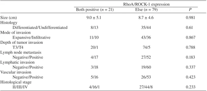
Table 2. Coexpression of RhoA and ROCK-1 proteins and clinicopathological parameters RhoA/ROCK-1 expression
Both positive (n = 21) Else (n = 79) P
Size (cm) 9.0 ± 5.1 8.7 ± 4.6 0.981
Histology
Differentiated/Undifferentiated 8/13 35/44 0.61
Mode of invasion
Expansive/Infiltrative 11/10 43/36 0.867
Depth of tumor invasion
T3/T4 20/1 74/5 0.788
Lymph node metastasis
Negative/Positive 4/17 27/52 0.183 Lymphatic invasion Negative/Positive 3/18 19/60 0.337 Vascular invasion Negative/Positive 5/16 26/53 0.423 Histological stage II/III/IV 4/16/1 27/44/8 0.233
Table 3. Correlation between RhoA/ROCK-1 protein expression and mode of recurrence
Recurrence pattern RhoA/ROCK-1 expression
Both positive (n = 21) Else (n = 79) P
Peritoneal 7 (33%) 18 (23%) 0.321
Hematogenic 5 (24%) 9 11%) 0.145
Nodal 1 ( 5%) 4 ( 5%) 0.955
Remnant stomach or others 2 ( 9%) 6 ( 8%) 0.772
Total 15 (71%) 37 (47%) 0.045
nificant correlation was detected between these expressions and clinicopathological parameters including age, sex, tumor size, histology, mode of invasion, lymph node metastasis, lymphatic-vascular space invasion and histological stage (data not shown).
The clinicopathological characteristics of 21 patients with tumors co-expressing RhoA and ROCK-1 were compared with those of the other 79 patients. The lymph node metastases were ob-served in 81% of the former group in comparison to 66% of the latter (P = 0.183, Table 2).
Mode of recurrence
Fifty-two patients died of gastric cancer during the follow-up period. Fifteen of 21 patients (71%) with the tumor co-expressing Rho and ROCK-1 died, while 37 of 79 patients (47%) with the
tu-Table 1. Correlation of RhoA and ROCK-1 ex-pression in 100 gastric cancers
ROCK-1 expression Positive Negative Total RhoA expression
Positive 21 18 39
Negative 9 52 61
Total 30 70 100
The association of RhoA and ROCK-1 expression: P < 0.001.
mor not expressing both Rho and ROCK-1 died as well (P = 0.045). Peritoneal metastatic recur-rences were frequent in both groups. However, the hematogenic metastases (to liver, lung or bone) tend to be more frequent in patients with Rho and ROCK-1 co-expressing tumors (9/79 [11%], P = 0.145, Table 3).
Prognosis of patients
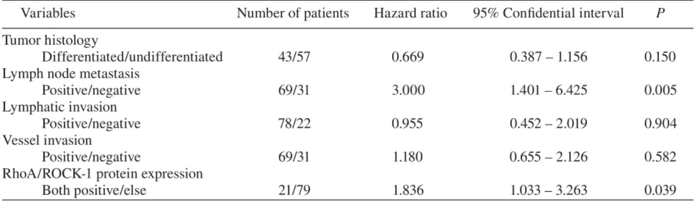
The overall 5-year survival rate of 100 patients with T3/T4 gastric cancer was 40%, and the disease-specific 5-year survival rate of these patients was 42%. The disease-specific 5-year survival rate of 21 patients with both RhoA- and ROCK-1-positive tumors (19%) was significantly lower than that of the remaining 79 patients (48%, P = 0.006, Fig. 2). Multivariate analysis was performed with the Cox proportional hazards model using the co-variates for histological type, lymph node metas-tasis, lymphatic invasion, venous invasion and co-expression of RhoA and ROCK-1 (Table 4). The analysis revealed that lymph node metastasis and RhoA/ROCK-1 expression independently contrib-uted to shortened survival.
Discussion
Rho family GTPases are key regulators of many different biological processes including cell motil-ity (Etienne-Manneville and Hall, 2002; Coleman et al., 2002). When bound to GTP, Rho family proteins recruit effector proteins that influence the architecture of the actin cytoskeleton. RhoA and RhoC activate the serine/threonine kinases ROCK-1 and ROCK-2. ROCK activation pro-motes the stabilization of filamentous actin (F-actin), phosphorylation of regulatory myosin light chains (MLCs), the increase in myosin ATPase activity and the coupling of actin-myosin force
generation and contractility. In fibroblasts grown in tissue culture, RhoA signals through ROCK-1 and ROCK-2 to promote the formation of stress fibers and focal adhesions that lead to contractile actin-myosin bundles (Leung et al., 1996; Uehata et al., 1997; Riento, 2003). Moreover, genetic experiments in Drosohila revealed that elevated Rho signaling in epithelial cells resulted in a loss of epithelial characteristics and increased invasive migratory behavior (Speck et al., 2003).
Co-overexpression of Rho and ROCK pro-teins in cancer cells has been reported in ovarian cancer, pancreatic cancer, testicular cancer and bladder cancer (Suwa et al., 1998; Kamai et al.,
Fig. 2. The disease-specific survival curves of patients with T3/T4 gastric cancer according to RhoA/ROCK-1 protein expression. The disease-specific survival curve of 21 patients with both RhoA- and ROCK-1-positive tumors (Rho/ROCK ON group) was significantly worse than that of remaining 79 patients (Rho/ROCK OFF group, P = 0.006).
Table 4. Multivariate survival analysis by the Cox proportional hazards model
Variables Number of patients Hazard ratio 95% Confidential interval P
Tumor histology
Differentiated/undifferentiated 43/57 0.669 0.387 – 1.156 0.150
Lymph node metastasis
Positive/negative 69/31 3.000 1.401 – 6.425 0.005
Lymphatic invasion
Positive/negative 78/22 0.955 0.452 – 2.019 0.904
Vessel invasion
Positive/negative 69/31 1.180 0.655 – 2.126 0.582
RhoA/ROCK-1 protein expression
2003, 2004). Among Rho family and ROCK pro-teins, RhoA and ROCK-1 proteins were reported to play important roles in carcinogenasis, cancer cell adhesion, metastasis, invasion and angiogen-esis (Itoh et al., 1999; Matsumoto et al., 2001; Igishi et al., 2003; Horiuchi et al., 2003; Shikada et al., 2003). On the other hand, RhoB plays a tumor-suppressive role in human lung cancer (Mazieres et al., 2004). Thus, to investigate the possible prognostic value, we analyzed RhoA and ROCK-1 protein expressions in advanced gastric cancer immunohistochemically.
In studies on gastric cancer concerning the expression levels of RhoA, RhoB, RhoC, Rac1, Rac2, Rac3 and Cdc42, increased expressions of RhoA and Rac1 were related to a higher TNM stage and undifferentiated type in gastric carcino-ma (Pan et al., 2004). In the current study, 21% of tumors were positive for both Rho and ROCK-1: they seemed to have a higher tendency for lymph node metastasis, lymphatic invasion, and vascular invasion. They did not show a significantly higher propensity of tumor invasion. However, we found that the prognosis of double-positive patients was significantly poorer than that of the other patients. Also, multivariate survival analysis revealed that the double positive tumor was an independent prognostic factor for advanced gastric cancer as well as lymph node metastasis. To our knowl-edge, this is the first report to suggest that patients with RhoA/ROCK-1 double positive gastric can-cer have a significantly poor prognosis.
References
1 Aznar S, Fernandez-Valeron P, Espina C, Lacal JC. Rho GTPase: potential candidates for anticancer therapy. Cancer Lett 2004;206:181–191.
2 Benitah SA, Valeron PF, van Aelst L, Marshall CJ, Lacal JC. Rho GTPases in human cancer: an unre-solved link to upstream and downstream transcrip-tional regulation. Biochim Biophys Acta 2004;1705: 121–132.
3 Chrzanowska-Wodnicka M, Burridge K. Rho-stimulat-ed contractility drives the formation of stress fibers and focal adhesions. J Cell Biol 1996;133: 1403–1415.
4 Coleman ML, Marshall CJ, Olson MF. RAS and RHO GTPases in G1-phase cell-cycle regulation. Nat Rev Mol Cell Biol 2004;5:355–366.
5 Croft DR, Sahai E, Mavria G, Li S, Tsai J, Lee WM, et al. Conditional ROCK activation in vivo induces tumor cell dissemination and angiogenesis. Cancer Res 2004;64:8994–9001.
6 Davies Sp, Reddy H, Caivano M, Cohen P. Specificity and mechanism of action of some commonly used pro-tein kinase inhibitors. Biochem J 2000;351:95–105. 7 Etienne-Manneville S, Hall A. Rho GTPases in cell
biology. Nature 2002;420:629–635.
8 Horiuchi A, Imai T, Wang C, Ohira S, Feng Y, Nikaido T, et al. Up-regulation of small GTPases, RhoA and RhoC, is associated with tumor progres-sion in ovarian carcinoma. Lab Invest 2003;83: 861–870.
9 Igishi T, Mikami M, Murakami K, Matsumoto S, Shigeoka Y, et al. Enhancement of cisplatin-induced cytotoxicity by ROCK inhibitor through suppression of focal adhesion kinase independent mechanism in lung carcinoma cells. Int J oncol 2003;23:1079– 1085.
10 Itoh K, Yoshioka K, Akedo H, Uehata M, Ishizaki T, Narumiya S. An essential part for rho-associated ki-nase in the transcellular invasion of tumor cells. Nat Med 1999;5:221–225.
11 Japanese Research Society for Gastric Cancer. Japa-nese Classification of Gastric Carcinoma. First Eng-lish Edition. Tokyo: Kanehara; 1995.
12 Kamai T, Tsujii T, Arai K, Takagi K, Asami H, Ito Y, et al. Significant association of Rho/ROCK pathway with invasion and metastasis of bladder cancer. Clin Cancer Res 2003;9:2632–2641.
13 Kamai T, Yamanishi T, Shirataki H, Takagi K, Asami H, Ito Y, et al. Overexpression of RhoA, Rac1, and Cdc42 GTPase is associated with progression in testicular can-cer. Clin Cancer Res 2004;10:4799–4805.
14 Kaneko K, Satoh K, Masa mune A, Satoh A, Shimosegawa T. Expression of ROCK-1 in human pan-creatic cancer: its down-regulation by morpholino oligo antisense can reduce the migration of pancreatic cancer cells in vitro. Pancreas 2002;24:251–257.
15 Leung T, Chen XQ, Manser E, Lim L. The p160 RhoA-binding kinase ROK alpha is a member of a kinase family and is involved in the reorganization of the cyto-skeleton. Mol Cell Biol 1996;16:5313–5327.
16 Liu N, Bi F, Pan Y, Sun L, Xue Y, Shi Y, et al. Re-versal of the malignant phenotype of gastric cancer cells by inhibition of RhoA expression and activity. Clin Cancer Res 2004;10:6239–6247.
17 Malliri A, Collard JG. Role of Rho-family proteins in cell adhesion and cancer. Curr Opin Cell Biol 2003;15:583–589.
18 Matsumoto Y, Tanaka K, Harimaya K, Nakatani F, Matsuda S, Iwamoto Y. Small GTP-binding protein,
Rho, both increased and decreased cellular motility, activation of Matrix Metalloproteinase 2 and inva-sion of human osteosarcoma calls. Jpn J Cancer Res 2001;492:429–438.
19 Mazieres J, Antonia T, Daste G, Muro-Cacho C, Berchery D, Tillement V, et al. Loss of RhoB expres-sion in human lung cancer progresexpres-sion. Clin Cancer Res 2004;10:2742–2750.
20 Nakajima M, Hayashi K, Egi Y, Katayama K, Amano Y, Uehata M, et al. Effect of Wf-536, a novel ROCK inhibitor, against metastasis of B16 melanoma. Cancer Chemother Pharmacol 2003a;52:319–324.
21 Nakajima M, Katayama K, Tamechika I, Hayashi K, Amano Y, Uehata M, et al. WF-536 inhibits metastatic invasion by enhancing the host cell barrier and inhibit-ing tumour cell motility. Clin Exp Pharmacol Physiol 2003b;30:457–463.
22 Pan Y, Bi F, Liu N, Xue Y, Yao X, Zheng Y, et al. Expression of seven main Rho family members in gastric carcinoma. Biochem Biophys Res Commun 2004;315:686–691.
23 Riento K, Ridley AJ. Rocks: multifunctional kinases in cell behavior. Nat Rev Mol Cell Biol 2003;4: 446–456. 24 Sahai E, Marshall CJ. Differing modes of tumour
cell invasion have distinct requirements for Rho/ ROCK signaling and extracellular proteolysis. Nat Cell Biol 2003;5:711–719.
25 Shikada Y, Yoshino I, Okamoto T, Fukuyama S, Kameyama T, Maehara Y. Higher expression of RhoC is related to invasiveness in non-small cell lung carci-noma. Clin Cancer Res 2003;9:5282–5286.
26 Somlyo AV, Phelps C, Dipierro C, Eto M, Read P, Barrett M, et al. Rho kinase and matrix
metallopro-teinase inhibitors cooperate to inhibit angiogenesis and growth of human prostate cancer xenotrans-plants. FASEB J 2003;17:223–234.
27 Speck O, Hughes SC, Noren NK, Kulikauskas RM, Fehon RG. Moesin functions antagonistically to the Rho pathway to maintain epithelial integrity. Nature 2003;421:83–87.
28 Suwa H, Ohshio G, Imamura T, Watanabe G, Arii S, Imamura M, et al. Overexpression of the rhoC gene correlates with progression of ductal adenocarci-noma of the pancreas. Br J Cancer 1998;77:147–152. 29 Takamura M, Sakamoto M, Genda T, Ichida T,
Asakura H, Hirohashi S. Inhibition of intrahepatic metastasis of human hepatocellular carcinoma by Rho-associated protein kinase inhibitor Y-27632. Hepatology 2001;33:577–581.
30 Uehata M, Ishizaki T, Satoh H, Ono T, Kawahara T, Morishita T, et al. Calcium sensitization of smooth muscle mediated by a Rho-associated protein kinase in hypertension. Nature 1997:389:990–994.
31 Yoshinaga K, Inoue H, Tanaka F, Mimori K, Utsunomiya T, Mori M. Platelet-derived endothelial cell growth factor mediates Rho-Associated coiled-coli domain kinase messenger RNA expression and pro-motes cell motility. Ann Surg Oncol 2003;10: 582–587. 32 Wojciak-Stothard B, Potempa S, Eichholtz T, Ridley
AJ. Rho and Rac but not Cdc42 regulate endothelial cell permeability. J Cell Sci 2001;114:1343–1355.
Received January 29, 2007; accepted February 13, 2007 Corresponding author: Kenjiro Taniguchi