Yonago Acta medica 2001;44:125–130
Abbreviations: CT, computed tomography; MRI, magnetic resonance imaging
Malignant Oncocytoma of the Parotid Gland: Report of a Case and
Review of Literature
Katsuyuki Kawamoto, Kei Nakahara and Hiromi Takeuchi
Department of Oto-Rhino-Laryngology, Tottori University Faculty of Medicine, Yonago 683-0826 Japan
We reported a case of malignant oncocytoma arising from the right parotid gland through neck metastasis. In 1992, the patient was treated by surgical removal of a lymph-node mass, diagnosed as malignant unclassified tumor. In 1995, a right parotid gland tumor and right neck lymph-node masses occurred. We suspected the patient of having a malignant tumor of the right parotid gland through neck metastasis, especially recurrence of the previous clear-cell carcinoma. In July 1995, a right-side total parot-idectomy and a neck dissection were carried out. A definite diagnosis was based on histological findings of the resected tumor as malignant oncocytoma: large eosinophilic granular cells were detected, and electron-microscopically, cytoplasms of tumor cells were rich in mitochondria with no encapsulation, perineural and intravascular invasion, and metastases to the cervical lymph nodes. Additional therapy such as chemotherapy and irradiation were not applied. To date, he has had no evidence of disease for 6 years.
Key words: malignant oncocytoma; parotid gland; parotidectomy
Malignant oncocytoma is an extremely rare tumor, especially when it arises in the major salivary glands. To date, about 50 cases have been reported in the literature. Of them, most arose in the parotid gland: 34 cases of parotid gland malignant oncocytoma have been report-ed since the first report by Bauer and Bauer (1953). The oncocyte is a large eosinophilic granular cell which can be seen in the glandular tissue, especially in the major salivary gland.
There are no characteristic examinations except for histological examinations for this disease due to its low incidence. Histological examinations of specimens obtained by needle aspiration biopsy and surgical removal are thought to be useful for diagnosis. The most desirable therapy is a curative operation, while irradiation and chemotherapy are thought to exert poor effects. Several cases of distant metastasis to the lung, liver and brain were re-ported, in which patients suffered a fatal out-come (Date et al., 1999). Long-term
observa-tion is necessary in treating patients with malig-nant oncocytoma which spreads by distant metastasis.
Patient report
In September 1992, a 70-year-old Japanese man was referred to our clinic at the University Hos-pital with a 1-month history of a slowly enlarg-ing mass in the right side of the neck. The mass was about 4 × 3 cm in size: we highly suspected its malignancy, and carried out a right-side neck dissection within the month. Histological examination revealed the specimen as a malig-nant unclassified tumor, which was highly sus-pected of being a clear-cell carcinoma. After surgery, we performed a detailed examination with magnetic resonance imaging (MRI) and scintigraphy, and observed no abnormal find-ings in general. Additional treatments such as chemotherapy or irradiation were not carried
out. The primary site of this tumor was un-known at that time.
In June 1995, he noticed a painless swelling in the right parotid gland region, without facial nerve paralysis. Examination by computed to-mography (CT) with sialography showed 2 masses about 1.8 cm and 1.4 cm in diameter at the inferior and dorsal portion of the right parotid gland, which were suspected of being hypervascular tumors. CT with sialography showed another mass about 1.2 cm in diameter in the right side of the neck, beyond the range of the previous operation (Figs. 1A–C). We sus-pected the patient of having a malignant tumor of the right parotid gland and neck metastasis: especially, recurrence of the previous clear-cell carcinoma. In July 1995, a right-side total parotidectomy and a neck dissection were carried out under general anesthesia. The 2 extirpated tumors were elastic hard, about 2 × 2 cm in size. Macroscopically, the cut surface was black in color and homogenous. Micro-scopically, the tumor cells showed a solid,
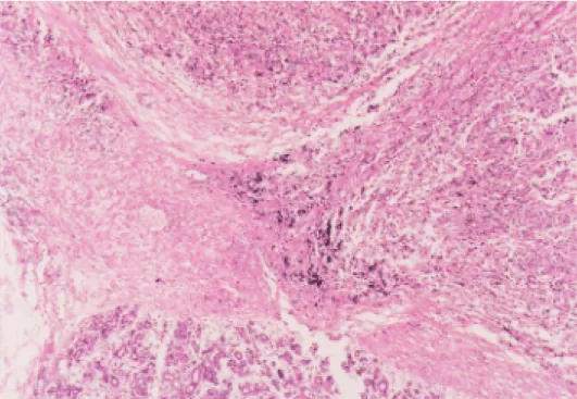
trabecular or alveolar pattern (Fig. 2). The nuclei of the tumor cells were round in shape and relatively uniform in size. No encapsu-lation, perineural and intravascular invasion were observed. The cytoplasm showed eosino-philia on hematoxylin and eosin stain (Fig. 3). Observed under an electron microscope, the cytoplasm of tumor cells had rich amounts of
Fig. 1. Computed tomographic images showing
tumors (arrows in A and B) in the parotid gland and neck metastasis lymph node (arrow in C).
A
B
(upper right)Malignant oncocytoma of the parotid gland
Fig. 2. The tumor cells show a solid, trabecular or alveolar pattern. Hematoxylin and eosin, × 40.
Fig. 3. Nuclei of the tumor cells are uniform in size and cytoplasm showing eosinophilia. Hematoxylin and
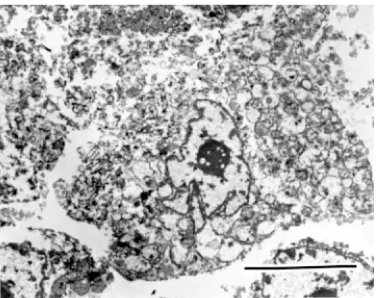
mitochondria (Fig. 4). The present findings were almost the same as in the previous histological features, but the number of clear cells was higher in 1992. The final diagnosis of the specimen was malignant oncocytoma with metastasis of the cervical lymph nodes. The postoperative course was uneventful, with no additional chemotherapy or irradiation admin-istered. The patient’s recovery has been follow-ed up for 6 years with no signs of recurrence.
Discussion
The oncocyte is a large eosinophilic granular cell which can be seen in the glandular tissue, especially in the major salivary gland. It is thought that the oncocyte increases with age in the parotid gland. A benign oncocytic tumor is called an oncocytoma, or mitochondrioma due to electron-microscopic features. Oncocytomas
Fig. 4. Ultrastructure features of the tumor cells. The tumor cell is rich in mitochondria. × 3900. Scale bar = 1 µm.
of the salivary glands are rare benign tumors, comprising less than 1% of all salivary tumors (Ellis et al., 1996). Further, malignant onco-cytomas in the head and neck region are much rarer, reported in only about 50 cases in the literature. To date, 34 cases of malignant onco-cytoma in the parotid gland were reported since the first report by Bauer and Bauer (1953). Nakada et al. (1998) reviewed in detail the literature on malignant oncocytomas except for those in the parotid gland: those tumors have been found in the submandibular gland, palate, maxillar sinus, ethmoid sinus, nasal cavity, kidneys, thyroid, mediastrinum and lungs.
There are several criteria fot determining the histological features of malignant oncocyto-ma: i) scattered mitoses and a focal cellular pleomorphism with local extension outside the capsule into adjacent soft tissue, bone or para-parotid lymph nodes; ii) perineural, lymphatic or intravascular invasion and iii) regional or
Malignant oncocytoma of the parotid gland
distant metastases (Gray et al., 1976). In the present case, absence of encapsulation, peri-neural and intravascular invasion, and meta-stases to the cervical lymph nodes were the criteria in establishing a diagnosis of malignant oncocytoma. In our case, though considering retrospectively, histological findings in 1992 and 1995 were the same as our results, but clear cells in the tumor cells were more frequent in 1992. The cervical lymph-node mass treated in 1992 was possibly based on a parotid gland malignant oncocytoma, which was not yet a proven discovery in 1992.
In the 34 cases of malignant oncocytoma of the parotid gland, the age of the affected pa-tients ranged from 30 to 91 years with a mean of 58 years, and the male to female ratio was 2:1 (Mahnke et al., 1998). Ardekian et al. (1999) reported that the main clinical symptom of malignant oncocytoma in the salivary gland was pain in 82.9% of the cases.
Sugimoto et al. (1993) reported that onco-cytomas commonly presented as a parotid mass and pain as well as facial nerve paralysis, and that such symptoms were involved with 1/3 of the patients. The present patient’s main symp-tom was a slowly progressive cervical mass with no pain and no facial nerve paralysis.
Except for histological examinations, no characteristic features have been reported on examinations by CT, MRI and echography. Scintigraphy with technetium showed an up-take ratio similar to benign oncocytoma and other malignant tumors in the head and neck (Kawamoto et al., 2000). Histological exami-nation by needle aspiration biopsy is thought to be useful for diagnosis (Laforga et al., 1994; Rajan et al., 1994).
Almost all the patients have been treated with surgery because other therapies such as irradiation and chemotherapy are thought to produce poor effects. Date et al. (1999) report-ed recurrent cases in which surgical resection and irradiation were performed: additional surgical neck dissections were needed.
Recurrences occurred in 25% to 52% of the cases (Mahnke et al., 1996). Intervals between surgery and recurrence ranged from 10 months to 10 years (Sugimoto et al., 1993). Distant
metastases into the lungs, liver and brain occur-red in several patients who suffeoccur-red fatalities (Date et al., 1999). Ardekian et al. (1999) as-serted that malignant oncocytomas appear to be good in short-term survival, but poor in long-term survival because of distant metastasis. Therefore, we determined that total parotidec-tomy and neck dissection were necessary in treating the malignant oncocytoma in the parot-id gland with neck metastasis. Irradiation and chemotherapy are thought to cause poor effects at present. Long observation is necessary after therapy, because the long-term survival rate is poor. In the present patient, parotidectomy and neck dissection were carried out, and he has had no evidence of recurrence for 6 years.
References
1 Ardekian L, Manor R, Peled M, Laufer D. Malig-nant oncocytoma of the parotid gland: case report and analysis of the literature. J Oral Maxillofac Surg 1999;57:325–328.
2 Bauer W, Bauer J. Classification of glandular tumors of salivary glands. Study of 143 cases. Arch Pathol Lab Med 1953;55:328–346. 3 Date T, Ueda T, Shirane M, Yamane T.
Malig-nant oncocytoma arising from a minor salivary gland of the buccal mucosa: a case report. Jibi-inkoka Rinsho 1999;102:117–120 (in Japanese). 4 Dimitrios K, Segas J, Papadimitriou K,
Koutsomanis P, Adamopoulos G. Malignant oncocytoma of the parotid with oncocytic change of the contralateral gland. Am J Otolaryngol 1995;16:200–204.
5 Ellis GL, Auclair PL. Tumors of the salivary glands. In Ellis GL, Auclair PL, eds. Atlas of tumor pathology. Washington D.C.: Armed Forces Institute of Pathology; 1996. p.318–324. 6 Gray SR, Cornog JL, Seo IS. Oncocytic
neo-plasms of salivary glands; a report of fifteen cases including two malignant oncocytomas. Cancer 1976;38:1306–1317.
7 Kawamoto K, Takeuchi H, Nakahara K, Hanamoto M. Oncocytoma of parotid gland: a case report. Jibi To Rinsho 2000;46:152–156(in Japanese).
8 Laforga JB, Aranda FI. Oncocytic carcinoma of parotid gland: fine needle aspiration and histol-ogic findings. Diagnostic Cytopathology 1994; 11:376–379.
9 Mahnke CG, Jänig U, Werner A. Metastasizing malignant oncocytoma of the submandibular gland. J Laryngol Otol 1998;112:106–109.
10 Nakada M, Nishizaki K, Akagi H, Masuda Y, Yoshino T. Oncocytic carcinoma of the subman-dibular gland: a case report and the literature review. J Oral Pathol Med 1998;27:225–228. 11 Rajan PB, Wadehra V, Hemming JD, Hawkesford
JE. Fine needle aspiration cytology of malignant oncocytoma of the parotid gland: a case report. Cytopathology 1994;5:110–113.
12 Sugimoto T, Wakizono S, Uemura T, Tsuneyoshi
M, Enjoji M. Malignant oncocytoma of the parotid gland: a case report with an immuno-histochemical and ultrastructural study. J Laryngol Otol 1993;107:69–74.
Received May 7, 2001; accepted May 28, 2001 Corresponding author: Dr. Katsuyuki Kawamoto