Title Isolation of secondary metabolites from medicinal plants andinvasive alien species and their biological activities( 本文 (Fulltext) )
Author(s) ANTONI PARDEDE
Report No.(Doctoral Degree) 博士(工学) 工博甲第533号 Issue Date 2018-03-25 Type 博士論文 Version ETD URL http://hdl.handle.net/20.500.12099/75260 ※この資料の著作権は、各資料の著者・学協会・出版社等に帰属します。
ISOLATION OF SECONDARY METABOLITES FROM MEDICINAL PLANTS AND INVASIVE ALIEN SPECIES AND THEIR
BIOLOGICAL ACTIVITIES
ANTONI PARDEDE
ISOLATION OF SECONDARY METABOLITES FROM MEDICINAL PLANTS AND INVASIVE ALIEN SPECIES AND THEIR
BIOLOGICAL ACTIVITIES
MATERIALS ENGINEERING DIVISION GRADUATE SCHOOL OF ENGINEERING
GIFU UNIVERSITY JAPAN
ANTONI PARDEDE
December 2017
Preface
The studies presented in this thesis have been carried out under the guidance of Professor Mamoru Koketsu at Materials Engineering Division, Graduate School of Engineering, Gifu University, during 2015-2018.
The studies are concerned with isolation of secondary metabolites from medicinal plants and invasive alien species and their biological activities.
February 2018
Summary
Plants are the tremendous source for the discovery of secondary metabolites with biological activity developments. In this study, the isolation of secondary metabolites from medicinal plants and invasive alien species and their biological
activities were investigated.
This thesis consists of 4 chapters. The first chapter describes antioxidant and antileukemic activity of chemical constituents from bark of Mangifera casturi. The second chapter describes flavonoid rutinosides from Cinnamomum parthenoxylon leaves and their hepatoprotective activity. The third chapter is
isolation of secondary metabolites from Stenochlaena palustris stems and structure activity relationship (SAR) of 20-hydroxyecdysone derivatives on antitermite activity. Finally, the fourth chapter describes chemical constituents of
Coreopsis lanceolata stems and their antitermite activity against Coptotermes curvignathus.
In chapter 1, the antioxidant and antileukemic activity of chemical components from bark of Mangifera casturi was investigated. Research findings
have shown that several extracts of Mangifera casturi have potent bioactivity. The methanol extract of Mangifera casturi bark was partitioned successively to yield hexane (Hex) fraction (6.7%), ethyl acetate (EtOAc) fraction (24.1%) and
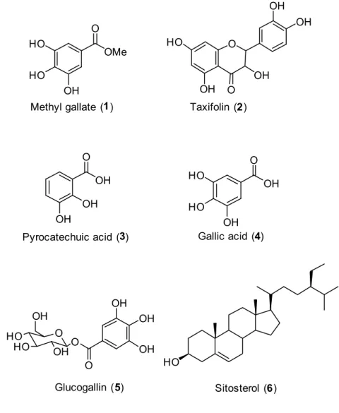
n-butanol (n-BuOH) fraction (28.1%). Five compounds were isolated from EtOAc fraction and one compound was isolated from n-hexane fraction. These
compounds were identified as methyl gallate (1), taxifolin (2), pyrocatechuic acid (3), gallic acid (4), glucogallin (5), and β-sitosterol (6), respectively; they were
confirmed by spectroscopic analysis and ultra-performance liquid chromatography electrospray ionization time-of-flight mass spectrometry (UPLC-ESITOFMS). All compounds were isolated from bark of Mangifera casturi for the first time. The EtOAc fraction as well as the isolated gallic acid (4) showed potent antioxidative and antileukemic activity against Human Leukemia HL-60 cells.
In chapter 2, flavonoid rutinosides were isolated from Cinnamomum parthenoxylon leaves and their hepatoprotective and antioxidant activity were
evaluated. The EtOAc fraction of C. parthenoxylon leaves showed potent hepatoprotective activity on tert-butyl hydroperoxide (t-BHP) induced
cytotoxicity in human hepatoma (HepG2) cells and also higher antioxidant activity. UPLC-ESITOFMS analysis revealed that flavonoid rutinosides; rutin (6), nicotiflorin (9), and isorhoifolin (2) are major constituents in the EtOAc fraction. The catechol group on B ring in the structure of rutin holds potential for hepatoprotective and antioxidant activity.
In chapter 3, the effects of fractions of Stenochlaena palustris stems and isolated constituents on termite feeding behavior and mortality were studied. Treatment of the ethyl acetate (EtOAc) fraction on paper discs greatly led to the death of Coptotermes curvignathus within 6 days. The phytochemical investigation of S. palustris stems led to the isolation of major constituents and
20-hydroxyecdysone exerted the highest termicidal activity, followed by stenopalustroside A and ajugasterone C. Moreover, the structure activity relationships of synthetic derivatives from 20-hydroxyecdysone and ajugasterone C suggested that a 2,3-diol of them has considerable effect on their antitermite properties against C. curvignathus.
In chapter 4, the chemical constituents were isolated from Coreopsis lanceolata stems and their antitermite activity was evaluated. Coreopsis lanceolata is an Asteraceous plant having high fertility and resistance to
pathogenic organisms. The working purpose is the discovery of termite resistant constituents from C. lanceolata stems. Our phytochemical research led to the isolation of major components. Their antitermite effects were evaluated with the no-choice test against Coptotermes curvignathus. Of the isolates,
5-phenyl-2-(1-propynyl)-thiophene and 1-phenylhepta-1,3,5-triyne showed strong potent antitermite activity. Our findings suggested that C. lanceolata appears to be an
antitermite material.
In conclusions, the isolation of secondary metabolites from medicinal plants and invasive alien species and their biological activities was investigated. The gallic acid compound isolated from bark of M. casturi showed potent antioxidant activity and antileukemic activity against human leukemia HL-60 cells. Further, rutin compound isolated from C. parthenoxylon leaves showed high hepatoprotective and antioxidant activity among the investigated flavonoid rutinosides. More attention must be given because S. palustris stems possessed the
excellent potential as the natural resource of ecdysteroids. The application of phenyl-2-(1-propynyl)-thiophene, 1-phenylhepta-1,3,5-triyne and 20-hydroxyecdysone compound isolated from C. lanceolata stems and S. palustris stems, respectively, to be used as antitermite materials.
Table of Content
Preface i Summary ii Table of Content v Abbreviations viii General Introduction 1Chapter 1. Antioxidant and antileukemic activity of chemical components
9 from bark of Mangifera casturi
1.1. Introduction 9
1.2. Materials and Methods 11
1.2.1. General experimental procedures 11
1.2.2. Plant material 11
1.2.3. Extraction and isolation of chemical components from bark of 11
Mangifera casturi
1.2.4. Spectral data of isolated compounds 14
1.2.5. Antioxidant activity 15
1.2.6. Antileukemic activity 16
1.2.7. UPLC-ESITOFMS procedures 16
1.3. Results and Discussion 17
1.3.1. Isolation of chemical component from bark of Mangifera
17
casturi
1.3.2. Antioxidant and antileukemic activity 24
1.3.3. UPLC-ESITOFMS analysis 29
1.4. Conclusions 31
References 32
and their hepatoprotective and antioxidant activity
2.1. Introduction 35
2.2. Materials and Methods 37
2.2.1. General experimental procedures 37
2.2.2. Extraction and isolation of chemical constituents from
38
Cinnamomum parthenoxylon leaves
2.2.3. Spectral data of isolated compounds 41
2.2.4. Hepatoprotective activity 44
2.2.5. Antioxidant activity 44
2.2.6. UPLC-ESITOFMS procedures 45
2.3. Results and Discussion 45
2.3.1. Isolation of chemical constituents from Cinnamomum
45
parthenoxylon leaves
2.3.2. UPLC-ESITOFMS analysis 56
2.3.3. Hepatoprotective and antioxidant activity 58
2.4. Conclusions 61
References 62
Chapter 3. Isolation of secondary metabolites from Stenochlaena palustris
65
stems and structure activity relationship of 20-hydroxyecdysone
derivatives on antitermite activity
3.1. Introduction 65
3.2. Materials and Methods 67
3.2.1. General experimental procedures 67
3.2.2. Extraction and isolation of chemical constituents from
68
Stenochlaena palustris stems
3.2.3. Spectral data of isolated compounds 70
3.2.4. Synthesis of 20-hydroxyecdysone derivatives 71
3.2.5. UPLC-ESITOFMS procedures 76
3.2.6. A no-choice test for antitermite activity 77
3.3.1. Isolation of chemical constituents from Stenochlaena palustris
78 stems
3.3.2. Antitermite activity of isolated compounds from Stenochlaena
87
palustris stems
3.3.3. UPLC-ESITOFMS analysis 88
3.3.4. Structure activity relationship (SAR) of 20-hydroxyecdysone
90 derivatives on antitermite activity
3.4. Conclusions 94
References 95
Chapter 4. Chemical constituents of Coreopsis lanceolata stems and their
97
antitermite activity against Coptotermes curvignathus
4.1. Introduction 97
4.2. Materials and Methods 100
4.2.1. General experimental procedures 100
4.2.2. Extraction and isolation of chemical constituents from
100
Coreopsis lanceolata stems
4.2.3. Spectral data of isolated compounds 103
4.2.4. A no-choice test for antitermite activity 104
4.3. Results and Discussion 105
4.3.1. Isolation of chemical constituents from Coreopsis lanceolata
105 stems
4.3.2. Antitermite activity of isolated compounds from Coreopsis
112 lanceolata stems 4.4. Conclusions 116 References 117 Acknowledgements 119 Curriculum Vitae 122 List of Publications 123 List of Presentations 124
Abbreviations
δ Chemical shift (ppm)
2D NMR Two dimention NMR
Ac2O Acetic anhydrous
AcOH Acetic acid
CC Column chromatography
CH3CN Acetonitrile
CH2Cl2 Dicloromethane
CHCl3 Chloroform
COSY Correlation spectroscopy
d doublet
DEPT Distortionless enhancement by polarization transfer
DMSO Dimethyl sulfoxide
EtOAc Ethyl acetate
Fr Fraction
HMBC Heternuclear multiple bond connectivity HMQC Heteronuclear multiple quantum coherence
HREIMS High resolution electron ionization mass spectrometry HRESITOFMS High resolution electrospray ionization time-of-flight mass
spectrometry
Hz Hertz
IR Infrared Spectroscopy
m/z Mass to charge ratio
MeOH Methanol
MS Mass spectroscopy
Na2SO4 Sodium sulfate
NaHCO3 Sodium hydrogen carbonate n-BuOH Normal butanol
n-Hex normal hexane
NMR Nuclear magnetic resonance
PTLC Preparative thin layer chromatography
q Quartet
r.t Room temperature
RH Relative humidity
s Singlet
SiO2 Silica gel
t triplet
TLC Thin layer chromatography
UPLC-ESITOFMS Ultra performance liquid chromatography electrospray ionization time-of-flight mass spectrometry
ZnCl2 Zinc chloride
General Introduction
Plants are surrounded by an enormous number of potential enemies in their natural habitats. Nearly all ecosystems contain a wide variety of bacteria, fungi, mites, insects, nematodes and other herbivorous animals. By their nature, plants cannot avoid these pathogens and herbivores simply by moving away; they must protect themselves in other ways. Plants produce large numbers of organic compounds that appear to have functioned in defend plants against a variety of herbivores and pathogenic microbes. These organic compounds are known as secondary metabolites (Mazid et al., 2011).
A simple classification of secondary metabolites includes tree main groups: terpenes (such as volatiles compound, carotenoids and sterols), phenolics (such as phenolic acids, coumarins, lignans, stilbenes, flavonoids, tannins and lignins) and nitrogen containing compounds such as alkaloids. A number of separation techniques with various stationary and mobile phases of column chromatography, spray reagents and spectroscopic analysis have been described as having the ability to separate and identify secondary metabolites (Agustini-Costa et al., 2012). The secondary metabolites from plants possesses a wide spectrum of biological properties, such as antileukemia, antioxidant, antibacterial, hepatoprotective, antitermite and insecticidal activities (George et al., 2011; Kakumu et al., 2014; Ahmad et al., 2015).
Republic of Indonesia since its independence on August 17, 1945 has the motto “Unity in Diversity” which is expressing the condition of Indonesia.
Diversity in the motto refers to ethnics, religions, languages, cultures and natural resources including plants, wildlife, mineral etc (Damayanti et al., 2011).
The Indonesian plants were utilized as medicine that has been in traditional recipes bequeathed by the ancestors of Indonesian people. Indonesian plants are a remarkable opportunity for the development of secondary metabolites for biological activities. More researches are necessary to reveal the secondary metabolites in the Indonesian plants and biological assay system such as anticancer, antidiabetic, antibacterial, antioxidant, antitermite and insecticidal properties. The secondary metabolites such as terpens, phenolics and alkaloids reported had been responsible for the biological activities (Ninomiya et al., 2013; Adfa et al., 2016; Sholikhah, 2016; Dos Santos et al., 2017; Joshi et al., 2017).
Recently, isolation of secondary metabolites of terpens, phenolics and alkaloids has been reported from various parts of the Indonesian plants. Three clerodane diterpenes, kolavenic acid, polyalthialdoic acid and 16α-hydroxy-cleroda-3,13(14)Z-dien-15,16-olide isolated from Polyalthia longifolia leaves induce apoptotic death in the human leukemia HL-60 cells. The resuts revealed that the P. longifolia is important as a chemopreventive medicinal plant (Sari et al.,
2013). Further, Kakumu et al. (2014) have described a phytochemical study on
Toona sinensis and isolated several polyphenolic constituents. The gallic acid and
loropetalin D isolated from Toona sinensis showed the strong activity against human leukemia HL-60 cells. Two alkaloid compounds (N-trans-feruloyltyramine
and salicifoline chloride) successively isolated from branches of Enicosanthum membranifolium (Efdi et al., 2007). The coumarins compounds also isolated from
the results, scopoletin exhibited the strongest termiticidal activity against
Coptotermes formosanus Shiraki. The structures of chemical constituents isolated
from Indonesian plants (Polyalthia longifolia, Toona sinensis, Enicosanthum
O OR1 OH HO OH O O OR3 OH HO OH R2 R2= OH, R3 = H R2= OH, R3= -rhamnose R2 = H, R3= -rhamnose Quercetin Quercitrin Afzelin R2= H, R3= O GO OH OH HO R2= H, R3= O GO OH OH GO Astragalin 2"-O-gallate Loropetalin D R1= H R1= Et Gallic acid Ethyl gallate O OH OH OH OH HO R4 R4 = H R4= (+)-catechin (+)-Catechin Procyanidin B3 G = OH OH OH H HOOC H CHO COOH H O O OH
Kolavenic acid Polyalthialdoic acid 16-Hydroxy-cleroda-3,13(14)
Z-dien-15,16-olide H N O HO OH OCH3 N-trans-Feruloyltyramine N OH HO Cl Solicifoline chloride O O H3CO HO Scopoletin O
Figure 1. The structures of the chemical constituents isolated from Indonesian
plants (Polyalthia longifolia, Toona sinensis, Enicosanthum
Due to the diversity of secondary metabolites and biological activities of the Indonesian plants, we are interested in isolation and bioactivity of the secondary metabolites of Indonesian plants. For this purpose, we have collected three Indonesian plants (Mangifera casturi bark, Cinnamomum parthenoxylon leaves
and Stenochlaena palustris stems). In addition, Coreopsis lanceolata stems was collected around Gifu city, Japan. Coreopsis lanceolata is native in North
America and now widely distributed in Asia and Oceania regions. Previously, because this plant produces beautiful flowers in spring, it was planted for decoration on the roadside and bankside in Japan. However, C. lanceolata has
high fertility; it is recognized as the invasive alien species today. The C.
lanceolata stems had been selected in this research base on our previously
phytochemical study. We identified rare flavonoid including a flavonone, chalcones and aurones were major constituents of the Coreopsis lanceolata flowers and they displayed potent antileukemic activity (Pardede et al., 2016).
Mangifera casturi (Anacardiaceae) is an endemic plant from South
Kalimantan Indonesia. It is called as mangga kasturi or mangga Borneo in Indonesia, which has several applications in traditional medicine (Suhartono et al., 2012).
Cinnamomum parthenoxylon tree belongs to the Lauraceae family. It is
called kayu gadis and has been used by local people as spices in foods, fragrances, fumigants, and traditional medicines in Indonesia (Wang et al, 2013; Kawatra and Rajagopalan, 2015). Cinnamomum parthenoxylon is a large, evergreen tree
slightly fragrant. The leaves occur singly, upper layer is glabrous, whereas the lower layer is brilliantly green and slightly fragrant (Sein and Mitlöhner, 2011).
Stenochlaena palustris is an edible fern which is commonly referred to as
kelakai in South Kalimantan Indonesia, belongs to Blechnaceae family. The
reddish, young sterile fronds of the fern are harvested from the wild, consumed as vegetable and commonly sold on local market (Chai et al., 2012).
Coreopsis lanceolata is a kind of perennial plants that belongs to the Asteraceae family. This plant has high fertility, produces beautiful flowers and
plants for decoration on the homested (Trader et al., 2006).
The purposes of this research are to isolate the secondary metabolites from
Mangifera casturi bark, Cinnamomum parthenoxylon leaves, Stenochlaena palustris stems and Coreopsis lanceolata stems and investigated their biological
activity such as antileukemia, antioxidant, hepatoprotective and antitermite activity. In addition, in the chapter 3, synthesis of 20-hydroxyecdysone derivatives and their structure activity relationship (SAR) on antitermite activity also investigated in this study.
References
Adfa, M., Rahmad, R., Ninomiya, M., Yudha, S. S., Tanaka, K., Koketsu, M. 2016. Antileukemic activity of lignans and phenylpropanoids of
Cinnamomum parthenoxylon. Bioorg. Med. Chem. Lett. 26:761-764.
Adfa, M., Yoshimura, T., Komura, K., Koketsu, M. 2010. Antitermite activities of coumarin derivatives and scopoletin from Protium javanicum Burm. f. J. Chem. Ecol. 36:720-726.
Agustini-costa, T. D. S., Vieira, R. F., Bizzo, H. R., Silviera, D., Gimenes, M. A., 2012. Secondary metabolites. Chromatograp and its applications. p. 131. Ahmad, S., Sukari, M. A., Ismail, N. 2015. Phytochemicals from Mangifera
pajang Kosterm and their biological activities.Compl. Alter. Med. 15:83.
Chai, T. T., Panirchellvum, E., Ong, H. C., Wong, F. C. 2012. Phenolic contents and antioxidant properties of Stenochlaena palustris, an edible medicinal fern. Biochem. 53:439-446.
Damayanti, E. K., Hikmat, A., Zuhud E. A. M. 2011. Indonesia tropical medicinal plants diversity: problems and challenges in identification. International workshop “linking biodiversity and computer vision technology to enhance sustainable utilization of Indonesian tropical medicinal plants. p. 1-17. Dos Santos, AR., Pires, C., Marques, F. A., Lobão, A. Q., Maia, B. H. L. N. S.
2017. Isoquinoline alkaloids isolated from three Guatteria species. Biochem. Syst. Ecol. 73:1-2.
Efdi, M., Itoh, T., Akao, Y., Nozawa, Y., Koketsu, M., Ishihara H. 2007. The isolation of secondary metabolites and in vitro potent anti-cancer activity of clerodermic acid from Enicosanthum membranifolium. Bioorg. Med. Chem.
15:3667-3671.
George, S., Benny, P. J., Sunny, K., Cincy, G. 2011. Antibiotic activity of 2, 3-dihydroxybenzoic acid isolated from Flacourtia inermis fruit against
multidrug resistant bacteria. Asian J. Pharm. Clin. Res. 4:126-130.
Joshi, P., Vishwakarma, RA., Bharate., SB. 2017. Natural alkaloids as P-gp inhibitors for multidrug resistance reversal in cancer. Eur. J. Med. Chem. 138: 273-292.
Kakumu, A., Ninomiya, M., Efdi, M., Adfa, M., Hayashi, M., Tanaka, K., Koketsu, M. 2014. Phytochemical analysis and antileukemic activity of poliphenolic constituents of Toona sinensis. Bioorg. Med. Chem. Lett.
Kawatra, P., Rajagopalan, R. 2015. Cinnamon: Mystic powers of a minute ingredient. Pharmacognosy Res. 7:1-6.
Mazid, A., Khan, T. A., Muhammad, F. 2011. Role of secondary metabolites in defense mechanisme of plants. Biol. Med. 3: 232-249.
Ninomiya, M., Nishid, K., Watanabe, K., Koketsu, M. 2013. Structure-activity relationship studies of 5,7-dihydroxyflavones as naturally occurring inhibitors of cell proliferation in human leukemia HL-60 cells. J. Nat. Med.
67:460-467.
Pardede, A., Mashita, K., Ninomiya, M., Tanaka, K., Koketsu, M. 2016. Flavonoid profile and antileukemic activity of Coreopsis lanceolata flowers. Bioorg. Med. Chem. Lett. 26:2784-2787.
Sari, D. P., Ninomiya, M., Efdi, M., Santoni, A., Ibrahim, S., Tanaka, K., Koketsu, M. 2013. Clerodane diterpenes isolated from Polyalthia longifolia induce
apoptosis in human leukemia HL-60 cells. J. Oleo Sci. 62:843-848.
Sein, C. C., Mitlöhner, R. 2011. Cinnamomum parthenoxylon (Jack) Meisn.
ecology and silviculture in Vietnam. Center for International Forestry Research p. 1-2.
Sholikhah, E. N. 2016. Indonesian medicinal plants as sources of secondary metabolites for pharmaceutical industry. J. Med. Sci. 48:226-239.
Suhartono, E., Viani, E., Rahmadhan, M. A. 2012. Total flavanoid and antioxidant activity of some selected medicinal plants in South Kalimantan of Indonesia.
Asia Pacific Chem. Biol. Environ. Eng. 4:235-239.
Trader, B. W., Gruszewski, H. A., Scoggins, H. L., Veilleux, R. E. 2006. Somaclonal variation of Coreopsis regenerated from leaf explants.
Hortscience 41:749-752.
Wang, Y. H., Avula, B., Nanayakkara N. P., Zhao, J., Khan, I. A. 2013. Cassia
Cinnamon as a source of coumarin in Cinnamon-Flavored food and food
supplements in the United States. J. Agric. Food Chem. 61:4470-4476.
Chapter 1
Antioxidant and antileukemic activity of chemical components
from bark of
Mangifera casturi
1.1. Introduction
Mangifera casturi (Anacardiaceae) is an endemic plant from South
Kalimantan, Indonesia. It is called as mangga kasturi or mangga Borneo in Indonesia, which has several applications in traditional medicine (Suhartono et al., 2012). The picture of Mangifera casturi part is show in Figure 2.
a b
c d
Figure 2. Part of Mangifera casturi, fruits (a), tree (b), leave (c) and grounded
Chemical components from plants such as phenols, flavonoids, and terpenoids have shown a wide array of biological activities such as antioxidant, anticancer, antimicrobial, antiinflammatory and antidiabetic properties (George et al., 2011; Kakumu et al., 2014; Ahmad et al., 2015; Pardede et al., 2016).
The extract and chemical components from the bark and fruit of Mangifera
indica showed antioxidant and aggressive activity against breast cancer cells
(Rodeiro et al., 2006; Rivera et al., 2011; Afifa et al., 2014; Meneses et al., 2015). The crude extracts and isolated compounds from Mangifera pajang could be confirmed as anticancer, antimicrobial, and free radical scavenging agents (Ahmad et al., 2015). There are only a few phytochemical studies on Mangifera
casturi. The methanol extract and lupeol (terpenoids compound) of Mangifera casturi fruit are reported as significant antioxidative, antiinflammatory and
immunomodulatory activities (Suhartono et al., 2012; Sutomo et al., 2013; Fakhrudin et al., 2013). The chemical structure of lupeol is show in Figure 3.
However, there has been no investigation on Mangifera casturi bark. In this
study, the chemical components were isolated from Mangifera casturi, and
confirmed their antioxidative and antileukemic activity.
1.2. Materials and Methods
1.2.1. General experimental procedures
1H and 13C NMR spectra were recorded on a JEOL ECA 600/400 spectrometer with tetramethylsilane (TMS) as an internal standard. MS spectra were obtained using a JEOL JMS-700/GI spectrometer and the waters UPLCMS system (Aquity UPLC XevoQTof). Column chromatography (CC) was performed on a neutral silica gel (Silica Gel 60 N, spherical, neutral, 40-50 μm) (KANTO Chemical Co., Inc.). Preparative thin-layer chromatography (PTLC) was performed on silica gel 60 F254 (1 mm layer thickness Merck).
1.2.2. Plant material
The bark of Mangifera casturi was collected from Pengaron, South Kalimantan,
Indonesia.
1.2.3. Extraction and isolation of chemical components from bark of Mangifera casturi
Dried bark of M. casturi (1 kg) was macerated with methanol at room
temperature (3 x 2.5 L). The extract was filtered and evaporated in vacuo to yield
n-hexane, EtOAc, and n-BuOH to yield n-Hex fraction (6.4 g), EtOAc fraction
(23.0 g), and n-BuOH fraction (26.8 g).
A portion of EtOAc fraction (23.0 g) was separated by silica gel column chromatography (CC) eluted with, n-hexane - EtOAc, EtOAc - MeOH, which
yielded 15 fractions (E1 - E15). Fraction E6 (4.0 g) was subjected to CC on silica gel (n-hexane - EtOAc) (EtOAc - MeOH) in a stepwise manner (10/0 to 0/10) to
give 10 subfractions (E6.1 - E6.10). Subfraction E6.6 (2.1 g) was subjected to CC on silica gel eluted with CH2Cl2 - CHCl3, then CHCl3 - EtOAc to obtain 8 subfractions (E6.6.1 - E6.6.8). Subfraction E6.6.8 (15.0 mg) was further separated by PTLC (n-hexane - EtOAc 3:7) to obtain compound 1 (10 mg). Subfraction E6.6.5 (399 mg) was separated by PTLC (CHCl3: EtOAc = 3:7) to obtain compound 2 (76 mg), subfraction of E6.6.5 was further separated by PTLC (EtOAc 100%) to obtain compound 3 (12 mg). Subfraction E6.6.7 (428.8 mg) was subjected to CC on silica gel eluted with CHCl3 – EtOAc, then EtOAc - MeOH in a stepwise manner (10/0 to 0/10) to obtain compound 4 (136.1 mg). Fraction E10 was subjected to CC on silica gel (CHCl3 - MeOH) also in a stepwise manner of increasing polarity (10/0 to 0/10), yielding 5 fractions (E10.1 - E10.5). Compound 5 (140.8 mg) was isolated from fraction E10.3 (646.2 mg) by re-crystallization.
A portion of n-Hex fraction (6.4 g) was subjected to CC on silica gel
(n-hexane - EtOAc) (EtOAc - MeOH) in a stepwise manner (10/0 to 0/10) to give 6 fractions (H1 - H6). Fraction H4 (595 mg) was subjected to CC on silica gel ( n-hexane - EtOAc) to obtain compound 6 (54.2 mg) (Scheme 1).
1.2.4. Spectral data of isolated compounds Methyl gallate (1)
White powder, HRESITOFMS m/z 183.0274 [M-H]- (calcd. for C
8H7O5, 183.0293). 1H NMR (400 MHz, CD3OD): δ 6.99 (2H, s, H-2 and H-6), 3.77 (3H, s, OCH3); 13C NMR (100 MHz, CD3OD): δ 167.7, 145.2, 138.4, 120.1, 108.7, 50.9.
Taxifolin (2)
Yellow crystals, HRESITOFMS m/z 303.0479 [M-H]- (calcd. for C
15H11O7, 303.0505). 1H NMR (600 MHz, CD3OD): δ 6.97 (1H, d, J = 1.9 Hz, H-2'), 6.84 (1H, dd, J = 8.2 and 2.0 Hz, H-6'), 6.80 (1H, d, J = 7.6 Hz, H-5'), 5.88 (1H, d, J = 2.1 Hz, H-6), 5.84 (1H, d, J = 2.6 Hz, H-8), 4.87 (1H, d, J = 11.6 Hz, H-2), 4.47 (1H, d, J = 11.0 Hz, H-3); 13C NMR (150 MHz, CD3OD): δ 196.2, 170.1, 163.9, 163.1, 145.7, 144.9, 128.7, 119.6, 114.8, 114.5, 99.8, 96.7, 95.8, 83.6, 72.3. Pyrocatechuic acid (3)
White powder, HRESITOFMS m/z 153.0180 [M-H]- (calcd. for C7H5O4, 153.0188). 1H NMR (600 MHz, CD3OD): δ 7.41 (1H, d, J = 2.0 Hz, H-6), 7.34 (1H, dd, J = 2.0 Hz and 8.2 Hz, H-4), 6.70 (1H, dd, J = 2.7 Hz and 8.2 Hz, H-5); 13C NMR (150 MHz, CD3OD): δ 174.6, 147.8, 144.0, 129.2, 121.6, 116.34, 113.9.
Gallic acid (4)
White powder, HRESITOFMS m/z 169.0143 [M-H]- (calcd. for C7H5O5, 169.0137). 1H NMR (400 MHz, CD
3OD): δ 7.05 (2H, s, H-2 and H-6); 13C NMR (100 MHz, CD3OD): δ 169.2, 145.0, 138.1, 120.7, 108.9.
Glucogallin (5)
Yellow powder, HRESITOFMS m/z 331.0688 [M-H]- (calcd. for C
13H15O10, 331.0665). 1H NMR (400 MHZ, CD3OD): δ glucose moiety 5.64 (1H, d, J = 8.2 Hz, H-1'), 3.85 (1H, dd, J = 11.9 Hz and 1.6 Hz, H-6'α), 3.68 (1H, dd, J = 12.3 Hz and 4.8 Hz, H-6'β), 3.30 - 3.46 (4H, m, H-2', H-3', H-4', and H-5'), galloyl moiety
7.12 (2H, s, H2 and H6); 13C NMR (100 MHz, CD
3OD): δ 165.7, 145.2, 139.0, 119.4, 109.2, 94.6, 77.5, 76.8, 72.8, 69.7, 60.9.
β-Sitosterol (6)
Colorless needle crystals, HREIMS m/z 414.9071 [M]+ (indicating a molecular formula C29H50O). 1H NMR (400 MHz, CDCl3): δ 5.35 (1H, t, J = 2.7 Hz, H-5), 5.12 - 4.99 (2H, m, H-22 and H-23), 3.59 - 3.48 (1H, m, H-3), 2.17 - 2.31 (1H, m, H-20), 2.06 - 1.81 (10H, m), 1.71 - 1.44 (9H, m), 1.43 - 1.35 (4H, m), 1.13 - 1.07 (3H, m), 1.06 - 0.91 (6H, m, H-19 and H-29), 0.86 - 0.77 (9H, m, H24, H-26, and H-27), 0.73- 0.67 (3H, m, H-28); 13C NMR (100 MHz, CDCl3): δ 140.8, 121.8, 71.8, 56.8, 56.1, 50.3, 50.2, 45.9, 42.4, 42.3, 39.8, 37.3, 36.5, 36.2, 34.0, 31.9, 31.7, 29.2, 28.3, 26.1, 24.3, 23.1, 21.1, 19.9, 19.4, 19.1, 18.8, 12.0, 11.9. 1.2.5 Antioxidant activity
The antioxidant activity of fractions and isolated compounds from M. casturi bark was analysed using 1,1-diphenyl-2-picrylhydrazyl (DPPH) method.
The diluted working solutions of the fractions, isolated compounds and trolox (positive control) were prepared in methanol. The final concentration of fractions
and isolated compounds are 100 μg/mL and 10 μM, respectively. The reduction of the DPPH was followed by monitoring the decrease in absorbance (Abs) at 545 nm (Kato et al., 2016).
1.2.6. Antileukemic activity
CCK-8 assay: HL-60 cells were obtained from DS Pharma Biomedical Co.,
Ltd., (Osaka, Japan) and cultured in RPMI 1640 media (Wako Pure Chemical Industries, Ltd., Osaka, Japan) supplemented with 10% heat-inactivated fetal bovine serum (FBS) and 1% antibiotics, penicillin-streptomycin (Gibco®, Life Technologies, Thermo Fisher Scientific Inc., MA, USA). Cells were maintained at 37oC under a humidified atmosphere of 5% CO2. Cell counting kit-8 (CCK-8) was purchased from Dojindo Molecular Technologies, Inc. (Kumamoto, Japan). The cells (2.5 × 10⁴ cells/mL) were seeded in 96-well plates. After 24 h, sample solutions were added. Following 48-h incubation, CCK-8 solution (10 L) was added, and the plates were incubated for an additional 4 h. Visible absorption (490 nm) was measured using a microplate reader (Kakumu et al., 2014; Pardede et al., 2016).
1.2.7. UPLC-ESITOFMS procedures
The samples were dissolved in DMSO/H2O (1/1) at 20 mg/mL and filtered through 0.45 µm membrane filter (ADVANTEC®, Japan), and an aliquot (5 μL) of the sample was injected in the UPLC. Analysis was carried out by the Waters UPLC system (Aquity UPLC XevoQTof), using a UPLC BEH C18 analytical column (1.7 μm, 2.1 × 100 mm). The mobile phase contained solvent A (1% v/v
AcOH in distilled water) and solvent B (acetonitrile). The liner gradient system employed was: 0-30 min 90% solvent A to 70% solvent A and 10% solvent B to 30% solvent B; kept for 5 min; 35-45 min 70% solvent A to 50% solvent A and 30% solvent B to 50% solvent B. The column eluate was monitored at 260 nm UV absorbance. Negative mode was employed in ESITOFMS
1.3. Results and Discussion
1.3.1. Isolation of chemical components from bark of Mangifera casturi
Methanol extract (95.4 g) of the bark of Mangifera casturi, followed by
solvent partition (n-Hex, EtOAc and n-BuOH) gave the n-Hex fraction (6.4 g), EtOAc fraction (23.0 g) and n-BuOH fraction (26.8 g).
The n-Hex and EtOAc fraction were separated by CC on silica gel (SiO2) and purified using PTLC and recrystallization, to yield six compounds. Compound
1 - 5 were isolated from EtOAc fraction and compound 6 was isolated from n-Hex
fraction. The chemical structures of the six isolated compounds were elucidated by their spectroscopic analysis, MS and literature data.
Compound 2 was isolated as yellow crystals and its molecular formula was established as C15H12O7 from HRESITOFMS m/z 303.0479 [M-H]- (calcd. for C15H11O7, 303.0505). 1H NMR spectrum of 2 showed the presence of two meta-coupled doublet proton on the A ring at δH 5.84 (1H, d, J = 2.6 Hz, δC95.8) and 5.88 (1H, d, J = 2.1 Hz, δC 96.7), which were assigned to H-8 and H-6, respectively. The remaining aromatic protons at δH 6.84 (1H, dd, J = 8.2 and 2.0 Hz, δC 119.6), 6.80 (1H, d, J = 7.6 Hz, δC 114.7) and 6.97 (1H, d, J = 1.9 Hz, δC 114.5) were assigned to H-6', H-5' and H-2', respectively. The 13C NMR spectrum
displayed 11 carbon signals, 1 carbonyl carbon signal at δC 196.2 (C-4) (Figure 4 and Table 1). The HRESITOFMS, 1H and 13C NMR data confirmed that 2 were taxifolin (Rusak et al., 2005; Andersen and Markham, 2006; Bahia et al., 2010).
Table 1. 1H and 13C NMR data of compound 2.
No Compound 2 (CD3OD) Taxifolin (CD3OD)*
δC δH δC δH 2 83.6 4.87, d, J = 11.6 Hz 85.1 4.92, d, J = 11.0 Hz 3 72.3 4.47, d, J = 11.0 Hz 73.6 4.50, d, J = 11.0 Hz 4 196.2 198.4 5 163.1 164.3 6 96.7 5.88, d, J = 2.1 Hz 97.3 5.91, d, J = 2.0 Hz 7 170.1 168.7 8 95.8 5.84, d, J = 2.6 Hz 96.3 5.89, d, J = 2.0 Hz 9 163.9 164.5 10 99.8 101.8 1' 128.7 129.8 2' 114.5 6.97, d, J = 1.9 Hz 115.9 6.97, d, J = 2.0 Hz 3' 144.9 146.3 4' 145.7 147.1 5' 114.8 6.80, d, J = 7.6 Hz 116.1 6.81, d, J = 8.0 Hz 6' 119.6 6.84, d, J = 2.0, 8.2 Hz 120.9 6.86, dd, J = 2.0, 8.0 Hz
*Bahia et al., 2010. Quim Nova 33:1297-1300.
According to multiplicities in 1H and 13C NMR spectra of compounds 1, 3, 4 and 5, the compounds skeleton was similar (Figure 5 and Table 2). Compound 4 was isolated as white powder and its molecular formula was established as C7H6O5 from HRESITOFMS for the peak at m/z 169.0143 [M-H]- (calcd. for C7H5O5, 169.0137). The 1H NMR of compound 4 showed a signal of aromatic proton at δH 7.05 (2H, s), which were assigned to H-2 and H-6. The 13C NMR displayed a carboxylic group at δC 169.2 and 6 aromatic carbons at δC 145 (C-3 and C-5), 138.1 (C-4), 120.7 (C-1) and 108.9 (C-2 and C-6). The HRESITOFMS 1H and 13C NMR data confirmed that 4 were gallic acid (Mahajan and Nandini, 2010; Gangadhar et al., 2011). An additional methyl and glucose moieties to the carboxylic group in the compound 4 determined compound 1 as methyl gallate and 5 as glucogallin, respectively (Majeed at al., 2009; Mahajan and Nandini, 2010; Hisham et al., 2011; Liu et al., 2012). Futhermore, absence of the hydroxyl
group at (C-4 and C-5) and appear hydroxyl group at C-2 in the compound 4 determined compound 3 as pyrocatechuic acid (Benny et al., 2010; George at al., 2011).
(3)
O
OH
OH
OH
(4)
(1)
OH
HO
HO
O
OMe
1 2 3 4 5 6OH
HO
HO
O
OH
1 2 3 4 5 6 1 2 3 4 5 6OH
OH
OH
O
O
OH
O
OH
HO
HO
(5)
1' 2' 3' 4' 5' 6' 1 2 3 4 5 6Figure 5. Chemical structures of methyl gallate (1), pyrocatechuic acid (3),
Table 2. 1H and 13C NMR (solvent: CD3OD) of methyl gallate (1), pyrocatechuic acid (3), gallic acid (4) and glucogallin (5).
No Methyl Gallate (1) Pyrocatechuic acid (3) Gallic acid (4) Glucogallin (5) δC δH δC δH δC δH δC δH 1 120.1 147.8 120.7 119.4 2 108.7 6.99, s 116.2 108.9 7.05, s 109.2 7.12, s 3 145.2 113.8 145 145.2 4 138.4 121.6 7.34, dd, J = 2.0, 8.2 Hz 138.1 139 5 145.2 129.1 6.70, dd, J = 2.7, 8.2 Hz 145 145.2 6 108.7 6.99, s 144 7.41, d, J = 2.0 Hz 108.9 7.05, s 109.2 7.12, s C=O 167.7 174.5 169.2 165.7 OMe 50.6 3.77, s 1' 94.6 5.64, d, J = 8.2 Hz 2' 72.8 3.46 - 3.30, m 3' 76.8 3.46 - 3.30, m 4' 69.7 3.46 - 3.30, m 5' 77.5 6' β 6' α 60.9 3.68, dd, J = 11.9, 1. 6 Hz 3.85, dd, J = 11.9 Hz and 1.6 Hz
Compound 6 was isolated as colorless needle crystals. On the TLC, the spot of compound 6 was inactive at UV lamp 254 and 365 nm, which was primary characteristic of steroids compounds. The molecular formula was established as C29H50O from HREIMS m/z 414.9071 [M]+. The spectral data of compound 6 were identical to those previously reported (Kamboj and Saluja, 2011; Chaturvedula and Prakash, 2012; Mahdavi, 2014), 6 was confirm as as β-sitosterol (Figure 6 and Table 3).
Table 3. 1H and 13C NMR of compound (6). No β-sitosterol (CDCl3)* Compound (6) (CDCl3) δC δC δH 1 37.5 39.8 2.06 – 1.07, m 2 31.9 31.7 2.06 – 1.07, m 3 72.0 71.8 3.59 – 3.48, m 4 42.5 42.4 2.06 – 1.07, m 5 140.9 140.8 6 121.9 121.8 5.35, t, J = 2.7 Hz 7 32.1 31.9 2.06 – 1.07, m 8 32.1 34.0 2.06 – 1.07, m 9 50.3 50.3 2.06 – 1.07, m 10 36.7 37.3 11 21.3 21.1 2.06 – 1.07, m 12 39.9 42.3 2.06 – 1.07, m 13 42.6 45.9 14 56.9 56.8 2.06 – 1.07, m 15 26.3 24.3 2.06 – 1.07, m 16 28.5 28.3 2.06 – 1.07, m 17 56.3 56.1 2.06 – 1.07, m 18 36.3 36.5 2.06 – 1.07, m 19 19.2 19.1 1.06 – 0.91, m 20 34.2 36.2 2.31 – 2.17, m 21 26.3 26.1 2.06 – 1.07, m 22 46.1 50.2 5.12 – 4.99, m 23 23.3 23.1 5.12 – 4.99, m 24 12.2 12.0 0.86 – 0.77, m 25 29.4 29.2 2.06 – 1.07, m 26 20.1 19.9 0.86 – 0.77, m 27 19.6 19.4 0.86 – 0.77, m 28 19.0 18.8 0.73 – 0.67, m 29 12.0 11.9 1.06 – 0.91, m
*Chaturvedula and Prakash, 2012. Int. Curr. Pharm. J. 1: 239-242.
All compounds were isolated from bark of Mangifera casturi for the first time. The chemical structures of isolated compounds from bark of Mangifera casturi were given in Figure 7.
O HO O OH OH Taxifolin (2) Pyrocatechuic acid (3) O OH OH OH Gallic acid (4) O OH OH HO HO Methyl gallate (1) OH HO HO O OMe Glucogallin (5) OH OH OH O O OH O OH HO HO Sitosterol (6) HO OH OH
Figure 7. Chemical structures of isolated compounds from bark of Mangifera casturi.
1.3.2. Antioxidant and antileukemic activity
The methanol extract of Mangifera casturi bark was partitioned successively with n-Hex, EtOAc and n-BuOH. Antioxidant and antileukemic activity of the
each fraction were evaluated. EtOAc fraction had the highest antioxidant activity (96.4%) and also showed highest activity against human leukemia HL-60 with cell
survival rate of 52.6% (Figures 8 and 9). Furthermore, the antioxidant and antileukemic activities of isolated compounds were confirmed.
Figure 8. Antioxidant activity of fractions from Mangifera casturi (Means ±
SEMs, n = 3, 100 µg/mL). 0 20 40 60 80 100 120
MeOH n-Hex EtOAc n-BuOH
Antioxidant activity
(%
of contr
ol)
Fractions
Figure 9. Antileukemic activity of fractions Mangifera casturi (Means ± SEMs,
n = 3, 100 µg/mL).
The hydroxyl groups are important for free radical scavenging efficiency. In particular, the hydroxyl group at the para-position to the carboxylic group appears essential to maintain the scavenging activity (Lu et al., 2006), based on these data, the hydroxyl group at the para-position to the carboxylic group in the gallic acid (4) showed high antioxidant activity when compared to pyrocatechuic acid (3). Furthermore, additional methyl or glucose group into a carboxylic moiety of gallic acid (4) decreased antioxidant activity. This can be seen in gallic acid (4) when compared with methyl gallate (1) and glucogallin (5), indicating that the presence of methyl or glucose moiety reduces antioxidant activity (Figure. 10).
0 20 40 60 80 100
MeOH n-Hex EtOAc n-BuOH
Cell survival rate (% o f contr ol) Fractions
Figure 10. Antioxidant activity of isolated compounds from bark of Mangifera
casturi (Means ± SEMs, n = 3, 10 μM). Methyl gallate (1), taxifolin
(2), pyrocatechuic acid (3), gallic acid (4), glucogallin (5) and β-sitosterol (6).
In flavonoids, the C2 - C3 double bond with a 4-oxo functional group in the C ring is responsible for its antioxidant activity, this is through electron delocalization from the B ring. The antioxidant activity of taxifolin (2) is 57.5% because it lacks the C2 – C3 double bond, although taxifolin (2) has potential for maximum radical scavenging activity in the 3- and 5-hydroxyl group in A and C ring (Dai and Mumper, 2010).
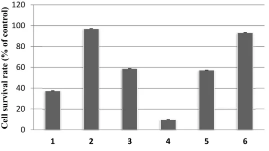
We evaluated the antileukemic activity of isolated compounds against HL-60 cells using the CCK-8 assay method. The isolated compounds, methyl gallate (1), taxifolin (2), pyrocatechuic acid (3), gallic acid (4), glucogallin (5) and
β-0 20 40 60 80 100 120 1 2 3 4 5 6 Trolox Antioxidant activity (% of contr ol)
sitosterol (6) at a final concentration of 50 μM exhibited cell survival rate 37.5%, 97.0%, 58.8%, 9.75%, 57.3% and 93.2%, respectively, shown in Figure 11.
Figure 11. Antileukemic activity of isolated compounds from bark of Mangifera
casturi (Means ± SEMs, n = 6, 50 µM). Methyl gallate (1), taxifolin
(2), pyrocatechuic acid (3), gallic acid (4), glucogallin (5) and β-sitosterol (6).
Comparing literature data, cell survival rate of quercetin was 29.7% (Kakumu et al., 2014) and that of taxifolin (2) was 97.0% in human leukemia HL-60 cells; it was observed that the structure of quercetin and taxifolin (2) was
similar; the major difference was the absence of C2 - C3 double bond in the structure of taxifolin (2). This makes it ineffective against human leukemia HL-60 cells (Rusak et al., 2005).
0 20 40 60 80 100 120 1 2 3 4 5 6 Cell survival rate (% o f contr ol)
The addition of a methyl and glucose group to the carboxylic moiety of gallic acid decreased its antileukemic activity (Kakumu et al., 2014). Gallic acid (4) strongly inhibited the cell proliferation with cell survival rate of 9.71%, when compared with methyl gallate (1) (37.5%) and glucogallin (5) (57.3%).
1.3.3. UPLC-ESITOFMS analysis
Analysis of the chemical components from EtOAc fraction of M. casturi
bark was carried out using UPLC-ESITOFMS. The chemical components were assigned as [M-H]- ions in Figure 12. The retention time of each compound in the EtOAc fraction were assigned 5.23, 5.74, 11.05, 15.72, and 21.95 min for glucogallin (5), gallic acid (4), pyrocatechuic acid (3), methyl gallate (1) and taxifolin (2), respectively. The HRESITOFMS spectrum of each retention time of compounds isolated from bark of Mangifera casturi were given in Figure 13.
Figure 12. UPLC-ESITOFMS chromatogram of the EtOAc fraction of
Mangifera casturi bark. Methyl gallate (1), taxifolin (2),
Figure 13. The HRESITOFMS spectrum of each retention time of compounds
1.4. Conclusions
In summary, methyl gallate (1), taxifolin (2), pyrocatechuic acid (3), gallic acid (4), glucogallin (5) and β-sitosterol (6) were isolated from bark of M. casturi for the first time (Figure 14). The antioxidant and antileukemic activities of isolated compounds were evaluated. The presence and position of hydroxyl, methyl and glucose group in the isolated compounds affects their antioxidant and antileukemic activities. M. casturi bark is a good source of gallic acid and the
derivatives. O HO O OH OH Taxifolin (2) Pyrocatechuic acid (3) O OH OH OH Gallic acid (4) O OH OH HO HO Methyl gallate (1) OH HO HO O OMe Glucogallin (5) OH OH OH O O OH O OH HO HO Sitosterol (6) HO OH OH
Figure 14. Chemical structures of isolated compounds from bark of Mangifera casturi.
References
Afifa, K., Kamruzzaman, M., Mahfuza, I. 2014. A comparison with antioxidant and functional properties among five mango (Mangifera indica L.) varieties in Bangladesh. Int. Food Res. J. 21:1501-1506.
Ahmad, S., Sukari, M. A., Ismail, N. 2015. Phytochemicals from Mangifera pajang Kosterm and their biological activities.Compl. Alter. Med. 15:83.
Andersen, Ø. M., Markham, K. R. 2006. Flavonoids chemistry, biochemistry and applications. Taylor & Francis Group, New York, pp 67-73.
Bahia, M.V., David, J. P., David, J. M. 2010. Occurrence of biflavones in leaves of Caesalpinia pyramidalis specimens. Quim Nova 33:1297-1300.
Benny, P. J., Shibumon., Sunny, K., Cincy, G. 2010. 2,3-Dihydroxybenzoic acid: An effective antifungal agent isolated from Flacourtia inermis fruit. Int. J.
Pharm. Clin. Res. 2:101-105.
Chaturvedula, V. S. P., Prakash, I. 2012. Isolation of stigmasterol and β-sitosterol from the dichloromethane extract of Rubus suavissimus. Int. Curr. Pharm. J.
1: 239-242.
Dai, J., Mumper, R. J. 2010. Plant phenolics: extraction, analysis, and their antioxidant and anticancer properties. Molecules 15:7313.
Fakhrudin, N., Putri, P. S., Sutomo., Wahyuono, S. 2013. Antiinflamatory activity of methanolic extract of Mangifera casturi in thioglycollate-induced leukocyte migration on mice. Trad. Med. J. 18:151-156.
Gangadhar, M., Bhavana, P., Sunil, Y., Datta, S. 2011. Isolation and characterization gallic acid from Terminilia bellerica and its effect on
carbohydrate regulatory system in vitro. Int. J. Res. Ayurveda Pharm.
2:559-562.
George, S., Benny, P. J., Sunny, K., Cincy, G. 2011. Antibiotic activity of 2,3-dihydroxybenzoic acid isolated from Flacourtia inermis fruit against multidrug resistant bacteria. Asian J. Pharm. Clin. Res. 4:126-130.
Hisham, D. M. N., Lip, J. M., Noh, J. M. 2011. Identification and isolation of methyl gallate as a polar chemical marker from Labisia pumila Benth. J.
Trop. Agric. Food Sci. 39:279-284.
Kakumu, A., Ninomiya, M., Efdi, M., Adfa, M., Hayashi, M., Tanaka, K., Koketsu, M. 2014. Phytochemical analysis and antileukemic activity of poliphenolic constituents of Toona sinensis. Bioorg. Med. Chem. Lett. 24:4286-4290.
Kamboj, A., Saluja, A. K. 2011. Isolation of stigmasterol and β-sitosterol from petroleum ether extract of aerial parts of Ageratum conyzoides (Asteraceae).
Int. J. Pharm. Sci. 3:94-96.
Kato, K., Ninomiya, M., Tanaka, K., Koketsu, M. 2016. Effect of functional groups and sugar composition of quercetin derivatives on their radical scavenging properties. J. Nat. Prod. 79:1808-1814.
Liu, Y., Gao, L., Liu, L. 2012. Purification and characterization of a novel galloyltransferase involved in catechin galloylation in the tea plant Camellia sinensis. J. Biol. Chem. 6:1-22.
Lu, Z., Nie, G., Belton, P. S. 2006. Structure activity relationship analysis of antioxidant ability and neuroprotective effect of gallic acid derivatives.
Neurochem. Int. 48:263-274.
Mahajan, A., Nandini, P. 2010. Simultaneous isolation and identification of phytoconstituents from Terminalia chebula by preparative chromatography.
J. Chem. Pharm. Res. 2:97-103.
Mahdavi, B. 2014. Chemical constituents of the aerial parts of Etlingera brevilabrum (Zingiberaceae). Der. Pharma. Chem. 6:360-365.
Majeed, M., Bhat, B., Jadhav, A. N. 2009. Ascorbic Acid and Tannins from
Emblica officinalis Gaertn Fruitss A Revisit. J. Agric. Food Chem.
57:220-225.
Meneses, M. A., Caputo, G., Scognamiglio, M. 2015. Antioxidant phenolic compounds recovery from Mangifera indica L. by-products by supercritical antisolvent extraction. J. Food Eng. 163:45-53.
Pardede, A., Mashita, K., Ninomiya, M., Tanaka, K., Koketsu, M. 2016. Flavonoid profile and antileukemic activity of Coreopsis lanceolata flowers. Bioorg. Med. Chem. Lett. 26:2784-2787.
Rivera, D. G., Delgado, R., Bougarne, N. 2011. Gallic acid indanone and mangiferin xanthone are strong determinants of immunosuppressive anti-tumour effects of Mangifera indica L. bark in MDA-MB231 breast cancer cells. Cancer Lett. 305:21-31.
Rodeiro, I., Cancino, L., Gonzales, J. E. 2006. Evaluation of the genotoxic potential of Mangifera indica L. extract (Vimang), a new natural product with antioxidant activity. Food Chem. Toxicol. 44:1707-1713.
Rusak, G., Gutzeit, O. H., Muller, J. L. 2005. Structurally related flavonoids with antioxidative properties differentially affect cell cycle progression and apoptosis of human acute leukemia cells. Nutr. Res. 25:141-153.
Suhartono, E. Viani, E., Rahmadhan, M. A. 2012. Total flavanoid and antioxidant activity of some selected medicinal plants in South Kalimantan of Indonesia.
Asia Pacific Chem. Biol. Environ. Eng. 4:235-239.
Sutomo., Wahyuono., Rianto., Setyowati. 2013. Isolation and identification of active compound of n-hexane fraction from kasturi (Mangifera casturi Kosterm) against antioxidant and immunomodulatory activity. J. Biol. Sci. 13:596-604.
Chapter 2
Flavonoid rutinosides from
Cinnamomum parthenoxylon leaves
and their hepatoprotective and antioxidant activity
2.1. Introduction
Cinnamomum parthenoxylon tree belongs to the Lauraceae family. It is
called kayu gadis and has been used by local people as spices in foods, fragrances, fumigants, and traditional medicines in Indonesia (Wang et al., 2013; Kawatra and Rajagopalan, 2015). The picture of Cinnamomum parthenoxylon part is show in
Figure 15.
a b
Figure 15. Cinnamomum parthenoxylon stem (a) and leaves (b).
The species from Cinnamomum genus are known to have various biological activities. For instance, Cinnamomum verum, Cinnamomum loureirii, Cinnamomum burmannii and Cinnamomum cassia showed high inhibitory
activities against various cancer cell lines and are also employed in the treatment of diabetes (Hong et al., 2002; Jia et al., 2009; Unlu et al., 2010; Cao et al., 2010; Daker et al., 2013; Lee et al., 2013). Previous studies indicated that several lignans and phenylpropanoids such as dehydroxycubebin, hinokinin, cubebin, 3-(3,4-methylenedioxyphenyl)-1,2-propanediol and safrole isolated from Cinnamomum
parthenoxylon woods have been reported to exert antileukemic activity against
human leukemia HL-60 and U937 cells (Adfa et al., 2016). The lignans and phenylpropanoids isolated from Cinnamomum parthenoxylon wood shown in
Figure 16.
Thus far, based on literature survey (Wei et al., 2017), no biological activities has been reported on the compounds isolated from Cinnamomum parthenoxylon leaves. Flavonoid is an important class of secondary metabolites
from plants and possesses a wide spectrum of pharmacological properties, such as antileukemic activity (Ninomiya et al., 2013; Pardede et al., 2016). Flavonoids have received much attention in human health due to its antioxidant activity (Kato et al., 2016), hepatoprotective activity as well as reducing oxidative stress (Kinjo et al., 2006). Hence, the hepatoprotective and antioxidant activity of flavonoid rutinosides isolated from C. parthenoxylon leaves was investigated.
2.2. Materials and Methods
2.2.1. General experimental procedures
All solvents and reagents were purchased from the suppliers and used without further purification. MS spectra were obtained using UPLCMS system (Aquity UPLC XevoQTof). 1H (400 MHz) and 13C (100 MHz) NMR spectra were recorded with a JEOL ECX400 spectrometer with tetramethylsilane as an internal standard. Silica gel column chromatography (CC) was performed on silica gel N-60 (40-50 μm). Thin-layer chromatography (TLC) spots on plates pre-coated with silica gel 60 F254 were detected with a UV lamp (254 nm). Fractionations for all CC were based on TLC analyses.
2.2.2. Extraction and isolation chemical constituents from Cinnamomum parthenoxylon leaves
Dried leaves of Cinnamomum parthenoxylon (3.1 kg) was macerated with methanol at room temperature. The extract was filtered and evaporated in vacuo to
yield methanol extract (214.6 g). The methanol extract was suspended in water and partitioned successively with n-hexane and EtOAc to yield n-hexane fraction
(25.4 g) and EtOAc fraction (39.8 g).
A portion of EtOAc fraction (15.0 g) was separated by silica gel CC eluted with CHCl3 - MeOH, which yielded 8 fractions (E1 - E8). Compound 1 (19.1 mg) and compound 2 (803.4 mg) was isolated from fraction E1 (873 mg) and E5 (5.2 g) by recrystallization, respectively. Fraction E3 was separated by silica gel CC eluted with CHCl3 - MeOH to give 9 subfractions (E3.1 - E3.9). E3.4 (127.3 mg) was further purified by silica gel CC eluting CHCl3 - MeOH (8: 2), to obtained compound 3 (20.8 mg). Fraction E4 (2.4 g) was separated by silica gel (CC) eluted CHCl3 – MeOH stepwise manner of polarity to give 7 subfraction (E4.1 - E4.7). E4.3 (413 mg) was further purified by silica gel CC eluting CHCl3 - MeOH, to obtained compound 4 (4.0 mg). Fraction E2 (691.5 mg) was divided by silica gel CC eluted CHCl3 - MeOH stepwise manner of polarity to give 6 subfraction (E2.1 - E2.6). E2.5 (271 mg) was applied by silica gel CC eluted with CHCl3 - MeOH (8 : 2), to give 6 subfraction (E2.5.1 - E2.5.6), furthermore subfraction E2.5.3 (30 mg) was purified using preparative thin layer chromatography (PTLC) eluted with (CHCl3 - MeOH 8 : 2), to gave compound 5 (3.6 mg). Fraction E7 (1.1 g) was applied by silica gel CC eluted with CHCl3 - MeOH (8 : 2) (7 : 3), to give 8 subfraction (E7.1 – E7.8). SubFr7.4 was separated using Sephadex LH-20 CC eluting (CHCl3 - MeOH 8 : 2),
to give 4 subfraction (E7.4.1 – E7.4.4). SubFrE7.4.2 was purified by PTLC (CHCl3 - MeOH 8 : 2) to yield compound 6 (29.4 mg). SubFrE7.4.1 was purified by Sephadex LH-20 eluting (CHCl3 - MeOH 8 : 2) to yield compound 9 (24.7 mg).
The n-hexane fraction was divided by silica gel CC eluting n-hexane -
CHCl3 - EtOAc step gradient polarity to yield 6 fractions (H1 to H6). Fraction H2 (3.2 g) was further purified by silica gel CC eluting n-hexane - CH2Cl2 - CHCl3 stepwise manner of polarity to afford compound 7 (144.3 mg) and compound 8 (96.1 mg), respectively (Scheme 2).
2.2.3. Spectral data of isolated compounds Scopoletin (1)
Pale yellow crystals, HRESITOFMS m/z 191.0328 [M-H]- (calcd. for C
10H7O4, 191.0344). 1H NMR (400 MHz, CDCl3): δ 7.84 (1H, d, J = 9.6 Hz, H-4), 7.19 (1H, s, H-5), 6.79 (1H, s, H-8), 6.18 (1H, d, J = 9.6 Hz, H-3), 3.90 (3H, s, OCH3); 13C NMR (100 MHz, CDCl3): δ 160.5, 151.0, 150.3, 145.1, 143.8, 112.49, 111.3, 109.1, 102.9, 55.9. Isorhoifolin (2)
Yellow powder, HRESITOFMS m/z 577.1555 [M-H]- (calcd. for C
27H29O14, 577.1557). 1H NMR (400 MHz, DMSO): δ 7.95 (2H, d, J = 9.2 Hz, H-2' and H-6'), 6.95 (2H, d, J = 9.2 Hz, H-3' and H-5'), 6.88 (1H, s, H-3), 6.80 (1H, d, J = 2.3 Hz,
H-8), 6.38 (d, J = 1.8 Hz, H-6), 5.24 (1H, d, J = 7.3 Hz, H-1'''), 5.14 (1H, s, H-1''), 5.36, 5.18, 4.75, 4.69, 4.50 (6H, sugar hydroxyls), 3.21 – 3.77 (10H, m, sugar protons), 1.21 (3H, d, J = 6.4 Hz, H-6'''); 13C NMR (100 MHz, DMSO): δ 182.5, 164.8, 163.1, 161.9, 161.6, 157.5, 129.1, 121.1, 116.6, 106.0, 103.7, 101.0, 99.8, 98.3, 95.0, 77.5, 76.8, 72.4, 71.0, 70.9, 70.6, 70.1, 68.9, 61.0, 18.6.
Epicatechin (3)
Yellow powder, HRESITOFMS m/z 289.0714 [M-H]- (calcd. for C
15H13O6, 289.0712). 1H NMR (400 MHz, Acetone-d6): δ 7.06 ( 1H, d, J = 1.8 Hz, H-2'), 6.84 (1H, dd, J = 8.5 Hz and 1.8 Hz, H-6'), 6.79 (1H, d, J = 8.2 Hz, H-5'), 6.03
(1H, d, J = 2.3 Hz, H-6), 5.92 (1H, d, J = 2.3 Hz, H-8), 4.88 (1H, s, H-2), 4.21 (1H, s, H-3), 2.87 (1H, dd, J = 16.7 Hz and 4.6 Hz, H-4β), 2.74 (1H, dd, J = 16.9
Hz and 3.2 Hz, H-4α); 13C NMR (100 MHz, Acetone-d6): δ 156.8, 156.7, 156.3, 144.6, 144.5, 131.4, 118.5, 114.7, 114.5, 99.0, 95.3, 94.9, 78.6, 66.1, 28.8.
Blumenol A (4)
Pale yellow powder, HRESITOFMS m/z 225.1479 [M-H]- (calcd. for C
13H21O3, 225.1491). 1H NMR (400 MHz, CDCl3): δ 5.83 (1H, dd, J = 15.8 Hz and 5.5 Hz, H-8), 5.70 (1H, d, J = 16.9 Hz, H-7), 4.44 (1H, quint, H-9), 2.84 (1H, d, J = 13.3
Hz, H-2 ax), 2.40 1(H, d, J = 12.8 Hz, H-4 ax), 2.29 (1H, m, H-5), 2.13 (1H, dd, J = 13.4 Hz and 2.1 Hz, H-4 eq ), 1.92 (H, dd, J = 13.8 Hz and 2.3 Hz, H-2 eq), 1.33
(3H, d, J = 6.4 Hz, H-10), 0.97 (3H, s, H-11), 0.94 (3H, s, H-12), 0.90 (3H, d, J = 6.6 Hz, H-13) 13C NMR (100 MHz, CDCl3): δ 211.3, 135.2, 131.9, 77.3, 68.4, 51.5, 45.2, 42.6, 36.4, 24.5, 24.4, 23.9, 16.0.
4-Hydroxybenzoic acid (5)
White powder, HRESITOFMS m/z 137.0265 [M-H]- (calcd. for C
7H5O3, 137.0239). 1H NMR (400 MHz, Acetone-d6): δ 7.92 (2H, d, J = 9.2 Hz, H-2 and H-6), 6.91 (2H, d, J = 9.2 Hz, H-3 and H-5); 13C NMR (100 MHz, Acetone-d6): δ 167.8, 162.5, 132.7, 123.1, 115.9.
Rutin (6)
Yellow powder, HRESITOFMS m/z 609.1476 [M-H]- (calcd. for C27H29O16, 609.1456). 1H NMR (400 MHz, CD3OD): δ 7.67 (1H, d, J = 2.3 Hz, H-2'), 7.62 (1H, dd, J = 8.0 Hz, and 2.3 Hz, H-6'), 6.87 (1H, d, J = 8.7 Hz, H-5'), 6.40 (1H, d,
(1H, d, J = 1.4 Hz, H-1'''), 3.80 - 3.27 (m), 1.12 (1H, d, J = 6.4 Hz, H-6'''); 13C NMR (100 MHz, CD3OD): δ 178.1, 164.7, 161.6, 158.1, 157.2, 148.5, 144.5, 134.2, 122.2, 121.8, 116.4, 114.8, 104.3, 103.3, 101.1, 98.7, 93.6, 76.8, 75.8, 74.4, 72.6, 70.9, 70.7, 70.0, 68.4, 67.1, 16.6.
Hexadecanoic acid methyl ester (7)
Yellow oil, 1H NMR (400 MHz, CDCl
3): δ 3.66 (s, 3H, OCH3), 2.29 (t, J = 13.8 Hz, H-14), 1.61 (1H, m, H-15), 1.27 (m, H-3), 0.88 (1H, t, J = 7.1 Hz, H-16); 13C NMR (100 MHz, CDCl3): δ 174.3, 51.4, 34.1, 32.0, 29.8, 29.7, 29.5, 29.4, 29.3, 29.2, 25.0, 22.8, 14.1.
12-Hexadecenoic acid methyl ester (8)
Yellow oil, 1H NMR (400 MHz, CDCl 3): δ 5.35 (m), 3.67 (s, OCH3), 2.29 (t, J = 7.8 Hz), 2.06 – 2.00 (m), 1.68 – 1.59 (m), 1.3 (m), 0.88 (m); 13C NMR (100 MHz, CDCl3): δ 174.3, 130.0, 129.8, 51.4, 34.2, 32.0, 29.8, 29.7, 29.5, 29.4, 29.3, 29.2, 25.0, 22.8, 14.2. Nicotiflorin (9)
Yellow powder, HRESITOFMS m/z 593.1489 [M-H]- (calcd. for C27H29O15, 593.1479). 1H NMR (400 MHz, CD 3OD): δ 8.11 (2H, d, J = 8.9 Hz, 2' and H-6'), 6.93 (2H, J = 8.9 Hz, H-3' and H-5'), 6.45 (1H, d, J = 2.1 Hz, H-8), 6.25 (1H, d, J = 2.1 Hz, H-6), 5.11 (1H, d, J = 7.4 Hz, H-1''), 4.56 (1H, d, J = 1.4 Hz, H-1'''), 3.67 (1H, dd, J = 3.4 Hz and 1.6 Hz, H-2'''), 3.56 (1H, dd, J = 9.5 Hz and 3.5 Hz, H-3'''), 3.52 - 3.41 (8H, m), 1.16 (1H, d, J = 6.2 Hz, H-6'''); 13C NMR (100 MHz,
CD3OD): δ 179.4, 166.1, 161.5, 163.0, 159.4, 158.6, 135.5, 132.4, 122.8, 116.1, 105.6, 104.6, 102.4, 100.1, 95.0, 78.2, 77.2, 75.8, 73.9, 72.3, 72.1, 71.4, 68.6, 67.7, 17.9.
2.2.4. Hepatoprotective activity
HepG2 cells were kindly provided from Division of Antioxidant Research (Life Science Research Center, Gifu University). Cells were cultured in DMEM media (Wako Pure Chemical Industries, Ltd., Osaka, Japan) supplemented with 10% heat-inactivated fetal bovine serum (FBS) and 1% antibiotics, penicillin-streptomycin (Gibco®, Life Technologies, Thermo Fisher Scientific Inc., MA and USA) and were maintained at 37oC under a humidified atmosphere of 5% CO2. HepG2 cells (2 × 10⁴ cells/mL, 100 L) were seeded in 96-well plates. After 24-h incubation, cells were pretreated with samples for 1 h, and were subsequently exposed to t-BHP (final concentration: 300 M) for 3 h. A 10 L amount of
CCK-8 solution was added into the culture, and the plates were incubated for an additional 3 h. Visible absorption (490 nm) was measured using a microplate reader (Emax precision microplate reader, Molecular Devices Japan, Tokyo, Japan).
2.2.5. Antioxidant activity
Protocol of DPPH radical scavenging assay was performed essentially as described previously (Kato et al, 2016). A 10 L amount of sample solutions and 190 L of DPPH solution (78 M in distilled H2O/MeOH = 5/3) were added to 96-well plates, resulting in a final concentration of 74 M for DPPH. The
solutions were vigorously mixed and allowed to stand. Visible absorption ( = 545 nm) was measured after 30 min using a microplate reader (Emax precision microplate reader, Molecular Devices Japan, Tokyo, Japan). Wells without the compounds were considered as negative controls. At least three replicates were performed for each compound and control.
2.2.6. UPLC-ESITOFMS procedures
The samples were dissolved in DMSO/H2O (1/1) at 20 mg/mL and filtered through 0.45 µm membrane filter (ADVANTEC®, Japan) and an aliquot (5 μL) of the sample was injected in the UPLC. Analysis was carried out by the Waters UPLC system (Aquity UPLC XevoQTof), using a UPLC BEH C18 analytical column (1.7 μm, 2.1 × 100 mm). The mobile phase contained solvent A (1% v/v AcOH in distilled water) and solvent B (acetonitrile). The liner gradient system employed was: 0 - 30 min 90% solvent A to 70% solvent A and 10% solvent B to 30% solvent B; kept for 5 min; 35 - 45 min 70% solvent A to 50% solvent A and 30% solvent B to 50% solvent B. The column eluate was monitored at 260 nm UV absorbance. Negative mode was employed in ESITOFMS.
2.3. Results and Discussion
2.3.1. Isolation of chemical constituents from Cinnamomum parthenoxylon leaves
The MeOH extract (214.6 g) of C. parthenoxylon leaves was partitioned successively with n-hexane and EtOAc to yield n-hexane fraction (25.4 g) and
EtOAc fraction (39.8 g). The EtOAc fraction demonstrated the higher radical scavenging activity (Figure 17) and showed high inhibition on t-BHP-induced
cytotoxicity in HepG2 cells (Figure 18). Therefore, we carried out isolation of compounds responsible for the activities from the EtOAc fraction.
Figure 17. DPPH scavenging activity of n-Hex and EtOAc fractions of Cinnamomum parthenoxylon leaves (Means ± SEMs, n = 3).
Figure 18. Hepatoprotective effect of EtOAc fraction of Cinnamomum
parthenoxylon leaves on t-BHP induced cytotoxicity in HepG2
cells (Means ± SEMs, n = 3). Cells were treated with EtOAc
fraction for 1 h, and then t-BHP was added at a final concentration of 300 µM and incubated for 3 h.
The fractions were separated by column chromatography (CC) on silica gel (SiO2) and purified using Sephadex LH-20 CC and PTLC, to yield nine compounds. The chemical structures of the isolated compounds were elucidated by their 1H, 13C and 2D NMR spectra of each compound and literature data.
The HRESITOFMS peak at m/z 191.0328 [M-H]- (calcd. for C
10H7O4, 191.0344) suggested C10H8O4 as the molecular formula of compound 1. Its 1H NMR displayed two doublet signals at δH 6.18 (1H, d, J = 9.6 Hz, H-3) and 7.84 (1H, d, J = 9.6 Hz, H-4), which are characteristic signals of a pyrone ring of the coumarin framework. Furthermore, aromatic singlet signals were observed at δH