Science Monographs, No. 39, pp. 405-446, 2009
Deep-sea Mysidaceans (Crustacea: Lophogastrida and Mysida) from the North- western North Pacifi c off Japan, with Descriptions of Six New Species
Kouki Fukuoka
Ishigaki Tropical Station, Seikai National Fisheries Research Institute, Fisheries Research Agency, 148‑446 Fukai-Ohta, Ishigaki, Okinawa, 907‑0451 Japan
E-mail: [email protected]
Abstract: Mysidaceans (Lophogastrida and Mysida) from deep waters off the northern Japan are reported.
Four species of Lophogastrida and 33 species of Mysida were identifi ed. A new genus, Neoamblyops, and six new species, Ceratomysis japonica, C. orientalis, Holmesiella bisaetigera, Mysimenzies borealis, Neoambly- ops latisquamatus, and Paramblyops hamatilis, are described.
Key words: Crustacea, Lophogastrida, Mysida, deep water, northern Japan, new genus, new species.
Introduction
Mysidaceans (Lophogastrida and Mysida) from deep waters off the Pacifi c coast of the north- ern Honshu, Japan, have been reported by W. Tattersall (1951), Birstein and Tchindonova (1958), Taniguchi (1969), Murano (1975, 1976), Fukuoka et al. (2005), and Fukuoka and Murano (2006).
To date, two species of Lophogastrida and 11 species of Mysida have been recorded (Table 1).
The present paper provides the taxonomic result of mysidacean specimens collected from deep waters off the northern Japan during a research project entitled “Research on Deep-sea Fauna and Pollutants off Pacifi c Coast of Northern Japan” as part of the “Study on Deep-Sea Fauna and Conservation of the Deep-Sea Ecosystem” conducted by the National Museum of Nature and Sci- ence, Tokyo. This paper reports four species of Lophogastrida and 33 species of Mysida including a new genus, Neoamblyops, and six new species, Ceratomysis japonica, C. orientalis, Holmesiella bisaetigera, Mysimenzies borealis, Neoamblyops latisquamatus, and Paramblyops hamatilis.
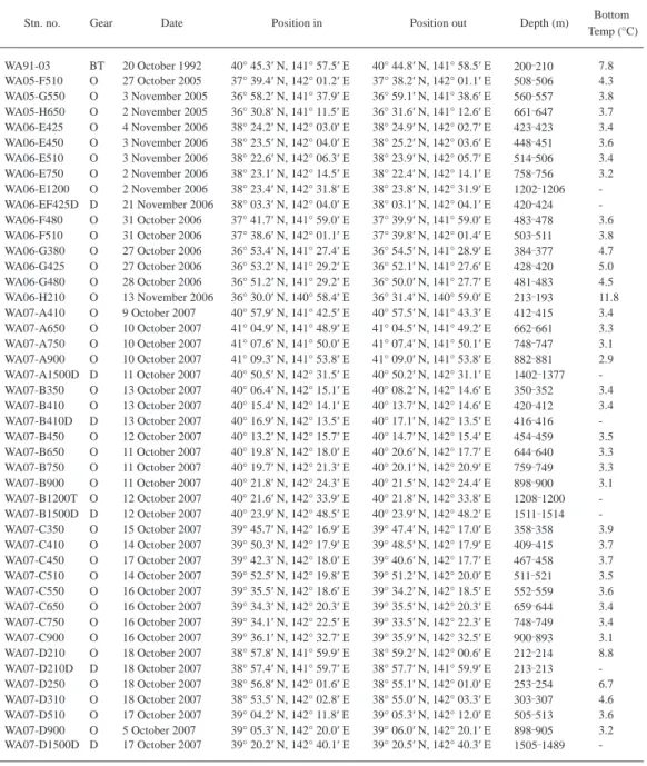
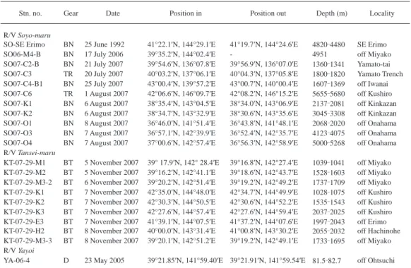
Body length was measured from the tip of the rostrum to the posterior end of the telson ex- cluding spines. In Gnathophausiidae specimens, the total length was measured from the posterior margin of the orbit to the posterior end of the telson excluding spines. Specimens examined in this study were stored in the National Museum of Nature and Science, Tokyo (NSMT). Sampling data are shown in Tables 2 3.
Taxonomic accounts
Order Lophogastrida Family Gnathophausiidae
Neognathophausia gigas (Willemoes-Suhm, 1875)
Gnathophausia gigas Willemoes-Suhm, 1875: 28 31, pl. 9, fi gs. 16 17, pl. 10, fi gs. 2 3; G. Sars, 1885: 33 35, pl. 3; Ortmann, 1906: 36 38, pl. 2, fi g. 1; Fage, 1941: 24‑27; Nouvel, 1943: 12 15, pl. 1, fi gs. 11 15; Banner, 1948a: 357 358, fi g. 1;
W. Tattersall, 1951: 26 28; W. Tattersall and O. Tattersall, 1951: 77 82, fi gs. 1 2; O. Tattersall, 1955: 36 38; Birstein and Tchindonova, 1958: 260 261; 1962: 57 58; Taniguchi, 1969: 44 45, fi g. 2; Kathman et al., 1986: 158, fi g. a h (p.
159); Ledoyer, 1995: 602 603; Liu and Wang, 2000: 68 70, fi g. 5.
Gnathophausia drepanephora Holt and W. Tattersall, 1905: 113 116, pl. 18.
Neognathophausia gigas: Petryashov, 1992: 47, fi g. 1 (1 5); 2005: 959, fi g. 2.
Material examined. 1 juvenile (24.5 mm), WA06-E750, NSMT-Cr 19613. 1 female (70.2 mm), WA06-E1200, NSMT-Cr 19614. 1 juvenile (damaged), WA07-A650, NSMT-Cr 19615. 1 female (44.7 mm), WA07-A750, NSMT-Cr 19616. 1 juvenile (49.2 mm), WA07-A900, NSMT-Cr 19617. 1 female (68.0 mm) and 1 juvenile (61.2 mm), WA07-B450, NSMT-Cr 19618. 2 juveniles (37.9, 45.0 mm), WA07-B650, NSMT-Cr 19619. 3 juveniles (31.6 37.8 mm), WA07-B750, NSMT-Cr 19620. 4 juveniles (31.1 40.9 mm), WA07-B900, NSMT-Cr 19621. 7 males (55.3 86.3 mm) and 6 females (54.0 84.2 mm), WA07-B1200T, NSMT-Cr 29622. 1 juvenile (27.4 mm), WA07-B1500D, NSMT-Cr 19623. 1 juvenile (46.4 mm), WA07-D900, NSMT-Cr 19624. 1 male (95.0 mm), SO07-O3, NSMT-Cr 19625. 1 female (75.6 mm), KT-07-29-M3-2, NSMT-Cr 19626.
1 female (85.2 mm) and 1 juvenile (22.2 mm), KT-07-29-M3-3, NSMT-Cr 19627. 1 male (71.3 mm) and 2 juveniles (22.5, 47.2 mm), KT-07-29-K3, NSMT-Cr 19628.
Distribution. Cosmopolitan (see Müller, 1993).
Family Lophogastridae
Lophogaster japonicus W. Tattersall, 1951
Lophogaster typicus (Japanese specimens): Ortmann, 1906: 23 26.
Lophogaster japonicus W. Tattersall, 1951: 19 20, fi gs. 1a, 2a; Murano, 1970c: 1 4, fi g. 1a d.
Material examined. 1 female (24.8 mm), WA06-H210, NSMT-Cr 19629. 2 males (23.3, 23.4
Table 1. List of species of Lophogastrida and Mysida recorded from deep waters off the Pacifi c coast of Tohoku District and Hokkaido, northern Japan.
Species References
Order Lophogastrida Family Ganthophausiidae
Neognathophausia gigas (Willemoes-Suhm, 1875) Taniguchi (1969) Family Eucopiidae
Eucopia grimardii Nouvel, 1942 Taniguchi (1969) Order Mysida
Family Petalophthalmidae
Ceratomysis spinosa Faxon, 1893 W. Tattersall (1951) Family Mysidae
Subfamily Boreomysinae
Boreomysis californica Ortmann, 1894 Taniguchi (1969)
Boreomysis incisa Nouvel, 1942 Birstein and Tchindonova (1958) Subfamily Mysinae
Tribe Erythropini
Caesaromysis hispida Ortmann, 1893 Murano (1977) Hyperamblyops nana Birstein and Tchindonova, 1958 Murano (1975) Katerythrops oceanae Holt and W. Tattersall, 1905 Taniguchi (1969) Meterythrops microphthalmus W. Tattersall, 1951 Murano (1977),
Fukuoka and Murano (2006) Meterythrops robustus Smith, 1879 Fukuoka and Murano (2006) Tribe Mysini
Parastilomysis paradoxa Ii, 1936 Fukuoka et al. (2005) Parastilomysis prominula Fukuoka, Bravo and Murano, 2005 Fukuoka et al. (2005) Parastilomysis separata Fukuoka, Bravo and Murano, 2005 Fukuoka et al. (2005)
mm), WA07-D210, NSMT-Cr 19630. 33 males (19.4 28.2 mm) and 32 females (18.9 28.0 mm), WA07-D210D, NSMT-Cr 19631. 1 male (23.0 mm) and 1 female (25.0 mm), WA07-D250, NSMT-Cr 19632. 2 females (23.5, 26.5 mm), WA07-D310, NSMT-Cr 19633.
Distribution. Japan (Ortmann, 1906; W. Tattersall, 1951; Murano, 1970c).
Family Eucopiidae Eucopia australis Dana, 1852
Eucopia australis Dana, 1852: 609 611; G. Sars, 1885: 55 62, pls. 9 10 (part); Faxon, 1895: 218 219; Hansen, 1905: 5, fi g.
Table 2. List of stations by R/V Wakataka-maru. BT, beam trawl with 2 m span opening; D, dredge; O, otter trawl.
Stn. no. Gear Date Position in Position out Depth (m) Bottom
Temp (°C)
WA91-03 BT 20 October 1992 40° 45.3′ N, 141° 57.5′ E 40° 44.8′ N, 141° 58.5′ E 200 210 7.8 WA05-F510 O 27 October 2005 37° 39.4′ N, 142° 01.2′ E 37° 38.2′ N, 142° 01.1′ E 508 506 4.3 WA05-G550 O 3 November 2005 36° 58.2′ N, 141° 37.9′ E 36° 59.1′ N, 141° 38.6′ E 560 557 3.8 WA05-H650 O 2 November 2005 36° 30.8′ N, 141° 11.5′ E 36° 31.6′ N, 141° 12.6′ E 661 647 3.7 WA06-E425 O 4 November 2006 38° 24.2′ N, 142° 03.0′ E 38° 24.9′ N, 142° 02.7′ E 423 423 3.4 WA06-E450 O 3 November 2006 38° 23.5′ N, 142° 04.0′ E 38° 25.2′ N, 142° 03.6′ E 448 451 3.6 WA06-E510 O 3 November 2006 38° 22.6′ N, 142° 06.3′ E 38° 23.9′ N, 142° 05.7′ E 514 506 3.4 WA06-E750 O 2 November 2006 38° 23.1′ N, 142° 14.5′ E 38° 22.4′ N, 142° 14.1′ E 758 756 3.2 WA06-E1200 O 2 November 2006 38° 23.4′ N, 142° 31.8′ E 38° 23.8′ N, 142° 31.9′ E 1202 1206 - WA06-EF425D D 21 November 2006 38° 03.3′ N, 142° 04.0′ E 38° 03.1′ N, 142° 04.1′ E 420 424 - WA06-F480 O 31 October 2006 37° 41.7′ N, 141° 59.0′ E 37° 39.9′ N, 141° 59.0′ E 483 478 3.6 WA06-F510 O 31 October 2006 37° 38.6′ N, 142° 01.1′ E 37° 39.8′ N, 142° 01.4′ E 503 511 3.8 WA06-G380 O 27 October 2006 36° 53.4′ N, 141° 27.4′ E 36° 54.5′ N, 141° 28.9′ E 384 377 4.7 WA06-G425 O 27 October 2006 36° 53.2′ N, 141° 29.2′ E 36° 52.1′ N, 141° 27.6′ E 428 420 5.0 WA06-G480 O 28 October 2006 36° 51.2′ N, 141° 29.2′ E 36° 50.0′ N, 141° 27.7′ E 481 483 4.5 WA06-H210 O 13 November 2006 36° 30.0′ N, 140° 58.4′ E 36° 31.4′ N, 140° 59.0′ E 213 193 11.8 WA07-A410 O 9 October 2007 40° 57.9′ N, 141° 42.5′ E 40° 57.5′ N, 141° 43.3′ E 412 415 3.4 WA07-A650 O 10 October 2007 41° 04.9′ N, 141° 48.9′ E 41° 04.5′ N, 141° 49.2′ E 662 661 3.3 WA07-A750 O 10 October 2007 41° 07.6′ N, 141° 50.0′ E 41° 07.4′ N, 141° 50.1′ E 748 747 3.1 WA07-A900 O 10 October 2007 41° 09.3′ N, 141° 53.8′ E 41° 09.0′ N, 141° 53.8′ E 882 881 2.9 WA07-A1500D D 11 October 2007 40° 50.5′ N, 142° 31.5′ E 40° 50.2′ N, 142° 31.1′ E 1402 1377 - WA07-B350 O 13 October 2007 40° 06.4′ N, 142° 15.1′ E 40° 08.2′ N, 142° 14.6′ E 350 352 3.4 WA07-B410 O 13 October 2007 40° 15.4′ N, 142° 14.1′ E 40° 13.7′ N, 142° 14.6′ E 420 412 3.4 WA07-B410D D 13 October 2007 40° 16.9′ N, 142° 13.5′ E 40° 17.1′ N, 142° 13.5′ E 416 416 - WA07-B450 O 12 October 2007 40° 13.2′ N, 142° 15.7′ E 40° 14.7′ N, 142° 15.4′ E 454 459 3.5 WA07-B650 O 11 October 2007 40° 19.8′ N, 142° 18.0′ E 40° 20.6′ N, 142° 17.7′ E 644 640 3.3 WA07-B750 O 11 October 2007 40° 19.7′ N, 142° 21.3′ E 40° 20.1′ N, 142° 20.9′ E 759 749 3.3 WA07-B900 O 11 October 2007 40° 21.8′ N, 142° 24.3′ E 40° 21.5′ N, 142° 24.4′ E 898 900 3.1 WA07-B1200T O 12 October 2007 40° 21.6′ N, 142° 33.9′ E 40° 21.8′ N, 142° 33.8′ E 1208 1200 - WA07-B1500D D 12 October 2007 40° 23.9′ N, 142° 48.5′ E 40° 23.9′ N, 142° 48.2′ E 1511 1514 - WA07-C350 O 15 October 2007 39° 45.7′ N, 142° 16.9′ E 39° 47.4′ N, 142° 17.0′ E 358 358 3.9 WA07-C410 O 14 October 2007 39° 50.3′ N, 142° 17.9′ E 39° 48.5′ N, 142° 17.9′ E 409 415 3.7 WA07-C450 O 17 October 2007 39° 42.3′ N, 142° 18.0′ E 39° 40.6′ N, 142° 17.7′ E 467 458 3.7 WA07-C510 O 14 October 2007 39° 52.5′ N, 142° 19.8′ E 39° 51.2′ N, 142° 20.0′ E 511 521 3.5 WA07-C550 O 16 October 2007 39° 35.5′ N, 142° 18.6′ E 39° 34.2′ N, 142° 18.5′ E 552 559 3.6 WA07-C650 O 16 October 2007 39° 34.3′ N, 142° 20.3′ E 39° 35.5′ N, 142° 20.3′ E 659 644 3.4 WA07-C750 O 16 October 2007 39° 34.1′ N, 142° 22.5′ E 39° 33.5′ N, 142° 22.3′ E 748 749 3.4 WA07-C900 O 16 October 2007 39° 36.1′ N, 142° 32.7′ E 39° 35.9′ N, 142° 32.5′ E 900 893 3.1 WA07-D210 O 18 October 2007 38° 57.8′ N, 141° 59.9′ E 38° 59.2′ N, 142° 00.6′ E 212 214 8.8 WA07-D210D D 18 October 2007 38° 57.4′ N, 141° 59.7′ E 38° 57.7′ N, 141° 59.9′ E 213 213 - WA07-D250 O 18 October 2007 38° 56.8′ N, 142° 01.6′ E 38° 55.1′ N, 142° 01.0′ E 253 254 6.7 WA07-D310 O 18 October 2007 38° 53.5′ N, 142° 02.8′ E 38° 55.0′ N, 142° 03.3′ E 303 307 4.6 WA07-D510 O 17 October 2007 39° 04.2′ N, 142° 11.8′ E 39° 05.3′ N, 142° 12.0′ E 505 513 3.6 WA07-D900 O 5 October 2007 39° 05.3′ N, 142° 20.0′ E 39° 06.0′ N, 142° 20.1′ E 898 905 3.2 WA07-D1500D D 17 October 2007 39° 20.2′ N, 142° 40.1′ E 39° 20.5′ N, 142° 40.3′ E 1505 1489 -
1; Ortmann, 1906: 53; Fage, 1942: 41 47, fi gs. 28a, 29, 30A, 31A B, 32A, 33; Nouvel, 1943: 26 27, pl. 1, fi gs. 17 19;
W. Tattersall, 1951: 33; O. Tattersall, 1955: 48 49, fi g. 4C D; Birstein and Tchindonova, 1958: 268; 1962: 60 61; Kath- man et al., 1986: 140, fi g. a f (p. 141); Liu and Wang, 2000: 75 76, fi g. 8.
Eucopia major Hansen, 1910: 21, pl. 1, fi g. 4; Nouvel, 1943: 28 29, pl. 1, fi gs. 20 25; W. Tattersall, 1951: 33.
Material examined. 1 adult female (53.0 mm), SO07-C6, NSMT-Cr 19634.
Distribution. Cosmopolitan (see Müller, 1993).
Eucopia grimaldii Nouvel, 1942
Eucopia grimaldii Nouvel, 1942a: 5 6, fi gs. 5 8; Fage, 1942: 47 56, fi gs. 31C, 32B; Nouvel, 1943: 40 43, pl. 2, fi gs. 36 40;
W. Tattersall and O. Tattersall, 1951: 106 108, fi g. 11; O. Tattersall, 1955: 54 56, fi g. 5; Birstein and Tchindonova, 1958: 266 267; 1962: 61; Murano, 1971: 46; Kathman et al., 1986: 144 145, fi g. c, e, g, i (p. 147); Petryashov, 2005:
962.
Eucopia unguiculata: Banner, 1948a: 359 360, fi g. 2; Taniguchi, 1969: 45 46, fi g. 3. [not E. unguiculata (Willemoes-Suhm, 1875)]
Material examined. 1 adult female (33.3 mm) and 1 juvenile (17.6 mm), WA07-A1500D, NSMT-Cr 19635. 1 adult male (36.6 mm), WA07-B750, NSMT-Cr 19636. 2 juveniles (17.2 mm, damaged), WA07-B1500D, NSMT-Cr 19637. 4 adult males (25.0 27.5 mm), WA07-D1500D, NSMT-Cr 19638. 7 adult males (22.2 31.3 mm) and 3 adult females (27.5 43.0 mm), SO07-C6, NSMT-Cr 19639. 1 adult male (22.0 mm), SO07-K1, NSMT-Cr 19640. 1 adult male (22.0 mm) and 1 adult female (27.1 mm), SO07-K2, NSMT-Cr 19641. 1 immature female (28.2 mm), KT-07- 29-E3, NSMT-Cr 19642. 1 adult male (22.3 mm), KT-07-29-K1, NSMT-Cr 19643. 2 adult males (20.8, 27.4 mm) and 3 adult females (21.0 34.5 mm), KT-07-29-K2, NSMT-Cr 19644. 1 adult male (26.2 mm), KT-07-29-M1, NSMT-Cr 19645. 2 adult females (broken), KT-07-29-M2,
Table 3. List of stations by R/Vs Soyo-maru, Tansei-maru and Yayoi. BN, benthos-net; BT, beam trawl with 3 m span opening;
D, dredge; TR, cage trap.
Stn. no. Gear Date Position in Position out Depth (m) Locality
R/V Soyo-maru
SO-SE Erimo BN 25 June 1992 41°22.1′N, 144°29.1′E 41°19.7′N, 144°24.6′E 4820 4480 SE Erimo
SO06-M4-B BN 17 July 2006 39°35.2′N, 144°02.4′E - 4951 off Miyako
SO07-C2-B BN 21 July 2007 39°54.6′N, 136°07.8′E 39°56.9′N, 136°07.0′E 1360 1341 Yamato-tai SO07-C3 TR 20 July 2007 40°03.2′N, 137°06.1′E 40°04.3′N, 137°05.8′E 1800 1820 Yamato Trench SO07-C4-B1 BN 25 July 2007 43°00.4′N, 139°57.2′E 43°00.7′N, 140°00.4′E 1607 1369 off Iwanai SO07-C6 TR 1 August 2007 42°06.6′N, 146°09.7′E 42°08.2′N, 146°15.2′E 5655 5680 off Kushiro SO07-K1 BN 6 August 2007 38°35.4′N, 143°04.5′E 38°34.0′N, 143°06.9′E 2137 2081 off Kinkazan SO07-K2 BN 6 August 2007 38°34.7′N, 143°32.9′E 38°30.6′N, 143°35.6′E 3045 3308 off Kinkazan SO07-O1 BN 8 August 2007 36°46.0′N, 141°51.4′E 36°43.8′N, 141°48.1′E 2068 2020 off Onahama SO07-O3 BN 7 August 2007 36°57.1′N, 142°39.9′E 36°52.4′N, 142°35.7′E 4123 4075 off Onahama SO07-O4 BN 7 August 2007 37°00.6′N, 142°57.4′E 36°56.3′N, 142°58.9′E 5000 5268 off Onahama R/V Tansei-maru
KT-07-29-M1 BT 5 November 2007 39° 17.9′N, 142° 28.4′E 39°16.8′N, 142°27.4′E 1039 1041 off Miyako KT-07-29-M2 BT 5 November 2007 39°16.2′N, 142°41.1′E 39°18.6′N, 142°43.7′E 1528 1603 off Miyako KT-07-29-M3-2 BT 6 November 2007 39°20.2′N, 142°51.4′E 39°19.2′N, 142°49.2′E 1737 1709 off Miyako KT-07-29-K1 BT 7 November 2007 42°35.0′N, 144°48.0′E 42°34.7′N, 144°49.9′E 1028 1075 off Kushiro KT-07-29-K2 BT 7 November 2007 42°30.3′N, 144°50.5′E 42°30.6′N, 144°52.2′E 1535 1543 off Kushiro KT-07-29-K3 BT 7 November 2007 42°27.6′N, 144°57.4′E 42°27.6′N, 144°59.4′E 2037 2025 off Kushiro KT-07-29-E3 BT 7 November 2007 41°39.1′N, 144°07.5′E 41°37.2′N, 144°07.6′E 1997 2043 off Erimo KT-07-29-H2 BT 8 November 2007 40°00.0′N, 143°31.4′E 41°00.8′N, 143°30.2′E 2055 2032 off Hachinohe KT-07-29-M3-3 BT 8 November 2007 39°20.1′N, 142°51.2′E 39°19.2′N, 142°49.1′E 1733 1695 off Miyako R/V Yayoi
YA-06-4 D 23 May 2005 39°21.85′N, 141°59.40′E 39°21.91′N, 141°59.54′E 81.5 82.7 off Ohtsuchi
NSMT-Cr 19646. 2 adult males (30.8 mm, broken) and 2 adult females (22.5, 35.5 mm), KT-07- 29-M3-2, NSMT-Cr 19647. 1 adult female (30.7 mm), KT-07-29-M3-3, NSMT-Cr 19648. 1 juve- nile (19.5 mm), KT-07-29-H2, NSMT-Cr 19649.
Distribution. Cosmopolitan (see Müller, 1993).
Order Mysida Family Petalophthalmidae Ceratomysis japonica sp. nov.
(Figs. 1 2)
Material examined. Holotype: adult female (28.0mm), WA06-G425, NSMT-Cr 19650. Para- types: 1 adult female (damaged, ca 22.6 mm), WA05-G550, NSMT-Cr 19651; 1 adult female (20.8 mm), WA05-H650, NSMT-Cr 19653; 1 adult female (30.0 mm), WA06-G425, NSMT-Cr 19652.
Description. Body spinous (Fig. 1A B). Seventh and eighth thoracic somites with dorsal me- dian spine with rounded apex. First abdominal somite with anterior simple spine and posterior bifi d spine along dorsal median line, those apices rounded; second to sixth somites with simple, dorsal, pointed spine on posterior part.
Carapace (Fig. 1A B) spinous; anterior margin almost straight; anterolateral angles produced into long spiniform process; posterior margin not emarginate, uncovering last 2 thoracic somites;
dorsal surface with 3 spines anterior to cervical sulcus, 2 pairs of spines posterior to cervical sul- cus, and 1 spine near posterior end, those apices rounded.
Antennular peduncle (Fig. 1A, C): second segment 1/2 of fi rst segment in length; third seg- ment slightly shorter than fi rst.
Antennal scale (Fig. 1C D) elongate, extending beyond distal margin of antennular peduncle by 2/3 of its length, 8 times as long as broad, armed with setae on entire margin except for basal 1/5 of lateral margin, and with 9 spines among setae on lateral margin. Antennal peduncle (Fig.
1D) slender, extending to distal 1/4 of scale; second segment 4.2 times as long as broad; third seg- ment 1.2 times longer than second. Antennal sympod (Fig. 1D) with anterolateral angle produced into long, spiniform process.
Eye (Fig. 1A, C) reduced to single plate without visual elements, each corner produced into spiniform process with pointed apex extending slightly beyond distal margin of proximal segment of antennular peduncle.
Labrum without acute process on anterior margin. Mandibular palp (Fig. 1E) with second segment armed with robust setae on lateral and mesial margins; third segment 1/2 length of third segment. Maxillule (Fig. 1F) with lateral lobe armed with 10 spines on distal margin and with 3 setae arranged irregularly on posterior surface. Maxilla (Fig. 1G): exopod large, extending to 1/2 of distal segment of endopod; second segment of endopod 1.5 times as long as broad.
Endopod of fi rst thoracopod (Fig. 2A) short, robust; ischium with mesial lobe; merus ex- panded mesially. Endopod of second thoracopod (Fig. 2B) robust; ischium with large mesial lobe.
Endopods of third to fi fth thoracopods (Fig. 2C D) long, slender, increasing in length from third to fi fth thoracopods; carpopropodus as long as merus, undivided into subsegments; dactylus very small. Endopods of sixth to eighth thoracopods (Fig. 2E G) long, robust; carpopropodus slightly shorter than merus, divided into 3 subsegments, proximal 2 subsegments short, distal subsegment 3.6 4.0 times longer than combined length of proximal 2 subsegments; dactylus 1/3 of carpopropo- dus in length, with terminal claw. First thoracopod without exopod; second to eighth thoracopods with fl agilliform exopod.
Marsupium of female composed of 7 pairs of oostegites.
All pleopods of female uniramous (Fig. 2H L); fi rst to fourth pleopods unsegmented; fi fth
pleopod 2-segmented, extending to proximal 1/7 of uropodal exopod, 1.4 times longer than fourth.
Uropodal endopod (Fig. 1A, H) long, slender, extending to distal 1/8 of telson, 16 times as long as broad, without spines on mesial margin. Uropodal exopod (Fig. 1A, H) slightly shorter
Fig. 1. Ceratomysis japonica sp. nov. Adult female (28.0 mm), holotype, NSMT-Cr 19650. A, body (dorsal); B, body (lateral); C, anterior part of body (dorsal); D, antenna (left, ventral); E, mandible (left, ventral); F, maxil- lule (left, posterior); G, maxilla (left, posterior); H, uropod (left, ventral); I, telson (dorsal).
Fig. 2. Ceratomysis japonica sp. nov. Adult female (28.0 mm), holotype, NSMT-Cr 19650. A, endopod of fi rst thoracopod (left, posterior); B, endopod of second thoracopod (left, posterior); C, endopod of third thoracopod (left, posterior); D, endopod of fourth thoracopod (left, posterior); E, endopod of sixth thoracopod (left, pos- terior); F, endopod of seventh thoracopod (left, posterior); G, endopod of eighth thoracopod (left, posterior);
H L, fi rst to fi fth pleopods (left, anterior).
than endopod, armed with 5 spines among setae on lateral margin, without suture near apex.
Telson (Fig. 1A, I) elongate, 4 times longer than sixth abdominal somite, 6 times as long as basal broadest part, with narrow truncate apex; lateral margin armed with spines on distal 6/7, spines on distal 2/3 arranged alternately 1 long spine and several short ones; apical spines un- known for missing; dorsal surface without spines.
Etymology. The specifi c name is derived from the type locality, Japan.
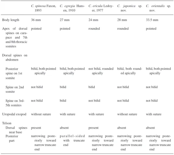
Remarks. Ceratomysis comprises three species, C. egregia Hansen, 1910 recorded from In- donesia (Hansen, 1910), C. ericula Ledoyer, 1977 from the Kerguelen Islands in the southern In- dian Ocean (Ledoyer, 1977, 1995), and C. spinosa Faxon, 1893 from the Gulf of Panama (Faxon, 1893), British Columbia and Japan (W. Tattersall, 1951).
Ceratomysis japonica is allied to C. spinosa with respect to the second to fi fth abdominal somites with a simple dorsal spine, the uropodal exopod with no suture, and the telson with a nar- row truncate apex. However, it is distinguished from C. spinosa by the absence of the dorsal spines of the telson and the apical shape of the dorsal spines on the carapace, last two thoracic somites and abdomen (Table 4).
Ceratomysis japonica is readily distinguishable from C. egregia and C. ericula by the differ- ent characters of the dorsal spines on the abdomen, the uropodal exopod and the telson (Table 4).
Table 4. Comparison of characters in the fi ve species in Ceratomysis.
C. spinosa Faxon, 1893
C. egregia Hans- en, 1910
C. ericula Ledoy- er, 1977
C. japonica sp.
nov.
C. orientalis sp.
nov.
Body length 36 mm 27 mm 24 mm 28 mm 33.5 mm
Apex of dorsal spines on cara- pace and 7th and 8th thoracic somites
pointed pointed rounded rounded pointed
Dorsal spines on abdomen Posterior spine on 1st
bifi d, both pointed apically
bifi d, both pointed apically
not bifi d, rounded apically
bifi d, both round- ed apically
bifi d, both pointed apically somite
Spine on 2nd somite
not bifi d bifi d bifi d not bifi d not bifi d
Spine on 3rd- 5th somites
not bifi d bifi d not bifi d not bifi d not bifi d
Uropodal exopod without suture with suture with suture without suture with suture Telson
Dorsal spines near base
present absent present absent absent
Posterior part
narrowing poste- riorly toward narrow truncate end
p a r a l l e l - s i d e d with truncate end
narrowing poste- riorly toward narrow truncate end
narrowing poste- riorly toward narrow truncate end
narrowing poste- riorly toward narrow truncate end
Ceratomysis orientalis sp. nov.
(Figs. 3 4)
Material examined. Holotype: adult female (33.5 mm), KT-07-29-K2, NSMT-Cr 19654.
Paratype: 1 immature male (22.7 mm), KT-07-29-K2, NSMT-Cr 19655.
Other material. 3 juveniles (13.3 mm, damaged), KT-07-29-K2, NSMT-Cr 19656.
Description. Body spinous (Fig. 3A B). Seventh and eighth thoracic somites with dorsal me- dian spine with pointed apex. First abdominal somite with anterior simple and posterior bifi d spines on dorsal median line, those spines with pointed apex; second to sixth somites with simple dorsal spine with pointed apex.
Carapace (Fig. 3A B) spinous; anterior margin straight; anterolateral angles produced into long spiniform process; posterior margin not emarginate, uncovering last 2 thoracic somites; dor- sal surface with 3 spines anterior to cervical sulcus, 2 pairs of spines posterior to cervical sulcus, and 1 spine near posterior end, those spines with pointed apex.
Antennular peduncle (Fig. 3C) robust. Lateral antennular fl agellum thick and slightly shorter than mesial one in male (Fig. 3C), slightly thicker than mesial one in female.
Antennal scale (Fig. 3C D) long, 2.2 times as long as antennular peduncle, 8 times as long as broad, armed with setae on entire margin except for proximal 2/5 of lateral margin naked, 7 spines present among setae on lateral margin. Antennal peduncle (Fig. 3D) long, extending to distal 1/5 of scale; second segment 4 times as long as broad; third segment 1.3 times longer than second.
Antennal sympod (Fig. 3D) with anterolateral angle protruded into immovable long spine.
Eye (Fig. 3C) reduced to single plate without visual elements, both angles produced into spiniform process.
Labrum without acute projection on anterior margin. Mandibular palp (Fig. 3E) with second segment armed with spine-like setae on mesial and lateral margins; third segment 2/3 of second segment in length. Lateral lobe of maxillule (Fig. 3F) armed with 12 spines on distal margin and 2 setae on posterior surface. Maxilla (Fig. 3G): exopod large, oval, extending to 1/2 of second seg- ment of endopod; second segment of endopod 1.6 times as long as broad.
Endopod of fi rst thoracopod (Fig. 3H) short, robust; ischium with mesial lobe; merus ex- panded mesially. Endopod of second thoracopod (Fig. 3I) robust; ischium with mesial lobe extend- ing to distal margin of merus; merus expanded mesially. Endopods of third to fi fth thoracopods (Fig. 4A C) long, slender, increasing in length from third to fi fth thoracopods; carpopropodus un- divided. Endopods of sixth to eighth thoracopods (Fig. 4D E) long; carpopropodus almost as long as merus, divided into 3 subsegments, proximal 2 subsegments short; dactylus 1/3 of carpopropo- dus in length, with terminal claw. First thoracopod without exopod; second to eighth thoracopods with fl agelliform exopod.
Marsupium of female composed of 7 pairs of oostegites.
All pleopods of female uniramous; fi rst to fourth pleopods (Fig. 4F) unsegmented, increasing in length from fi rst to fourth pleopod; fi fth pleopod (Fig. 4G) long, extending to proximal 1/5 of uropodal exopod, 2.1 times longer than fourth pleopod, 2-segmented, proximal segment with long lobe at distolateral angle.
Uropodal endopod (Fig. 4H) 1.1 times as long as exopod, without spines on mesial margin.
Uropodal exopod (Fig. 4H) lanceolate, with suture in distal 1/8; proximal segment armed with 8 spines among setae on lateral margin.
Telson (Fig. 4I) long, 4.3 times as long as sixth abdominal somite, 1.3 times longer than uropodal exopod, 6.3 times as long as maximum breadth at base, slightly broadened from proximal 1/3 to distal 1/7, then narrowing toward narrow truncate apex, without dorsal spines.
Etymology. The specifi c name is derived from Latin “oriens” referring to type locality.
Fig. 3. Ceratomysis orientalis sp. nov. A, C I, adult female (33.5 mm), holotype, NSMT-Cr 19654; B, immature male (22.7 mm), paratype, NSMT-Cr 19655. A, body (lateral); B, body (dorsal); C, anterior part of body (dor- sal); D, antenna (right, ventral); E, mandible (right, posterior); F, maxillule (right, posterior); G, maxilla (right, posterior); H, endopod of fi rst thoracopod (right, posterior); I, endopod of second thoracopod (right, posteri- or).
Fig. 4. Ceratomysis orientalis sp. nov. Adult female (33.5 mm), holotype, NSMT-Cr 19654. A, endopod of third thoracopod (right, posterior); B, endopod of fourth thoracopod (right, posterior); C, endopod and basal plate of exopod of fi fth thoracopod (right, posterior); D, endopod of seventh thoracopod (right, posterior); E, endo- pod of eighth thoracopod (right, posterior); F, fourth pleopod (right, anterior); G, fi fth pleopod (right, anterior);
H, uropod (right, dorsal); I, telson (dorsal).
Remarks. Ceratomysis orientalis closely resembles C. spinosa in that the carapace, last two thoracic somites and abdomen have dorsal spines with a pointed tip, the bifi d spine is present only on the fi rst abdominal somite, and the telson has a narrow truncate apex. However, C. orientalis differs from C. spinosa in its uropodal exopod with a suture near the apex and telson without dor- sal spines (Table 4).
Hansenomysis armata Birstein and Tchindonova, 1958 (Fig. 5)
Hansenomysis armata Birstein and Tchindonova, 1958: 271 273, fi gs. 4 5.
Material examined. 2 adult females (20.0 mm, damaged) and 2 juveniles (7.7, 9.3 mm), SO07-K2, NSMT-Cr 19657.
Distirbution. Previously known from the Kurile-Kamchatka Trench (Birstein and Tchindon- ova, 1958).
Remarks. In Hansenomysis, two species, H. armata and H. menziesi Bacescu, 1971, are char- acterized by a carapace with mid-dorsal spines and a posterior projection, and an antennal scale with spines among the setae on the lateral margin. The present specimens differ from H. menziesi in that the carapace shows a dorsal spine on the anterior margin in H. menziesi, whereas there are four more spines between the anterior end and the posterior projection in the present specimens;
all abdominal somites have paired robust spines posterolaterally in H. menziesi, whereas the pres- ent specimens are lacking such spines; and the proximal segment of the uropodal exopod is armed with spines and fi ne setae on the distal two-thirds of the lateral margin in H. menziesi, but is armed with spines only on the distal two-fi fths of the lateral margin in the present specimens.
The present specimens were thus identifi ed as H. armata. However, there is a certain amount of variation observed in the number of dorsal spines in the present specimens. The type specimen
Fig. 5. Hansenomysis armata Birstein and Tchindonova, 1958. Adult female (20.0 mm), NSMT-Cr 19657. A, cara- pace (lateral); B, uropod and telson (dorsal).
of H. armata has dorsal spines on the mid-dorsal line with one spine immediately front of the cervical sulcus and two spines on the posterior part of the cervical sulcus (Birstein and Tchindon- ova, 1958). In the present specimens, one female (damaged, ca. 15.0 mm) is consistent with the type specimen in this feature. However, another female (20.0 mm) is furnished with seven spines, three on the anterior part of the cervical sulcus and four on the posterior part (Fig. 5A). In all other respects, these specimens were consistent with each other.
Hansenomysis armata was previously recorded as a single damaged female specimen by Birstein and Tchindonova (1958). An illustration of the telson is fi rst provided in Fig. 5B.
?Hansenomysis fyllae (Hansen, 1887) (Fig. 6)
Arctomysis fyllae Hansen, 1887: 210 213, pl. 7, fi g. 5.
Hansenomysis fyllae: Stebbing, 1893: 268; Hansen, 1908: 96 98, pl. 4, fi g. 4a k; Zimmer, 1909: 41 43, fi gs. 68 70; W. Tat- tersall, 1911: 25 26; 1951: 43; W. Tattersall and O. Tattersall, 1951: 114 119, fi gs. 14 16.
Material examined. 1 immature male (14.5 mm), KT-07-29-M3-2, NSMT-Cr 19658.
Distirbution. Previously known from the northeastern North Atlantic (Hansen, 1887, 1908;
W. Tattersall, 1911; W. Tattersall and O. Tattersall, 1951).
Remarks. Hansenomysis fyllae is characterized by its carapace without dorsal spines, the an- tennal scale having approximately six spines among the setae on the lateral margin, and the proxi- mal segment of the uropodal exopod having approximately six spines among the setae on the lat- eral margin.
The present specimen is consistent with H. fyllae in the characteristics of its
Fig. 6. ?Hansenomysis fyllae (Hansen, 1887). Immature male (14.5 mm), NSMT-Cr 19658. A, anterior part of body (dorsal); B, telson (dorsal); C, distal part of telson (dorsal).
carapace, antennal scale and uropod. The armature of the telson is also consistent with the previous descriptions. However, this specimen differs from the previous descriptions in that its telson shows setae on the dorsal surface (Fig. 6B C). This feature has not been previously reported in this spe- cies. This paper therefore recorded this specimen tentatively as H. fyllae.
Hansenomysis japonica Bravo and Murano, 1997
Hansenomysis japonica Bravo and Murano, 1997: 228 232, fi gs. 1 2, 3A D.
Material examined. 1 adult female (15.0 mm), WA05-F510, NSMT-Cr 19659.
Distirbution. Previously known from Sagami Bay, Japan (Bravo and Murano, 1997).
Hansenomysis rostrata Birstein and Tchindonova, 1970
Hansenomysis rostrata Birstein and Tchindonova, 1970: 280 284, fi gs. 1 3.
Material examined. 1 adult male (38.5 mm), SO-SE Erimo, NSMT-Cr 19660. 1 adult female (44.8 mm), SO06-M4-B, NSMT-Cr 19661.
Distribution. Previously known from south-east off Etorofu Island, Kurile Islands (Birstein and Tchindonova, 1970).
Family Mysidae Subfamily Boreomysinae
Birsteiniamysis inermis (Willemoes-Suhm, 1874)
Petalophthalmus inermis Willemoes-Suhm, 1874: 15.
Petalophthalmus armiger Willemoes-Suhm, 1875: 41 (only female).
Boreomysis scyphops G. Sars, 1879b: 429 430; Hansen, 1908: 99 100, fi g. 1.
Boreomysis suhmi Faxon, 1893: 218.
Boreomysis distinguenda Hansen, 1908: 100, fi g. 2; W. Tattersall, 1913: 869.
Boreomysis inermis: W. Tattersall, 1951: 46 47; O. Tattersall, 1955: 75 76; Birstein and Tchindonova, 1958: 282 283, fi g. 9;
1962: 62; Kathman et al., 1986: 108, fi gs. a e (p. 109); Ledoyer, 1995: 603.
Birsteiniamysis scyphops: Tchindonova, 1981: 28 29.
Birsteiniamysis inermis: Tchindonova, 1981: 28 29; Petryashov, 2005: 963 964, fi g. 4 (6 11).
not Boreomysis inermis: Hansen, 1910: 26 27, pl. 2, fi g. 4 (=Boreomysis hanseni Holmquist, 1956).
Material examined. 1 adult male (39.5 mm), 2 immature males (25.0, 28.0 mm), 1 adult fe- male (45.0 mm), 1 immature female (28.2 mm) and 2 juveniles (damaged), KT-07-29-K2, NSMT- Cr 19662.
Distribution. Known from the northern North Pacifi c (W. Tattersall, 1951; Birstein and Tchin- donova, 1958), the northern North Atlantic (W. Tattersall, 1951), and Antarctic (O. Tattersall, 1955; Birstein and Tchindonova, 1962; Ledoyer, 1995; Petryashov, 2005).
Boreomysis arctica (Krøyer, 1861)
Mysis arctica Krøyer, 1861: 34, 42, fi g. 5.
Boreomysis arctica: G. Sars, 1869: 330 332; Zimmer, 1909: 53 54, fi gs. 71 74; W. Tattersall, 1951: 49 51, fi g. 8; W. Tattersall and O. Tattersall, 1951: 132 135, fi gs. 19B, 21B, 22; Birstein and Tchindonova, 1958: 284, fi g. 10; Lagardère and Nouvel, 1980: 385 386; Wittmann et al., 2003: 1263.
Arctomysis arctica: Czerniavsky, 1887: 7 8.
Material examined. 1 adult male (27.9 mm), KT-07-29-K2, NSMT-Cr 19663.
Distribution. Known from the North Atlantic (W. Tattersall and O. Tattersall, 1951; Lagardère and Nouvel, 1980; Wittmann et al., 2003) and the northern North Pacifi c (W. Tattersall, 1951).
Boreomysis californica Ortmann, 1894
Boreomysis californica Ortmann, 1894: 106, fi gs. 4 14; Banner, 1948a: 367 369, pl. 4, fi g. 5a i; W. Tattersall, 1951: 52 55, fi gs. 9 10; Birstein and Tchindonova, 1958: 294; 1962: 64, fi g. 5; Taniguchi, 1969: 46 47, fi g. 4; Petryashov, 2005:
965.
Boreomysis media Hansen, 1912: 190, pl. 1, fi g. 2a b; Illig, 1930: 419.
Boreomysis kincaidi Banner, 1948a: 361 365, pl. 2, fi g. 3a j.
Material examined. 1 adult female (16.3 mm), WA07-D1500D, NSMT-Cr 19664. 1 imma- ture male (6.8 mm), SO07-O1, NSMT-Cr 19665. 1 immature male (16.2 mm) and 1 juvenile (dam- aged), KT-07-29-K2, NSMT-Cr 19666. 1 immature male (15.5 mm) and 1 juvenile (13.0 mm), KT-07-29-M1, NSMT-Cr 19667.
Distribution. Widely distributed in the Pacifi c (W. Tattersall and O. Tattersall, 1951; Birstein and Tchindonova, 1958; Taniguchi, 1969), Indian (Hansen, 1912), and Antarctic Oceans (Birstein and Tchindonova, 1962; Petryashov, 2005).
Boreomysis incisa Nouvel, 1942
Boreomysis incisa Nouvel, 1942b: 4 5, fi gs. 9 11; 1943: 57 58, pl. 3, fi gs. 77, 84; Birstein and Tchindonova, 1958: 290 291, fi g. 16.
Material examined. 1 immature male (21.3 mm) and 2 immature females (22.3, 24.2 mm), SO07-C6, NSMT-Cr 19668.
Distribution. Known from the East Atlantic (Nouvel, 1942b, 1943) and the western North Pacifi c (Birstein and Tchindonova, 1958).
Boreomysis sp.
(Fig. 7)
Material examined. 1 immature male (13.8 mm), KT-07-29-M1, NSMT-Cr 19669.
Remarks. The present specimen is characterized by its fl attened eyes (Fig. 7B), which, in Boreomysis, is known only in B. latipes Birstein and Tchindonova, 1958. However, it differs from B. latipes in its antennal scale and second thoracopodal endopod. The apical lobe of the antennal scale of this specimen is three times longer than the lateral spiniform process (Fig. 7D), whereas that of B. latipes is almost the same in length with the lateral spiniform process (Birstein and Tchindonova, 1958). The carpopropodus of the second thoracopodal endopod is normal in this specimen (Fig. 7E), whereas it is projected mesially in B. latipes (Birstein and Tchindonova, 1958).
The present specimen may be an undescribed species, but is recorded here as Boreomysis sp.
for an immature.
Subfamily Mysinae Tribe Erythropini
Amblyops abbreviata (M. Sars, 1869)
Pseudomma abbreviatum M. Sars, 1869: 262.
Amblyopsis abbreviat: G. Sars, 1869: 328 330.
Amblyops abbreviata: G. Sars, 1872: 5 12, pl. 6; Zimmer, 1904: 39, fi gs. 108 111; 1909: 112 113, fi gs. 223 228; Banner, 1948a: 382 383; W. Tattersall, 1951: 128 130, fi g. 44; W. Tattersall and O. Tattersall, 1951: 247 251, fi gs. 56 57; Ban- ner, 1954: 581; Birstein and Tchindonova, 1958: 319; Kathman et al., 1986: 96, fi gs. a m (p. 97).
Material examined. 2 adult males (9.9 mm, damaged), KT-07-29-K2, NSMT-Cr 19670. 1 adult male (damaged), 1 immature male (9.7 mm) and 1 immature female (damaged), KT-07- 29-M1, NSMT-Cr 19671.
Distirbution. Known from boreal waters of both the Atlantic and Pacifi c Oceans (W. Tatter- sall, 1951; W. Tattersall and O. Tattersall, 1951).
Remarks. Banner (1948a) and W. Tattersall (1951) observed a difference in the shape of the process on the anteromedian margin of the eye between Atlantic and Pacifi c specimens of A. ab- breviata; that of the Pacifi c specimens is longer and more acute in the lateral view (W. Tattersall, 1951). The present specimens are similar to the Pacifi c specimens in this feature.
Amblyops ewingi Bacescu, 1967 (Fig. 8)
Amblyops ewingi Bacescu, 1967: 153 157, fi gs. 3 4.
Material examined. 1 adult female (damaged), KT-07-29-E3, NSMT-Cr 19672.
Distribution. Previously known only from the Peru Trench (Bacescu, 1967).
Remarks. Amblyops ewingi was established on the basis of a single female specimen col- lected from the Peru Trench (Bacescu, 1967). The present occurrence is the second record and far from the type locality.
Fig. 7. Boreomysis sp. Immature male (13.8 mm), NSMT-Cr 19669. A, anterior part of body (dorsal); B, eye (left, lateral), e=eye, r=rostral plate; C, antenna (right, ventral); D, apical part of antennal scale (right, dorsal); E, distal part of endopod of second thoracopod (right, posterior); F, uropod and telson (dorsal).
Amblyops magna Birstein and Tchindonova, 1958 (Fig. 9)
Amblyops magna Birstein and Tchindonova, 1958: 319 320, fi g. 36.
Material examined. 1 adult female (27.6 mm), SO07-O4, NSMT-Cr 19673. 1 immature fe- male (20.6 mm), SO-SE Erimo, NSMT-Cr 19674.
Distribution. Previously known from the Kurile-Kamchatka Trench (Birstein and Tchindon- ova, 1958).
Amblyopsoides ohlinii (W. Tattersall, 1951)
Amblyops crozetii: Ohlin, 1901: 371; Zimmer, 1904: 39 40, fi gs. 114 116.
Amblyops crozeti: Zimmer, 1909: 114, fi gs. 229 231.
Amblyops n. sp. Hansen, 1908: 108.
Amblyops ohlinii W. Tattersall, 1951: 130 132, fi g. 45.
Amblyopsoides ohlinii: O. Tattersall, 1955: 108.
Amblyops ohlini: Birstein and Tchindonova, 1970: 285 287, fi g. 5.
Material examined. 1 adult female (25.5 mm), KT-07-29-H2, NSMT-Cr 19675. 1 immature female (15.9 mm), KT-07-29-K3, NSMT-Cr 19676. 1 adult female (32.3 mm), KT-07-29-M3-2, NSMT-Cr 19677.
Distribution. Known from the northern North Atlantic (Ohlin, 1901; W. Tattersall, 1951) and the northern North Pacifi c (Birstein and Tchindonova, 1970).
Holmesiella affi nis Ii, 1937
Holmesiella affi nis Ii, 1937: 200 205, fi gs. 31 45; 1964: 348 351, fi g. 88; Murano, 1970a: 260; 1970b: 141, fi gs. 6 9; 1976:
25 26, fi g. 4; Liu and Wang, 2000: 154 156, fi g. 45.
Fig. 8. Amblyops ewingi Bacescu, 1967. Adult female (damaged), NSMT-Cr 19672. A, proximal part of uropodal endopod (ventral); B, telson (dorsal).
Material examined. 1 adult female (25.3 mm), WA05-G550, NSMT-Cr 19678. 1 adult female (21.5 mm), WA06-E510, NSMT-Cr 19679. 2 adult males (22.3, 24.0 mm), WA06-F480, NSMT-Cr 19680. 1 adult male (28.0 mm), WA06-F510, NSMT-Cr 19681. 1 adult male (24.0 mm), WA06- G380, NSMT-Cr 19682. 2 adult males (20.0, 22.8 mm) and 2 adult females (18.0, 20.5 mm), WA06-G425, NSMT-Cr 19683. 2 adult males (23.5, 24.5 mm), WA06-G480, NSMT-Cr 19684.
Distribution. The East China Sea (Ii, 1937, 1964), the South China Sea (Liu and Wang, 2000), and Pacifi c waters off central Japan (Murano, 1970a, b, 1976).
Remarks. Murano (1970b) recognized two races in this species; the large- and small-sized races, which differ in the size of the body and eyes, the shape of the rostrum, antennal scale and telson, and the number of segments of both rami of the fourth male pleopod.
The present specimens correspond to the large-sized race in their body length, however, they have fewer segments of the fourth male pleopod than those described by Murano (1970b): eight- segmented endopod and seven-segmented exopod in the present specimens, and 14-segmented endopod and 11-segmented exopod in Murano’s specimens (1970b); the number of spines on the lateral margin of the telson is also different from Murano’s illustration of the large-sized race: 15 to 26 spines in the present specimens, and 12 spines in Murano’s specimens (1970b).
This species has a wide range of body size from 15.0 to 28.0 mm (Ii, 1937; Murano, 1970b;
present study), and has been observed to show intraspecifi c variation in morphology in several of its features.
Holmesiella anomala Ortmann, 1908
Holmesiella anomala Ortmann, 1908: 6 8, pl. 1, fi gs. 1 13; W. Tattersall, 1933: 5 6; Banner, 1948a: 395 399; W. Tattersall, 1951: 106 109, fi gs. 32 33; Birstein and Tchindonova, 1958: 304 305; Murano, 1976: 23 24, fi g. 3; Murano and Kry- gier, 1985: 705, fi gs. 11 13; Kathman et al., 1986: 168, fi g. a j (p. 169).
Fig. 9. Amblyops magna Birstein and Tchindonova, 1958. Adult female (27.6 mm), NSMT-Cr 19673. A, anterior part of body (dorsal); B, antennal scale (right, dorsal); C, antennal peduncle (right, dorsal); D, uropod (left, ventral); E, telson (dorsal).
Material examined. 1 adult male (31.5 mm) and 1 adult female (31.8 mm), WA07-A410, NSMT-Cr 19685. 1 adult female (39.0 mm), WA07-A750, NSMT-Cr 19686. 1 adult male (36.0 mm), WA07-A900, NSMT-Cr 19687. 1 immature male (19.3 mm) and 2 immature females (18.4, 20.3 mm), WA07-B410, NSMT-Cr 19688. 1 immature male (19.3 mm), WA07-B410D, NSMT-Cr 19689. 1 adult male (30.5 mm), WA07-B450, NSMT-Cr 19690. 1 adult male (damaged) and 2 adult females (32.8, 33.0 mm), WA07-B650, NSMT-Cr 19691. 4 adult males (28.2-31.7 mm) and 2 adult females (35.8, 37.4 mm), WA07-B750, NSMT-Cr 19692. 1 adult male (25.8 mm) and 4 adult females (29.2 35.0 mm), WA07-B900, NSMT-Cr 19693. 1 adult male (28.3 mm) and 7 adult females (31.0 35.5 mm), WA07-B1200T, NSMT-Cr 19694. 4 adult males (26.0 32.5 mm) and 2 adult females (27.2, 34.0 mm), WA07-C450, NSMT-Cr 19695. 5 adult males (31.7 33.2 mm), WA07-C510, NSMT-Cr 19696. 1 adult male (25.5 mm), WA07-C550, NSMT-Cr 19697. 2 adult females (40.5, 45.7 mm), WA07-C650, NSMT-Cr 19698. 1 adult female (broken), WA07-C750, NSMT-Cr 19699. 2 adult females (32.5, 35.2 mm), WA07-C900, NSMT-Cr 19700. 2 adult males (30.0, 32.3 mm) and 3 adult females (28.0 34.2 mm), WA07-D510, NSMT-Cr 19701. 2 immature males (20.4, 27.0 mm), 1 immature female (30.7 mm) and 8 juveniles (9.5 11.8 mm), KT-07- 29-K2, NSMT-Cr 19702.
Distribution. Known from the northern North Pacifi c (Ortmann, 1908; W. Tattersall, 1933, 1951; Banner, 1948a; Birstein and Tchindonova, 1958; Murano, 1976; Murano and Krygier, 1985).
Holmesiella bisaetigera sp. nov.
(Figs. 10 11)
Material examined. Holotype: adult male (36.2 mm), WA06-E510, NSMT-Cr 19703.
Description. Integument smooth. First to fi fth abdominal somites subequal in length; sixth somite 1.9 times longer than fi fth.
Carapace produced anteriorly into trapezoidal rostral plate (Fig. 10A); anterolateral corner rounded; posterior margin emarginate, exposing last thoracic somite dorsally.
Antennular peduncle (Fig. 10A) robust; third segment slightly longer than fi rst segment, with developed appendix masculina.
Antennal scale (Fig. 10A B) lanceolate, extending beyond anterior end of antennular pedun- cle by 1/6 of its length, 4.8 times as long as broad, with suture near apex; lateral margin naked, terminating into spiniform process; apical lobe slightly longer than lateral process. Antennal pe- duncle (Fig. 10B C) extending to proximal 2/5 of scale, with third segment as long as second seg- ment. Antennal sympod (Fig. 10B) without spiniform processes at distolateral angle.
Eye (Fig. 10A) developed, globular, 1.1 times as long as broad in dorsal view; cornea occupy- ing 3/5 of eye.
Labrum without frontal processes. Mandibular palp (Fig. 10D) with second segment armed densely with setae on lateral margin; third segment almost 1/2 of second segment in length. Maxil- lule (Fig. 10E) with lateral lobe armed with 13 spines on distal margin and 5 plumose setae on posterior surface; mesial lobe with long setae on mesial margin. Maxilla (Fig. 10F) with exopod extending beyond distal margin of proximal segment of endopod and tapering toward apex; endo- pod with second segment 1.5 times as long as broad.
Endopod of fi rst thoracopod (Fig. 10G) short, robust; ischium and merus expanded mesially.
Endopod of second thoracopod (Fig. 10H) long. Endopod of third thoracopod missing. Endopods
of fourth to eighth thoracopods (Figs. 10I J, 11A B) long, robust; merus relatively broad; carpo-
propodus divided into 3 subsegments, proximal subsegment 1/2 3/5 of merus in length, 1.5 1.6
Fig. 10. Holmesiella bisaetigera sp. nov. Adult male (36.2 mm), holotype, NSMT-Cr 19703. A, anterior part of body (dorsal); B, antenna (right, dorsal); C, antennal peduncle (right, ventral); D, mandible (right, posterior);
E, maxillule (right, posterior); F, maxilla (right, posterior); G, endopod of fi rst thoracopod (right, anterior); H, endopod of second thoracopod (right, posterior); I, endopod of fourth thoracopod (right, posterior); J, endopod of sixth thoracopod (right, posterior); K, male genital appendage (right, lateral).
Fig. 11. Holmesiella bisaetigera sp. nov. Adult male (36.2 mm), holotype, NSMT-Cr 19703. A, endopod of seventh thoracopod (right); B, endopod of eighth thoracopod (right); C, fi rst pleopod (right, anterior); B, second pleo- pod (right, anterior); E, third pleopod (right, anterior); F, fourth pleopod (right, anterior); G, fi fth pleopod (right, anterior); H, uropod and telson (dorsal); I, uropodal endopod (right, ventral).
times as long as distal 2 subsgements combined, articulated obliquely from middle subsegment, distal subsegment slightly longer than middle subsegment, articulated transversely from middle subsegment; dactylus with long terminal claw. Exopods of all thoracopods fl agelliform, with basal plate with rounded anterolateral angle.
Male genital appendage (Fig. 10K) armed with 12 long, curved setae on posterodistal mar- gin.
All pleopods of male biramous. First pleopod (Fig. 11C) with endopod reduced to unseg- mented lobe, exopod 9-segmented. Second, third and fi fth pleopods (Fig. 11D E, G) with both rami 8-segmented. Exopod of fourth pleopod (Fig. 11F) 8-segmented. Endopod of fourth pleopod (Fig. 11F) elongate, extending beyond apex of uropodal endopod, 9-segmented; seventh to ninth segments lengthened; seventh segment 1.4 times as long as sixth segment, armed with short seta on lateral and mesial corners of distal margin; eighth segment 1.9 times as long as sixth, armed with short seta on each corner; ninth segment 3 times longer than sixth, armed with 1 short and 2 long, unequal terminal setae, longer seta 1.1 times as long as ninth segment and 1.5 times as long as shorter one.
Uropodal endopod (Fig. 11H I) extending beyond posterior margin of telson by 2/5 of its length, armed with 7 spines on mesial ventral margin from statocyst region to proximal 2/5, spines increasing in length and intervals becoming distantly posteriorly; exopod (Fig. 11H) 1.3 times longer than endopod.
Telson (Fig. 11H) subequal to sixth abdominal somite in length, 1.7 times as long as broadest part near base, triangular with truncate apex; lateral margin armed on distal 3/5 with 17 spines in- creasing in length posteriorly; distal margin armed with 2 pairs of spines and pair of median plu- mose setae, lateral pair of spines long, subequal to distalmost lateral spines in length, mesial pair of spines short, less than 1/5 of lateral pair in length.
Table 5. Comparison of characters in the three species of Holmesiella and Pteromysis amemiyai Ii, 1964.
H. affi nis Ii, 1937 H. anomala Ort- mann, 1908
H. bisaetigera sp.
nov.
P. amemiyai Ii, 1964
Antennular pedun- cle
proximal part of 3rd segment over- hanging 2nd seg- ment in dorsal view
normal normal proximal part of 3rd
segment over- hanging 2nd seg- ment in dorsal view
Endopod of 4th male pleopod
1.3 times as long as exopod; armed with 1 short and 1 long terminal set- ae
2.0 2.6 times as long as exopod;
armed with 1 short and 1 long terminal setae
2.5 times as long as exopod; armed with 1 short and 2 long terminal set- ae
1.4 times as long as exopod; armed with 1 short and 1 long terminal set- ae
Apical long spines of telson
1/5 1/4 of telson length; 1.7 2.2 times as long as distalmost lateral spines
1/8 1/6 of telson length; 1.4 2.0 times as long as distalmost lateral spines
less than 1/10 of tel- son length; sub- equal to distal- most lateral spines
1/5 of telson length;
1.7 1.8 times as long as distalmost lateral spines
References Ii (1937, 1964), Mu- rano (1970b)
Ortmann (1908), W.
Tattersall (1951), Murano and Kry- gier (1985)
present study Ii (1964), Murano (1976)
Etymology. The specifi c name is from combination of Latin “bi”, two, and “saetiger”, having bristles, referring to two long terminal setae of the endopod of the fourth male pleopod.
Remarks. The present species differs from the generic diagnosis of Holmesiella Ortmann, 1908 in the number of the long terminal setae of the endopod of the fourth male pleopod, but well agrees with it in the other characters. Therefore, the present study revises the generic diagnosis of Holmesiella in this respect and assigns the present species to this genus.
Holmesiella bisaetigera is readily distinguishable from H. affi nis and H. anomala by having two long spiniform terminal setae on the endopod of the fourth male pleopod (Table 5). Addition- ally, H. bisaetigera is distinguished from H. affi nis by the character of the antennal peduncle, the endopod of the fourth male pleopod, and the telson (Table 5).
Holmesiella is related to Pteromysis Ii, 1964 by a combination of the antennal scale with a naked lateral margin, the eyes with globular cornea, the triangular telson with spines on the distal three-fi fths of the lateral margin, and the fourth pleopod of the male, which has an elongate endo- pod. H. bisaetigera is distinguishable from P. amemiyai Ii, 1964 by the shape of the antennal pe- duncle and the length of the apical long spines of the telson (Table 5).
Revised generic diagnosis of Holmesiella. Eyes developed, with globular cornea; antennal scale with naked lateral margin terminating in spiniform process; carpopropodus of third to eighth thoracopodal endopods divided into 3 subsegments; both rami of all male pleopods multi-seg- mented except fi rst pleopodal endopod which is unsegmented; endopod of fourth male pleopod elongate, armed with 1 or 2 long spiniform terminal setae; uropodal endopod armed with spines on mesial margin; telson triangular with narrow truncate apex, lateral margin armed with spines on distal 3/5, and distal margin armed with 2 pairs of spines and pair of median plumose setae.
Meterythrops microphthalmus W. Tattersall, 1951
Meterythrops microphthalma W. Tattersall, 1951: 113 116, fi g. 36: Banner, 1954: 580 581 (part?); Birstein and Tchindonova, 1958: 305; Ii, 1964: 319; Murano, 1971: 47; 1977: 171 176; Jo et al., 1998: 40 41, fi g. 7.
Meterythrops robusta: Taniguchi, 1969, 47 48, fi g. 5 [not Meterythrops robustus Smith, 1879].
Meterythrops microphthalmus: Fukuoka and Murano, 2006: 1645 1651, fi gs. 2 3, 12B C.
Material examined. 1 adult female (21.2 mm), SO07-C3, NSMT-Cr 19704. 3 immature fe- males (13.2 14.0 mm) and 1 juvenile (7.2 mm), SO07-C4-B1, NSMT-Cr 19705.
Distribution. Known from the cold-water region of the North Pacifi c (see Fukuoka and Mu- rano, 2006).
Meterythrops robustus Smith, 1879
Meterythrops robusta Smith, 1879: 93 98, pl. 12; Hansen, 1908: 106 107; Zimmer, 1909: 85 87, fi gs. 168 172; Stephensen, 1912: 79 80; 1933: 12; W. Tattersall, 1933: 8; 1939: 283; Banner, 1948a: 377 379 (part?); W. Tattersall, 1951: 113, fi g.
35; Banner, 1954: 580 581 (part?); Birstein and Tchindonova, 1958: 305 306; Daly and Holmquist, 1986: 1208; Kath- man et al., 1986: 188, fi g. a h (p. 189).
Parerythrops robusta: G. Sars, 1879a: 98 102, tab. 39.
Meterythrops robustus: Fukuoka and Murano, 2006: 1655 1658, fi gs. 5 6, 12E F.
not Meterythrops robusta: Holt and W. Tattersall, 1905: 143 [=Parerythrops obesa (Sars, 1864)]; Taniguchi, 1969: 47 48, fi g.
5 [=Meterythrops microphthalmus].
Material examined. 2 adult females (11.5, 14.2 mm), WA9204, NSMT-Cr 19706. 1 adult fe- male (damaged, ca 11.2 mm), WA06-F510, NSMT-Cr 19707. 1 adult female (14.3 mm), WA07- C350, NSMT-Cr 19708. 2 adult females (13.8, 14.3 mm), WA07-C410, NSMT-Cr 19709.
Distribution. Circumpolar (see Fukuoka and Murano, 2006).
Meterythrops sp.
(Fig. 12)
Meterythrops sp.: Murano, 1977: 177, fi g. 24; Fukuoka and Murano, 2006: 1659 1661, fi g. 7.
Material examined. 1 immature female (24.5 mm), SO07-C4-B1, NSMT-Cr 19710.
Distribution. Known from the Japan Sea (Murano, 1977).
Remarks. Murano (1977) reported Meterythrops sp. based on two damaged female speci- mens obtained from the stomach of a tadpole sculpin collected with a trawl net from the fl oor of the Japan Sea at a depth of 1035 m. Murano (1977) recognized that the specimens belonged to the genus Meterythrops and distinguished them from other species of this genus. Nevertheless, he re- tained them as undescribed species due to the absence of a male specimen. The present specimen is also an immature female.
Mysimenzies borealis sp. nov.
(Figs. 13 14)
Material examined. Holotype: adult male (27.3 mm), SO07-O3, NSMT-Cr 19711. Paratypes:
2 adult males (20.0, 20.5 mm) and 2 adult females (26.1, 30.6 mm), SO07-O3, NSMT-Cr 19712;
2 adult males (broken) and 2 adult females (damaged), SO07-O4, NSMT-Cr 19713.
Description. Integument not hispid. Thoracic somites without sternal processes. First to fi fth abdominal somites subequal in length; sixth somite twice as long as fi fth.
Carapace produced anteriorly into triangular rostral plate with rounded apex extending to 1/2 of fi rst segment of antennular peduncle (Fig. 13A B); anterolateral corner rounded; posterior mar- gin emarginate, leaving last thoracic somite exposed dorsally.
Antennular peduncle of male (Fig. 13A) more robust than that of female (Fig. 13B); fi rst seg- ment with sensory fossette in middle of dorsal side and longitudinal lobe in middle of ventral side
Fig. 12. Meterythrops sp. Murano, 1977. Immature female (24.5 mm), NSMT-Cr 19710. A, antenna (right, ventral);
B, uropodal endopod (right, ventral); C, telson (dorsal).
Fig. 13. Mysimenzies borealis sp. nov. A, C J, adult male (27.3 mm), holotype, NSMT-Cr 19711; B, adult female (26.1 mm), paratype, NSMT-Cr 19712. A, B, anterior part of body (dorsal); C, fi rst and second segments of antennular peduncle (left, lateral); D, antenna (left, ventral); E, labrum (lateral); F, mandible (left, ventral); G, maxillule (left, posterior); H, maxilla (left, posterior); I, endopod of fi rst thoracopod (left, posterior); J, endo- pod of second thoracopod (left, posterior).
Fig. 14. Mysimenzies borealis sp. nov. A B, I, adult female (damaged), paratype, NSMT-Cr 19713; C G, adult male (27.3 mm), holotype, NSMT-Cr 19711; H, adult female (26.1 mm), paratype, NSMT-Cr 19712. A, third tho- racopod (right, posterior); B, fi fth thoracopod (right, anterior); C, male genital appendage (left, lateral); D, fourth pleopod of male (left, posterior); E, distal part of endopod of fourth male pleopod (left, posterior); F, fi fth pleopod of male (left, posterior); G, uropod and telson of male (dorsal); H, telson of female (dorsal); I, distal part of telson (dorsal).
(Fig. 13C); third segment 4/5 length of fi rst in male, and 3/5 length of fi rst in female.
Antennal scale (Fig. 13A B, D) extending beyond antennular peduncle by 1/2 of its length in male and by 2/5 of its length in female, 4 times as long as broad, without apical lobe; lateral margin almost straight, naked, terminating in spiniform process. Antennal peduncle (Fig. 13D) extending to 1/2 of scale. Antennal sympod (Fig. 13D) with spiniform process at anterolateral angle.
Eye (Fig. 13A C) reduced to triangular processes without visual elements, extending to distal 1/3 of fi rst segment of antennular peduncle.
Labrum (Fig. 13E) with short, spiniform, frontal process. Mandibular palp (Fig. 13F) with second segment cylindrical; third segment 2/5 of second segment in length. Lateral lobe of maxil- lule (Fig. 13G) armed with 14 spines on distal margin and 9 setae on posterior surface. Endopod of maxilla (Fig. 13H) with second segment 1.2 times as long as broad.
Endopod of fi rst thoracopod (Fig. 13I) short, robust. Endopod of second thoracopod (Fig.
13J) long, robust; carpopropodus 1.2 times as long as merus. Endopods of third and fi fth thoraco- pods (Fig. 14A B) long, slender; carpus 3/4 length of merus in third thoracopod and 4/5 length in fi fth thoracopod, undivided constriction present in distal 1/5; propodus short, 1/7 length of carpus in third thoracopod and 1/8 length in fi fth thoracopod. Endopods of other thoracopods broken or missing. Exopods of all thoracopods with 18- or 19-segmented fl agella.
Male genital appendage (Fig. 14C) broadened distally.
Marsupium of female composed of 3 pairs of oostegites.
First to third and fi fth pleopods of male (Fig. 14F) and all pleopods of female reduced to un- segmented single lobe. Fourth pleopod of male (Fig. 14D E) developed, biramous; exopod 10-seg- mented, proximal segment long, all segments except distal 2 segments armed with tiny seta on lateral and mesial angles of distal margin, penultimate segment armed with short seta on each distal angle, ultimate segment short, with 2 short, unequal terminal setae; endopod shorter and thicker than exopod, 9-segmented, proximal segment as long as succeeding 4 segments combined, second to fourth segments armed with tiny seta on each distal angle, sixth and seventh segments armed with long setae on each distal angle, penultimate segment armed on mesial angle with modifi ed seta which is setulose on distal 2/3 except for distal naked part, ultimate segment armed with 2 unequal, modifi ed setae, shorter one similar to modifi ed seta on penultimate segment in structure, longer one twice as long as shorter one.
Uropodal endopod (Fig. 14G) extending to distal margin of telson, setose on entire margin, without spines on mesial margin; exopod (Fig. 14G) 1.3 times longer than endopod, setose on margins.
Telson (Fig. 14G I) 1.1 times as long as sixth abdominal somite, 3.2 3.3 times as long as broadest part at base, with truncate apex; lateral margin with 8 11 denticles on entire length; distal margin with median small denticle, and 3 pairs of spines decreasing in length mesially and less than 1/10 of telson length.
Etymology. The specifi c name is from Latin “boreas”, north, referring to the type locality.
Remarks. Mysimenzies Bacescu, 1971 was established for M. hadalis Bacescu, 1971 which was described on the basis of a single female specimen collected from the Peru Trench. The re- lated genus Marumomysis Murano, 1999 was established later. Marumomysis differs from Mysim- enzies in the character of the antennular peduncle without a sensory fossette in the proximal seg- ment and the fi rst thoracopod with a fl agelliform exopod (Murano, 1999; San Vincente, 2007).
The present species differs from Marumomysis in the presence of a fossette in the proximal
segment of the antennular peduncle and in the male pleopods. In Marumomysis, all pleopods (fi rst,
second and fourth pleopods of M. hakuhoae unknown for missing: Murano, 1999) have multi-
segmented endopods and exopods except the fi rst pleopodal endopod, which is unsegmented,
whereas in the present species the fi rst to third and fi fth pleopods have only reduced unsegmented
endopods and the fourth pleopod has a multi-segmented endopod and exopod.
This species agrees with the generic diagnosis of Mysimenzies except for the presence of the fl agelliform exopod in the fi rst thoracopod and is therefore placed tentatively in Mysimenzies.
Mysimenzies borealis is distinguished from M. hadalis as follows. The antennal scale of M.
borealis does not produce into an apical lobe, whereas that of M. hadalis does (Bacescu, 1971).
The carpopropodus of the third to eighth thoracopodal endopods is divided into two segments (carpus and propodus) by a transverse articulation in M. borealis, whereas it is divided into three subsegments by a proximal oblique articulation and a distal transverse articulation in M. hadalis (Bacescu, 1971). Mysimenzies borealis has an exopod in the fi rst thoracopod, which M. hadalis lacks (Bacescu, 1971).
Bacescu (1971) placed this genus in the tribe Erythropini tentatively, and noted that the exact systematic position of this genus can be elucidated only when the morphology of the male is known. However, other specimens of M. hadalis have not been reported. The structure of the male pleopods of M. borealis differs from the character of the tribe Erythropini. The fi nding of the male specimen of M. hadalis makes clear the systematic position of Mysimenzies.
Neoamblyops gen. nov.
Type species. Neoamblyops latisquamatus sp. nov., by monotypy.
Diagnosis. Eyes united to single plate without visual elements. Antennal scale exceptionally broad, lateral margin naked on proximal 1/2 and without spiniform process. Endopods of third to eighth thoracopods long, slender. Marsupium of female composed of 3 pairs of oostegites. All pleopods of male biramous; endopod reduced to unsegmented lobe in fi rst pleopod, multi-seg- mented in second to fi fth pleopods; all exopods multi-segmented; fourth pleopod with modifi ed setae on endopod and exopod. All pleopods of female reduced to unsegmented single lobe. Telson with posterior cleft, unarmed with spines on lateral margin, armed with 2 spines on each posterior end; cleft broadly rounded, smooth except pair of plumose setae on anterior end.
Etymology. The generic name is from combination of Greek “neo“, new, and the genus Am- blyops; gender masculine.
Remarks. Neoamblyops is characterized by the eye, antennal scale and telson. In the tribe Erythropini, Euchaetomeropsis W. Tattersall, 1909, Heteroerythrops O. Tattersall, 1955 and sev- eral species of Hyperamblyops Birstein and Tchindonova, 1958 shares the antennal scale with a naked lateral margin without a terminal spiniform process. However, Neoamblyops is entirely dif- ferent from those in the reduced eyes without visual elements, elliptical antennal scale and bifi d telson.
The 14 genera of the tribe Erythropini are characterized by reduced eyes. However, Neoam- blyops is readily distinguishable from them by the telson with a bifi d apex.
Neoamblyops latisquamatus sp. nov.
(Figs. 15 16)
Material examined. Holotype: adult male (12.1 mm), KT-07-29-K2, NSMT-Cr 19714. Para- type: 1 immature female (11.7 mm), KT-07-29-K2, NSMT-Cr 19715.
Description. Integument smooth. Thoracic somites without sternal processes. First to fi fth abdominal somites subequal in length; sixth somite twice as long as fi fth.
Carapace (Fig. 15A B) produced anteriorly into low rounded rostral plate; anterolateral cor-
ner pointed; posterior margin emarginate, exposing 1/2 of last thoracic somite dorsally.
Antennular peduncle (Fig. 15A B) robust in male than in female; third segment of male with well developed appendix masculina.
Antennal scale (Fig. 15A B, D E) extending beyond anterior end of antennular peduncle by 1/10 of its length in male and by 1/7 of its length in female, 1.6 1.7 times as long as broad; lateral
Fig. 15. Neoamblyops latisquamatus gen. et sp. nov. A, C D, F J, adult male (12.1 mm), holotype, NSMT-Cr 19714; B, E, immature female (11.7 mm), paratype, NSMT-Cr 19715. A B, anterior part of body (dorsal); C, anterior part of body (lateral); D, antenna (left, dorsal); E, antenna (left, ventral); F, mandible (left, anterior);
G, maxillule (left, posterior); H, maxilla (left, posterior); I, endopod of fi rst thoracopod (left, posterior); J, second thoracopod (left, posterior).
Fig. 16. Neoamblyops latisquamatus gen. et sp. nov. Adult male (12.1 mm), holotype, NSMT-Cr 19714. A, third thoracopod (left, posterior); B, endopod of fourth thoracopod (left, posterior); C, endopod of fi fth thoracopod (left, posterior); D, seventh thoracopod (right, posterior); E, male genital appendage (left, lateral); F, fi rst pleopod (left, anterior); G, second pleopod (left, anterior); H, fourth pleopod (left, lateral); I, fi fth pleopod (left, anterior); J, uropod and telson (dorsal); K. distal part of telson (dorsal).