Features of and Mechanisms Underlying Insulitis In aly/aly Male Mice as an Animal Model of Autoimmune Pancreatitis :
Activation of CD11c+, CD4+, and Th2 Cells and Predominant Destruction of β-cells
Yoshiki SATO1), Hitoshi YOSHIDA1), Shigeki TANAKA2,1), Tomohiro NOMOTO1), Tadashi HONMA1), Tomoyuki IWATA1), Takahisa YAMAZAKI1), Akihiro YUKAWA1), Katsuya KITAMURA1), Tsunao IMAMURA3,1), Akitoshi IKEGAMI1) and Michio IMAWARI1)
Abstract : Diabetes mellitus (DM) is observed in patients with autoimmune pancreatitis (AIP). The development of DM in AIP is believed to be due to blood flow obstruction of the endocrine gland that accompanies pancreatitis, as well as injury to the islets caused by inflammation. The latter is called insuli- tis and the detailed mechanisms underlying its development are not yet clear.
The aim of the present study was to elucidate the mechanisms involved in the development of insulitis in AIP using aly mice as an animal model of AIP : results in aly/aly male mice, as the AIP group, were compared with those in aly/+ male mice as a control group. Mice in both groups were killed between 16 and 48 weeks of age, and pancreatitis and insulitis were evaluated histologi- cally. Inflammatory and endocrine cells were evaluated by immunofluorescence staining with anti-CD4, anti-CD8, anti-CD11b, and anti-CD11c antibodies, as well as immunohistochemical analyses using insulin and glucagon antibodies.
Plasma levels and the pancreatic content of interferon (IFN)-γ(as a Th1- secreted cytokine) and interleukin (IL)-4 (as a Th2-secreted cytokine) were determined. Pancreatitis was seen in aly/aly mice from 16 weeks of age and it developed gradually thereafter. Insulitis also developed gradually and was seen in mice after 24 weeks of age in association with a decrease in the number of islets. CD11c+ cells and CD4+ T cells were seen to infiltrate into the islets.
Although the number of β-cells decreased with time, the number of α-cells was maintained until mice were 48 weeks of age. IFN-γ content peaked in mice at 16 weeks of age and declined rapidly from 20 weeks. There were two peaks in IL-4 content, one at 16 weeks and the other at 32 weeks, suggesting an association between IL-4 content and advanced insulitis after 32 weeks. In conclusion, the results suggest that insulitis in AIP is induced predominantly by the infiltration of CD11c+ cells and CD4+ T cells into the islets, and progres- sion is facilitated by the imbalance of the activation of Th2 rather than Th1.
Furthermore, insulitis in AIP predominantly involves β-cells rather than α-cells.
1)Division of Gastroenterology, Department of Medicine, Showa University School of Medicine, 1-5-8 Hatanodai, Shinagawa-ku, Tokyo 142-8666, Japan.
2)Department of Acupuncture and Moxibustion, Tokyo Ariake University of Medical and Health Science.
3)Department of Gastroenterology, Toranomon Hospital.
Original
Key words : insulitis, autoimmune pancreatitis (AIP), aly mice, Th1/Th2, β-cell destruction
Introduction
The involvement of an autoimmune-like mechanism in the development of pancreatitis was first proposed in the 1960s1). Further investigations were not able to provide sufficient details of specific clinical characteristics or laboratory findings compared with other types of pancreatitis, and thus the condition was not recognized as an independent disease. Autoim- mune pancreatitis (AIP) was originally described in Japan2) and has been established as a specific disease entity. AIP is a unique form of pancreatitis characterized by evidence of the involvement of autoimmune mechanisms, such as hypergammaglobulinemia, increased serum levels of IgG and IgG4, the presence of autoantibodies, and an effective response to steroid therapy. Other organ involvement in pancreatitis may be observed in patients with AIP.
Sialadenitis and dacryocystitis are frequently observed, as are sclerosing cholangitis, retroperi- toneal fibrosis, and interstitial nephritis. Furthermore, the involvement of other organs in pancreatitis seems to extend systemically to various organs, suggesting that AIP is an IgG4- related systemic disease3,4), with “IgG4-related disease”5) recognized as a new entity. In AIP, disorders of both pancreatic exocrine and endocrine function are often induced, and many patients have either diabetes mellitus (DM) and/or impaired glucose tolerance (IGT)6-9). It has been reported that IGT and DM occur in 42%〜78% of cases of AIP10-13). In addition, in many of these patients steroid therapy not only improves AIP, but also ame- liorates DM and IGT14-18). The mechanism underlying the development of DM in AIP is considered to involve both blood flow obstruction of the endocrine gland (with accompany- ing formation of exocrine gland fibrosis) and injury to the pancreatic islets of Langerhanʼs caused by inflammation19). The latter is known as “insulitis”. Histological evidence suggests that the DM is due to direct pancreatic β-cell injury caused by lymphocytes, particularly T cells. The mechanisms underlying the development of DM in AIP may be similar to those in type 1 DM (T1DM). Although an autoimmune-like mechanism may be responsible for DM in AIP in addition to inflammation of the exocrine cells, so-called “pancreatitis” in AIP, the presence of pathogenic mechanisms remains unclear. In previous studies using aly mice as an animal model of AIP, we focused mainly on morbidity and other organ involvement in AIP, specifically exocrine gland injury20,21). In the present study, we elucidated the fea- tures of and pathogenic mechanisms involved in insulitis in AIP in aly mice.
Materials and Methods Materials
Twenty-four male aly/aly mice and 23 male aly/+ mice were purchased from CLEA Japan (Shizuoka, Japan). Mouse interleukin (IL)-4 and interferon (IFN)-γ immunoassays
were purchased from R&D Systems (Minneapolis, MN, USA). The polyclonal rabbit anti- glucagon antibody was obtained from Nichirei Bioscience (Tokyo, Japan), whereas the polyclonal guinea pig anti-insulin antibody was obtained from DAKO (Glostrup, Denmark). The extraction buffer22), monoclonal mouse anti-CD4, anti-CD8, anti-CD11b, and anti-CD11c antibodies were kindly provided by Drs. Susumu Ueda and Natsumi Takeyama (Nippon Institute for Biological Science, Ome, Japan).
aly mice
The characteristic features of adult aly/aly mice are the absence of lymph nodes and Peyerʼs patches in the jejunum and ileum. This is a mouse model of immune deficiency, with no lymphocyte function in vivo, even though T and B cells are present. These abnormalities are governed by the autosomal chromosome single recessive gene alymphoplasia (aly). Inflammatory cell permeation of lymphocytes histologically mainly occurs in the pancreas, and a pancreatic acinar cell obstacle is accepted, mechanisms of autoimmune abnormalities are thought to be associated with the onset and the development of pancreatitis, and we have been using it as a naturally developed animal model of pancreatitis. Although immune deficiency is seen in male aly/aly mice, similar disorders are not seen in aly/+ mice. There- fore, in the present study, we used aly/aly male mice as the AIP group and aly/+ male mice as the control (Ctr) group.
Pancreatitis and insulitis in aly mice
Both aly/aly and aly/+ male mice were purchased at 8 weeks of age. Mice were main- tained in a temperature-controlled environment (23℃) under a 12-h light-dark cycle. Mice had free access to high-pressure steam-sterilized food and cooled chlorine-containing water that had been boiled.
The pancreas was removed from mice in both groups at 16, 20, 24, 28, 32, and 46 weeks of age. Briefly, after mice had been anesthetized with pentobarbital, a blood sample was collected via heart puncture. Following collection of the blood sample, an abdominal incision was made and the pancreas was removed. Pancreatic tissues from both groups were compared at each time point. The animal protocols used in the present study were approved by the Committee for Animal Experimentation of Showa University.
Histologic examination
For light-microscopic examination, pancreatic tissues were fixed in 10% formaldehyde, embedded in paraffin wax and stained with hematoxylin-eosin (HE). Pancreatitis was evaluated as a disorder of pancreatic exocrine function, whereas insulitis was evaluated as a disorder of pancreatic endocrine function. The severity of the pancreatitis was determined quantitatively by evaluating the number of infiltrating inflammatory cells and vacuolization using ImageJ software (National Institutes of Health [NIH], Bethesda, MD, USA).
Immunofluorescent staining
Immunofluorescent staining with anti-CD4, anti-CD8, anti-CD11b, and anti-CD11c antibod- ies was used to determine inflammatory cells in pancreas. Briefly, samples of pancreatic tissue were fixed and quickly frozen at 70℃ using optimum cutting temperature (OCT)
compound before being sliced (7μm). The tissue slices were stored at 20℃ until they were stained using streptavidin-biotin peroxidase complex and mouse anti-CD4, anti-CD8, anti-CD11b, and anti-CD11c monoclonal antibodies.
Immunohistochemical staining
Endocrine cells (i.e. α- and β-cells) were evaluated immunohistochemically for insulin and glucagon. Paraffin blocks were stained for insulin using Simple Stain Rat MAX-PO
(MULTI)(Nichirei Corporation, Tokyo, Japan) and an insulin guinea pig polyclonal anti- body as the primary antibody, and for glucagon using Simple Stain Mouse MAX-PO (R)
(Nichirei Corporation) and a glucagon rabbit polyclonal antibody as the primary antibody.
Based on insulin staining, the number of β-cells and islets in a given area was determined using ImageJ software (NIH).
Serum levels and pancreatic content of IFN-γ and IL-4 level in aly mice
The pancreatic content of IFN-γ, as a T helper (Th) 1-secreted cytokine, and IL-4, as a Th2-secreted cytokine, was determined, as were plasma concentrations of IFN-γ and IL-4.
Briefly, pancreatic tissue samples were homogenized using extraction buffer containing vari- ous agents to protect against protein degradation, as well as chelating agents. Cytokine activity in the tissue samples was determined using a commercially available protein assay
(Bio-Rad) and expressed as pg/mg protein.
Serum levels of IFN-γ and IL-4 were determined by ELISA in plasma obtained by cen- trifuging the blood samples collected via heart puncture at specified times at 15,000 g for 10 min at 4℃ and using mouse anti-IFN-γ and anti-IL-4 antibodies.
Statistical analysis
Statistical analyses were performed using t-tests, with P<0.05 considered significant.
Results
Body and organ weight in aly mice
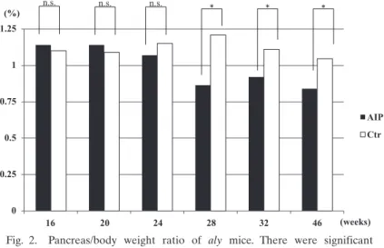
There were significant differences in the body weight of aly/aly (AIP) compared with aly/+ (Ctr) mice at 28, 32, and 46 weeks of age (Fig. 1 ; P<0.05), most likely due to the effect of pancreatitis. Similarly, the pancreas/body weight ratio differed significantly between AIP and Ctr mice at 28, 32, and 46 weeks of age (Fig. 2 ; P<0.05), again most likely due to pancreatitis.
Histological evaluation of pancreatic tissue Pancreatitis (HE staining)
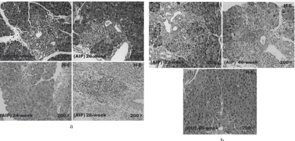
Histological examination of pancreatic sections from AIP mice revealed inflammatory cell infiltration and vacuolization in the circumference of small pancreatic ducts from 16 weeks of age, the absence of pancreatic acinar cells from 24 weeks of age, and, in addi- tion to inflammatory cell infiltration, fibrosis after 28 weeks of age. The inflammatory cell infiltration increased with age, with extensive infiltration and fibrosis observed in mice at 45 weeks of age. In contrast, histological examination of pancreatic sections from 45-week-old Ctr mice revealed normal tissue (Fig. 3a, b). The number of inflammatory cells that had
Fig. 1. Body weight of aly mice. There were significant differences between the aly/aly mice as a model of autoimmune pancreatitis (AIP) and aly/+ (control) mice at 28, 32, and 46 weeks of age. *P<0.05.
Fig. 2. Pancreas/body weight ratio of aly mice. There were significant differences between the aly/aly mice as a model of autoimmune pancreatitis (AIP) and aly/+ (control) mice at 28, 32, and 46 weeks of age. *P<0.05.
infiltrated the tissue and the degree of vacuolization were determined using ImageJ (NIH). Inflammatory cell infiltration and vacuolization both increased gradually in AIP mice after 16 weeks of age, with significant differences in both parameters observed between the AIP and Ctr groups (Fig. 4 ; P<0.05).
Insulitis (HE staining)
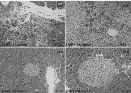
Insulitis is defined as inflammatory cell infiltration into the islets. In the present study, inflammatory cell infiltration into the islet was seen in the AIP group from 24 weeks of age.
In some tissues, it appeared that the entire islet had been replaced by inflammatory cells, which is equivalent to insulitis. The results of HE staining for islets were unclear (Fig. 5). In the AIP group, the number of recognizable islets decreased significantly compared with that in the Ctr group. Insulitis progressed with time, although it appeared to be slightly behind the development of pancreatitis.
Immunofluorescent staining
CD11c+ cells and CD4+ T cells predominantly infiltrated from the peripheral areas of islets to central areas. There were no CD11b+ or CD8+ cells detected in the pancreatic tissue (Fig. 6).
Immunohistochemical staining
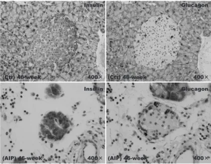
Immunohistochemical staining for insulin revealed the absence of islet β-cells in AIP mice from 24 weeks of age. Furthermore, morphological changes were observed, such as a reduc-
Fig. 3. Histological changes in the pancreatic tissue of aly/aly mice as a model of autoimmune pancreatitis
(AIP) and aly/+ (control) mice at (a) 16 and (b) 45 weeks of age. Pancreatitis (hematoxylin-eosin
[HE] staining). (a) Pancreatitis was observed in AIP mice from 16 weeks of age and progressed with time. (b) At 45 weeks of age, extensive inflammatory cell infiltration and fibrosis were observed in AIP mice. In contrast, in the control group, normal pancreatic tissue was evident at 45 weeks of age.
a
b
tion in the size of the islets and a greater irregularity in form, in addition to β-cell injury;
both these observations reflect the development of insulitis. In contrast, islets in the Ctr group were round and relatively large (Figs. 7, 9). ImageJ analysis of the number of β-cells
Fig. 4. Numbers of infiltrating inflammatory cells and vacuolization in the pancreatic tissue of aly/aly mice as a model of autoimmune pancreatitis (AIP) and aly/+ (control) mice. Infiltration of inflammatory cells, predominantly lymphocytes, was evident in the AIP and increased, along with vacuolization, in mice after 16 weeks of age. There were significant differences in the number of infiltrating cells and vacuolization counts between the AIP and control groups at 28, 32, and 46 weeks of age. Data are the mean
SEM. *P<0.05.
Fig. 5. Histological changes in the pancreatic tissue of aly/aly mice as a model of autoimmune pancreatitis (AIP) and aly/+ (control) mice
(hematoxylin-eosin staining). Insulitis (arrow) was evident in the AIP group at 24 weeks of age.
Fig. 6. Immunofluorescent staining with anti-CD4, anti-CD8, anti-CD11b, and anti-CD11c antibodies. Infiltration of CD11c+ cells and CD4+ cells was seen predominantly from peripheral areas of the islets to central areas.
Fig. 7. Immunohistochemical staining for insulin revealed the absence of β-cells in islets from aly/aly mice, as a model of autoimmune pancreatitis (AIP), at 24 weeks of age. Furthermore, morphological changes were seen in the islets, such as a reduction in size and irregular shape, reflecting the development of insulitis.
Fig. 8. Immunohistochemical staining for glucagon revealed no significant differences in α-cells between the aly/aly mice as a model of autoimmune pancreatitis (AIP) and aly/+ (control) mice, with little evidence of α-cell damage.
Fig. 9. Islets in aly/aly mice as a model of autoimmune pancreatitis (AIP)
and aly/+ (control) mice. In the control group, the islets appeared large and round ; in contrast, in the AIP group the islets were small and irregular in shape.
and islets confirmed a significant decrease in both in the AIP group compared with Ctr group. The decrease in the number of β-cells and islets in the AIP group increased with time (Fig. 10).
Immunohistochemical staining for glucagon revealed no significant differences in the number of α-cells, which exist around the edges of islets, between the AIP and Ctr groups, although there was evidence of some α-cells damage (Fig. 8). When the ratio of α-cells and β-cells within islets was evaluated between the AIP group and a control group, there was a significant difference in β-cells, but no difference in α-cells. Regarding for insulitis in AIP group, the β-cell was mainly received the obstacle.
Pancreatic content and serum levels of IFN-γ and IL-4
Pancreatic IFN-γ content peaked in the AIP group between 8 and 16 weeks of age, and rapidly declined after 24 weeks of age. This was similar to the results obtained in the Ctr group (Fig. 11). In contrast, there were two peaks in pancreatic IL-4 content in the AIP group, one in mice at 16 weeks of age and the second at 32 weeks of age (Fig. 12). Discussion
The term “insulitis” was first proposed by von Meyenburg in 1940. He reported on the postmortem findings in a woman who had died of diabetic ketoacidosis : specifically, there was a decrease in the number of islets in addition to morphological changes, such as acinar cell degradation and inflammatory cell infiltration into the islets. von Meyenburg termed inflammatory cell infiltration into the islets as insulitis23). In 1965, Gepts reported that
Fig. 10. The number of β-cells in the islets of aly/aly mice as a model of autoimmune pancreatitis (AIP) and aly/+
(control) mice. There were significantly fewer β-cells in islets from the AIP group, and their numbers decreased further with time. Data are the mean SEM. *P<0.05.
islets of many patients with T1DM exhibited signs of insulitis24), giving rise to the autoim- munity theory of T1DM. With the discovery of self-antibodies, such as islet antibodies, the coexistence of other autoimmune diseases, and a relationship with human leukocyte antigen
(HLA) antigens, it seemed clear that T1DM was an autoimmune disease. The existence of insulitis, which results from inflammatory cell (primarily lymphocyte) infiltration into the islets, became the basis of the autoimmunity theory. Foulis recognized insulitis in 78% of
Fig. 11. Interferon (IFN)-γ levels in pancreatic homogenates of aly/aly mice as a model of autoimmune pancreatitis
(AIP) and aly/+ (control) mice. Pancreatic levels of IFN-γ peaked in the AIP group at 16 weeks of age and declined rapidly after 20 weeks of age.
Fig. 12. Interleukin (IL)-4 levels in pancreatic homogenates of aly/aly mice as a model of autoimmune pancreatitis
(AIP) and aly/+ (control) mice. Pancreatic levels of IL-4 peaked twice in AIP mice : once at 16 weeks of age and then again at 32 weeks of age.
pancreatic tissues from patients with T1DM who died young, and interpreted these findings to mean that β-cell destruction was caused by an immunological mechanism25). Even today, T1DM is thought of as an autoimmune disease, and insulitis may be one of the autoim- munological mechanisms involved.
In AIP, disorders of pancreatic exocrine and endocrine function are often present, and DM and IGT are observed in many cases5-8); this is believed to be due to the effect of both blood flow obstruction of the endocrine gland accompanying formation of exocrine gland fibrosis and insulitis18). We have used aly mice as a mouse model of AIP in previous studies19,20). In the present study, we elucidated the morbidity and pathogenic mechanism of insulitis in AIP using aly mice.
We found that insulitis appeared after pancreatitis, increasing in severity with age, and there was evidence of inflammatory cell infiltration into the islets. On the basis of these observations, we assume that the inflammatory cell infiltration and fibrosis associated with pancreatitis causes islet ischemia, giving rise to insulitis secondary to pancreatitis. One would expect that the effects of inflammation associated with pancreatitis would affect all cells in the islets uniformly. However, when we analyzed the endocrine cells in the islets, we found minimal effects on α-cells. In contrast, the β-cells were markedly involved, as evidenced by reductions in β-cell numbers, modifications, and their eventual absence from the islets altogether. These findings are consistent with those of Tanaka, who found a decreased number of β-cells in islets from patients with AIP5,8). Furthermore, immunofluo- rescent staining of the pancreas revealed infiltration of CD11c+ cells and CD4+ T cells into the islets, implying the involvement of an autoimmune mechanism in the development of insulitis (see below). Therefore, although insulitis is considered to be an event secondary to advancing pancreatitis, the contribution of β-cells damage to this process appears consider- able.
Although pancreatic DM occurs as a result of decreased endocrine function of the pancreas with the progression of pancreatic disease, the obstacle not only of insulin but the secretion of glucagon is carried out in response to the influence of a primary disease.
Therefore, pancreatic DM presents with different characteristics and clinical findings com- pared with the usual DM, including impaired insulin secretion and glycemic control. So- called “pancreatic DM” is caused by chronic pancreatitis, pancreatic tumors, and acute pancreatitis, and it may develop after resection of the pancreas. The DM in AIP is also pancreatic DM, because glucose tolerance worsens in connection with pancreatitis. However, because insulitis is involved in the etiology of DM in AIP, insulitis is considered a different disease from the usual pancreatic DM in that it primarily involves damage to β-cells. In addition, in the present study, although there was a significant reduction in β-cells in AIP, the α-cell population was unaffected, so insulitis is considered to be inflammation specific to β-cells. Over the longer term, it is possible that AIP will progress to chronic pancreatitis, in which there are reductions in both β- and α-cells depending on the degree of fibrosis in
the pancreas ; this then mimics the pattern seen in pancreatic DM with increasing fibrosis of the pancreas. In the present study, the α-cell population was unaffected in the AIP group at 46 weeks of age. However, because the involvement of α-cells may appear in the mouse at much later stages (i.e. age), as in humans, we are currently undertaking experi- ments to evaluate pancreatic tissue in mice at an advanced age.
Immunofluorescent staining in the present study revealed infiltration of CD11c+ cells and CD4+ T cells in AIP. CD11c is a membrane antigen characteristic of leukocytes that is observed mainly in macrophages, monocytes, and dendritic cells as antigen-presenting cells
(APC). It participates in cell adhesion and phagocytosis. In terms of the development of insulitis, we assume that the mechanism involves antigen presentation by CD11c+ cells, followed by the migration of CD4+ T cells, which results in inflammation. CD4 is a typical membrane antigen of Th cells, which are classified into Th1 and Th2 cells on the basis of the types of cytokines they produce : Th1 cells produce IFN-γ and have cytotoxic activities, whereas Th2 cells produce IL-4 and are involved in antibody production25). Imbalances in the Th1/ Th2 ratio are indicative of autoimmune disease, with a shift to predominantly Th1 responses related to organ-specific autoimmune disease, and a shift to predominantly Th2 responses related to systemic autoimmune disease26). For example, T1DM, a disease that similarly causes insulitis, is an organ-specific autoimmune disease of pancreatic β-cells in which the DM arises because of the destruction of β-cells, which produce insulin. The mechanism involved is believed to be CD4+ T cell activation of cytotoxic (CD8+) T cells, which then damage the pancreatic β-cells. Using NOD mice, it has been shown that Th1 activation is involved in the activation of CD4+ T cells27-31). Evaluation of serum IFN-γ and IL-4 levels in the AIP group revealed an increase in IFN-γ concentrations in mice at the earlier ages, suggesting that Th1 responses were contributing to the development of pancreatitis. However, IFN-γ levels declined rapidly in AIP mice after 24 weeks of age to leve ls seen in the control group. Conversely, a double peak in pancreatic IL-4 levels was seen in the AIP group, at 16 and 32 weeks of age. We assume that this indicates the activation of Th2 responses after 32 weeks of age that contributed to the progression of insulitis. Staining for CD8+ cells as cytotoxic T cells was negative in the AIP group. On the basis of these findings, we believe that there are differences between insulitis of AIP and that of T1DM in terms of the underlying immunological mechanisms, although both diseases exhibit similar β-cell injury.
In conclusion, we believe the insulitis in AIP is secondary to advancing pancreatitis, but that endocrine disorders, such as DM, arise as a result of a decrease in β-cells. The mecha- nism may involve CD11c+ cells, which are AIP, triggering the migration of CD4+ T cells to induce an inflammatory response. Moreover, we also assume that a shift in the Th1/Th2 balance towards Th2 activation contributes considerably to the development of insulitis in AIP. Th2 responses are related to systemic autoimmune disease and we consider that this is the main cause of insulitis in AIP. In recent years, it has been proposed that AIP is an
IgG4-related systemic disease (an IgG4-related disease5)). This contention is supported by the results of the present study showing activation of Th2 responses in AIP. Although the DM that arises as a result of insulitis in AIP is similar to T1DM in terms of β-cell destruc- tion, it differs from so-called “pancreatic DM”, in which both α- and β-cells are involved.
Acknowledgements
The authors thank Drs Susumu Ueda, Natsumi Takeyama and Koji Uetsuka for their assistance.
References
1) Sarles H, Sarles JC, Muratore R and Guien C : Chronic inflammatory sclerosis of the pancreas-an autonomous pancreatic disease? Am J Dig Dis 6:688-698 (1961)
2) Yoshida K, Toki F, Takeuchi T, Watanabe S, Shiratori K and Hayashi N : Chronic pancreatitis caused by an autoimmune abnormality. Proposal of the concept of autoimmune pancreatitis. Dig Dis Sci 40:1561-1568 (1995)
3) Tanaka S and Yoshida H : Autoimmune pancreatitis. J Tokyo Ariake Univ Med Health Sci 1:63-81 (2010)(in Japanese)
4) Okazaki K, Kawa S, Kamisawa T, Ito T, Inui K, Irie H, Irisawa A, Kubo K, Notohara K, Hasebe O, Fujinaga Y, Ohara H, Tanaka S, Nishino T, Nishimori I, Nishiyama T, Suda K, Shiratori K, Shimosegawa T and Tanaka M : Japanese clinical guidelines for autoimmune pancreatitis. Pancreas 38:849-866 (2009)
5) Umehara H, Okazaki K, Masaki Y, Kawano Y, Yamamoto M, Saeki T, Matsui S, Yoshino T, Nakamura S, Kawa S, Hamano H, Kamisawa T, Shimosegawa T, Shimatsu A, Nakamura S, Ito T, Notohara K, Sumida T, Tanaka Y, Mimori T, Chiba T, Mishima M, Hibi T, Tsubouchi H, Inui K and Ohara H : Comprehensive diag- nostic criteria for IgG4-related disease (IgG4-RD), 2011. Mod Rheumatol 22:21-30 (2012)
6) Tanaka S and Kobayashi T : Pancreatic endocrine and exocrine function in the patients with autoimmune pancreatitis. Kan・Tan・Sui 43:193-201 (2001)(in Japanese)
7) Okazaki K, Sumimoto K, Fukui Y and Uchida K : Pancreatic exocrine and endocrine function in patients with autoimmune pancreatitis. Tan to Sui 32:499-503 (2011)(in Japanese)
8) Ito T, Igarashi H, Niina Y and Takayanagi R : Pancreatic diabetes in autoimmune pancreatitis. Diabetes J 38:
9-13 (2010)(in Japanese)
9) Tanaka S and Kobayashi T : Diabetes mellitus associated with autoimmune pancreatitis. Nippon Rinsho 60(増刊 9):821-833 (2002)(in Japanese)
10) Ito T, Nishimori I, Inoue N, Kawabe K, Gibo J, Arita Y, Okazaki K, Takayanagi R and Otsuki M : Treatment for autoimmune pancreatitis : consensus on the treatment for patients with autoimmune pancreatitis in Japan. J Gastroenterol 42(Suppl 18):50-58 (2007)
11) Kamisawa T, Egawa N, Inokuma S, Tsuruta K, Okamoto A, Kamata N, Nakamura T and Matsukawa M : Pancreatic endocrine and exocrine function and salivary gland function in autoimmune pancreatitis before and after steroid therapy. Pancreas 27:235-238 (2003)
12) Nishino T, Toki F, Oyama H, Shimizu K and Shiratori K : Long-term outcome of autoimmune pancreatitis after oral prednisolone therapy. Intern Med 45:497-501 (2006)
13) Nishimori I, Suda K, Ooi I and Ogawa M : Research on the clinical state of autoimmune pancreatitis. J Jpn Pancreas Soc 17:619-627 (2002)
14) Ito T, Nakano I, Koyanagi S, Miyahara T, Migita Y, Ogoshi K, Sakai H, Matsunaga S, Yasuda O, Sumii T and Nawata H : Autoimmune pancreatitis as a new clinical entity. Three cases of autoimmune pancreatitis with effective steroid therapy. Dig Dis Sci 42:1458-1468 (1997)
15) Kamisawa T, Shimosegawa T, Okazaki K, Nishino T, Watanabe H, Kanno A, Okumura F, Nishikawa T, Kobayashi K, Ichiya T, Takatori H, Yamakita K, Kubota K, Hamano H, Okamura K, Hirano K, Ito T, Ko SB and Omata M : Standard steroid treatment for autoimmune pancreatitis. Gut 58:1504-1507 (2009)
16) Tanaka S, Kobayashi T, Nakanishi K, Okubo M, Murase T, Hashimoto M and Takeuchi K : Corticosteroid-
responsive diabetes mellitus associated with autoimmune pancreatitis. Lancet 356:910-911 (2000)
17) Kamisawa T, Okazaki K, Kawa S, Shimosegawa T, Tanaka M, Research Committee for Intractable Pancreatic Disease and Japan Pancreas Society : Japanese consensus guidelines for management of autoimmune pancreati- tis : III, Treatment and prognosis of AIP. J Gastroenterol 45:471-477 (2010)
18) Tanaka S, Kobayashi T, Nakanishi K, Okubo M, Odawara M, Murase T, Hashimoto M, Watanabe G, Mat- sushita H, Inoko H and Takeuchi K : Corticosteroid-responsive diabetes mellitus associated with autoimmune pancreatitis: pathological examinations of the endocrine and exocrine pancreas. Ann N Y Acad Sci 958:152- 159 (2002)
19) Ito T, Kawabe K, Arita Y, Hisano T, Igarashi H, Funakoshi A, Sumii T, Yamanaka T and Takayanagi R: Evalu- ation of pancreatic endocrine and exocrine function in patients with autoimmune pancreatitis. Pancreas 34:254- 259 (2007)
20) Yoshida H, Tanaka S and Mitamura K : Animal models for autoimmune pancreatitis. Kan・Tan・Sui 43:179- 187 (2001)(in Japanese)
21) Yoshida H, Tanaka S, Yamazaki T, Yukawa A, Honma T, Kitamura K, Awai T, Hanawa K, Imamura T, Ikegami A and Imawari M : An animal model and immunopathy for autoimmune pancreatitis : elucidation of patho- physiology of the disease by utilizing aly/aly male mice. Med Frontline 62:1925-1934 (2007)(in Japanese)
22) Yoshida H, Nozu F, Lankisch TO, Mitamura K, Owyang C and Tsunoda Y : A possible role for Ca(2+)/ calmodulin-dependent protein kinase Ⅳ during pancreatic acinar stimulus-secretion coupling. Biochim Biopsys Acta 1497:155-167 (2000)
23) von Meyenburg H : Ueber “insulitis” bei diabetes. Schweiz Med Wochenschr 24:554-557 (1940)
24) Gepts W : Pathologic anatomy of the pancreas in juvenile diabetes mellitus. Diabetes 14:619-633 (1965)
25) Foulis AK, Liddle CN, Farquharson JA, Richmond JA and Weir RS : The histopathology of the pancreas in type 1 (insulin-dependent) diabetes mellitus : a 25-year review of deaths in patients under 20 years of age in the United Kingdom. Diabetologia 29:267-274 (1986)
26) Mosmann TR, Cherwinski H, Bond MW, Giedlin MA and Coffman RL : Two types of murine helper T-cell clone. I. Definition according to profiles of lymphokine activities and secreted protein. J Immunol 136:2348- 2357 (1986)
27) Sakaguchi S and Toda M : Autoimmune diseases and Th1 cells, Th2 cells. Inflamm Immun 7:231-236 (1999)
(in Japanese)
28) Hanafusa T and Imagawa A : Classification and pathogenesis of type 1 diabetes mellitus. Nippon Rinsho 66(増 刊3): 339-342 (2008)(in Japanese)
29) Isotani H and Hanafusa T : Molecular mechanisms of pancreatic beta-cell destruction in type 1 diabetes. Nip- pon Rinsho 60(増刊7):383-387 (2002)(in Japanese)
30) Morinani M and Itakura M : Th1/Th2 balance in diabetes mellitus. Surg Front 12:373-377 (2005)(in Japanese)
31) Satoh J and Toyoda T : Roles of cytokines in the pathogenesis of type 1 diabetes. J Clin Exp Med 188:385- 389 (1999)(in Japanese)
32) Rabinovitch A : An update on cytokines in the pathogenesis of insulin-dependent diabetes mellitus. Diabetes Metab Rev 14:129-151 (1998)
[Received February 14, 2012 : Accepted February 17, 2012]