Lysophosphatidic acid in medicinal herbs enhances prostaglandin E
2 1and protects against indomethacin-induced gastric cell damage in
2vivo and in vitro
34
Sheuli Afroza, Ayano Yagia, Kouki Fujikawaa, M Motiur Rahmana, Katsuya Moritoa,
5
Tatsuya Fukutaa, Shiro Watanabeb, Emi Kiyokagec, d, Kazunori Toidac, e, Taro
6
Shimizua, Tatsuhiro Ishidaa, Kentaro Kogurea, Akira Tokumuraf and Tamotsu
7
Tanakaa*
8 9
aInstitute of Biomedical Sciences, Tokushima University Graduate School, Tokushima 770-8505, 10
Japan
11
bInstitute of Natural Medicine, University of Toyama, 2 Supplementary Fig. 1630 Sugitani, 12
Toyama 930-0194, Japan
13
cDepartment of Anatomy, Kawasaki Medical School, 577 Matsushima, Kurashiki, Okayama 14
701-0192, Japan
15
dDepartment of Medical Technology, Faculty of Health Science and Technology, Kawasaki 16
University of Medical Welfare, 288 Matsushima, Kurashiki, Okayama 701-0193, Japan
17
eUltra-high Voltage Electron Microscopy Research Center, Osaka University, Ibaraki, Osaka 18
567-0047, Japan
19
fDepartment of Pharmaceutical Life Sciences, Faculty of Pharmacy, Yasuda Women’s 20
University, Hiroshima 731-0351, Japan
21 22
Corresponding author: *Tamotsu Tanaka, Institute of Biomedical Sciences, Tokushima
23
University Graduate School, Tokushima 770-8505, Japan, Tel: 633-7249; Fax:
+81-88-24
633-9572; E-mail: [email protected]
25
© 2018. This manuscript version is made available under the CC-BY-NC-ND 4.0 license http://creativecommons.org/licenses/by-nc-nd/4.0/ The published version is available via https://doi.org/10.1016/j.prostaglandins.2018.01.003
26
Abstract
27
Lysophosphatidic acid (LPA) is a bioactive phospholipid that induces diverse biological
28
responses. Recently, we found that LPA ameliorates NSAIDs-induced gastric ulcer in mice. Here,
29
we quantified LPA in 21 medicinal herbs used for treatment of gastrointestinal (GI) disorders.
30
We found that half of them contained LPA at relatively high levels (40–240 μg/g) compared to
31
soybean seed powder (4.6 μg/g), which we previously identified as an LPA-rich food. The LPA
32
in peony (Paeonia lactiflora) root powder is highly concentrated in the lipid fraction that
33
ameliorates indomethacin-induced gastric ulcer in mice. Synthetic 18:1 LPA, peony root LPA
34
and peony root lipid enhanced prostaglandin E2 production in a gastric cancer cell line, MKN74 35
cells that express LPA2 abundantly. These materials also prevented indomethacin-induced cell 36
death and stimulated the proliferation of MKN74 cells. We found that LPA was present in
37
stomach fluids at 2.4 µM, which is an effective LPA concentration for inducing a cellular
38
response in vitro. These results indicated that LPA is one of the active components of medicinal
39
herbs for the treatment of GI disorder and that orally administered LPA-rich herbs may augment
40
the protective actions of endogenous LPA on gastric mucosa.
41
Keywords: Lysophosphatidic acid; Medicinal herbs; Indomethacin; Prostaglandin E2; Cell 42
death; Cell proliferation
43
Abbreviations: GI, gastrointestinal; LPA, lysophosphatidic acid; PA, phosphatidic acid; TLC,
44
thin-layer chromatography; PL, phospholipid; MALDI-TOF MS, matrix-assisted laser desorption
45
ionization time-of-flight mass spectrometry; LC/MS/MS, liquid chromatography-tandem mass
spectrometry; CMC, carboxymethylcellulose; PLA2, phospholipase A2; PGE2, prostaglandin E2; 47
NSAIDs, non-steroidal anti-inflammatory drugs; COX, cyclooxygenase.
48
1. Introduction
49
Lysophosphatidic acid (LPA) is a bioactive phospholipid that induces diverse cellular
50
responses including proliferation, protection of cells from apoptosis, and migration of cells [1].
51
These cellular responses are mediated through six LPA-specific G-protein coupled receptors,
52
LPA1–6 [2]. Recent studies have revealed important actions of LPA in the mammalian 53
gastrointestinal (GI) tract [3, 4]. These include inhibition of diarrhea, regulation of intestinal
54
electrolyte transport, protection of intestinal cells from apoptosis, and wound healing [5-9].
55
Peptic ulcer is a major GI disorder that occurs due to an imbalance in mucosal offensive
56
(gastric acid secretion) and defensive (gastric mucosal integrity) factors [10, 11]. Infection with
57
Helicobacter pylori, smoking, drinking alcohol, and chronic ingestion of drugs are major causes
58
of peptic ulcer. Recently, the number of patients with non-steroidal anti-inflammatory drug
59
(NSAID)-induced gastric ulcer is increasing due to increased use of NSAIDs for pain treatment
60
and prevention of thrombosis [12]. NSAIDs cause gastric ulcer by inhibition of cyclooxygenase
61
(COX)-1 and COX-2, which produce a predominant mucosal defense factor, prostaglandin E2 62
(PGE2) [13]. Our recent study showed that orally administered synthetic LPA ameliorates 63
aspirin- and indomethacin-induced gastric ulcer in mice [14, 15]. We also showed that LPA
up-64
regulates COX-2 and enhances production of PGE2 via activation of LPA2 receptors, which are 65
located on the apical side of gastric mucosal cells [16]. However, further uncharacterized
66
mechanisms other than COX-2 induction are considered to function in the protective action of
67
LPA, because LPA protects the gastric mucosa from the acute toxicity of NSAIDs.
Medicinal herbs have been traditionally used for the treatment of many diseases, including
69
gastric ulcer [17]. Considering that some medicinal herbs and vegetables contain LPA
70
abundantly [18, 19], it is rational to postulate the existence of anti-ulcer medicinal herbs that
71
contains LPA as an active component. To examine this possibility, we determined the LPA
72
content of 21 herbs that are traditionally used for the treatment of GI disorders. We also aimed to
73
examine the effects of LPA and herbal lipids on NSAID-induced gastric ulcer. The results
74
showed that peony root lipid, which contain highly concentrated LPA, had an ameliorative effect
75
on NSAID-induced gastric ulcer and enhanced PGE2 production in gastric cells. We also showed 76
evidence that LPA/LPA2 signaling protects against acute cytotoxicity of NSAIDs in cultured 77
gastric cells.
78
2. Materials and Methods
79
2.1 Materials
80
Herbs used for the treatment of various digestive disorders were selected based on the
81
descriptions in the oldest Chinese traditional herbal medicine book, the Shennong Ben Cao Jing.
82
Coptis rhizome (Coptis japonica), moutan cortex (Paeonia suffruticosa), atractylodes rhizome
83
(Atractylodes japonica), atractylodes lancea rhizome (Atractylodes lancea), amomum seed
84
(Amomum xanthioides), peony root (Paeonia lactiflora), poria sclerotium (Poria cocos), and
85
phellodendron bark (Phellodendron amurense) were obtained from Yoshimi Seiyaku Co. Ltd.
86
(Osaka, Japan). Licorice root (Glycyrrhiza glabra), platycodon root (Platycodon grandiflorum),
87
bupleurum root (Bupleurum falcatum), zedoary rhizome (Curcuma zedoaria), fennel fruit
88
(Foeniculum vulgare), dried ginger rhizome (Zingiber officinale), and stripped, steamed, and
89
dried ginger rhizome (Zingiber officinale) were purchased from Nakaya Hikojuro Co. Ltd.
(Ishikawa, Japan). Sophora root (Sophora flavescens) and schisandra fruit (Schisandra chinensis)
91
were obtained from Kojima Kampo Co. Ltd. (Osaka, Japan). Pinellia tuber (Pinellia ternata),
92
cimicifuga rhizome (Cimicifuga simplex), panax rhizome (Panax japonicus), and corydalis tuber
93
(Corydalis turtschaninovii) were purchased from a local drug store.
94
2.2 Reagents
95
One-oleoyl-2-hydroxy-sn-glycero-3-phosphate (18:1 LPA),
1-heptadecanoyl-2-hydroxy-sn-96
glycero-3-phosphate (17:0 LPA), 1-palmitoyl-2-hydroxy-sn-glycero-3-phosphocholine (16:0
97
LPC), and arachidonic acid were purchased from Avanti Polar Lipids (Alabaster, AL, USA).
98
Peony root LPA was freshly prepared from peony root powder as described below. Pertussis
99
toxin (PTX) and calcium ionophore A23187 were purchased from Sigma-Aldrich (St. Louis, MO,
100
USA). Phos-tag was obtained from Wako Pure Chemical Industries (Osaka, Japan).
101
Carboxymethylcellulose (CMC), aspirin, and indomethacin were obtained from Kanto Chemical
102
Co. (Tokyo, Japan), Wako Pure Chemical Industries (Osaka, Japan), and Nacalai Tesque Inc.
103
(Kyoto, Japan), respectively. A prostaglandin E2 EIA kit was obtained from Cayman Chemical 104
Co. (Ann Arbor, MI). A lactate dehydrogenase (LDH) assay kit was obtained from Dojindo
105
Laboratories Co. Ltd. (Kumamoto, Japan). A bromo-2-deoxy-uridine (BrdU) cell proliferation
106
ELISA kit was purchased from Roche (Mannheim, Germany).
107
2.3 Animals
108
Five-week-old male ICR mice (35g body weight) were obtained from Charles River
109
Laboratories Japan, Inc. (Kanagawa, Japan). The animals were adapted to an animal room
110
maintained at 24 ± 2 °C and housed in a 12 h light/dark cycle. The care and handling of mice
were in accordance with the National Institute of Health guidelines. All experimental procedures
112
were approved by the Tokushima University Animal Care and Use Committee.
113
2.4 Extraction of lipid and isolation of LPA from herbs
114
Lipids were extracted from the medicinal herbs by the Bligh and Dyer method [20] with
115
acidification of the water/methanol phase, as described previously [19]. In brief, 1 g of herb
116
powder was mixed with 15.2 ml of a solvent consisting of chloroform/methanol/water in the
117
ratio of 1:2:0.8 (v/v/v) and centrifuged to collect the supernatant. The pellet was added to the
118
same amount of the mixed solvent consisting of chloroform/methanol/water and centrifuged. The
119
combined supernatant fraction was mixed with an appropriate volume of chloroform and water
120
to make solvent system consisting of chloroform/methanol/water in the ratio of 1:1:0.9 (v/v/v).
121
The resulting two-layer solution was mixed with 0.15 ml of 5 N HCl and centrifuged. Lipids
122
were obtained from the lower phase (chloroform phase). The LPA in the lipid extract was
123
isolated by TLC. The solvent system of the chromatography was chloroform/methanol/28%
124
aqueous ammonia (60:35:8, v/v/v). After development, the plate was dried for a few minutes
125
with blowing air and sprayed with primulin for visualization under UV light. LPA was identified,
126
extracted from the silica gel by the Bligh and Dyer method [20], and quantified by the
127
colorimetric method based on phospho-molybdenum-malachite green formation [21]. The weight
128
of phospholipids (μg/g) was determined from a weight of lipid phosphorus (µg inorganic
129
phosphorus/g) in a way recommended by American Oil Chemists’ Society [22].
130
2.5 Matrix-assisted laser desorption ionization time-of-flight mass spectrometry
(MALDI-131
TOF MS)
Molecular species of LPA in herbs were determined by MALDI-TOF MS as described
133
previously [15]. An aliquot of LPA was dissolved in 100 µl of methanol containing 0.1%
134
aqueous ammonia. This solution (10 µl) was mixed with 5 µl 0.1 mM 68Zn Phos-tag solution. A
135
small portion (0.5 µl) of this mixture was spotted on a sample plate. Immediately, 0.5 µl of 2, 4,
136
6-trihydroxyacetophenone (THAP) solution (10 mg/ml in acetonitrile) was layered onto the
137
mixture as a matrix solution. The sample plate was dried for a few minutes, and the
138
matrix/analyte co-crystal that formed was subjected to MALDI-TOF MS. MALDI-TOF mass
139
spectra were acquired using a Bruker Microflex mass spectrometer (Bruker Daltonics, Bremen,
140
Germany) in positive ion detection mode. The wavelength of the nitrogen-emitting laser and the
141
accelerating voltage were 337 nm and 20 kV, respectively. To enhance the reproducibility, 300
142
laser shots were averaged for each mass spectrum.
143
2.6 NSAIDs-induced gastric ulcer
144
Mouse models of aspirin- and indomethacin-induced gastric ulcer were developed as described
145
previously [14, 15]. In brief, aspirin (300 mg/kg body weight), indomethacin (22.9 mg/kg body
146
weight), or peony root lipid was suspended in 3% (w/v) CMC and sonicated for 1 min. Peony
147
root powder was suspended in water. Fasted mice were intragastrically administered peony root
148
lipid or powder suspension in a volume of 0.2 ml. After 0.5 h, 0.2 ml of the aspirin or
149
indomethacin suspension was administered intragastrically. The mice were anesthetized with
150
diethyl ether and sacrificed 3 or 5 h after the aspirin or indomethacin administration, respectively.
151
The isolated stomach was ligated at both ends, filled with 1.5 ml of 2% formalin, and immersed
152
in 2% formalin for 15 min. Then, the stomach was cut along the greater curvature, and the
153
lengths of lesions on the stomach wall were measured using a millimeter scale with a magnifying
154
glass. The total length of lesions was used as a lesion index.
2.7 Cell culture and reverse transcription-PCR (RT-PCR)
156
MKN74 cells, a human gastric cancer cell line, were obtained from the RIKEN Cell Bank
157
(Tsukuba, Japan). The MKN74 cells were grown in RPMI-1640 medium containing 10% fetal
158
bovine serum (FBS), 100 U/ml penicillin, and 100 μg/ml streptomycin in a humidified
159
atmosphere of 5% CO2 and 95% air at 37 ºC. Total RNA from MKN74 cells was prepared by 160
using an RNeasy Mini Kit (Qiagen, Hilden, Germany) according to the manufacturer's
161
instructions. The first-strand complementary DNA (cDNA) was synthesized from the purified
162
total cellular RNA with random hexamer primers using the SuperScript™ III synthesis system
163
for RT-PCR kit (Invitrogen, Carlsbad, CA). The cDNA was then subjected to PCR amplification
164
with primer sets and condition as described in supplementary Table 1. The PCR products were
165
examined by electrophoresis on 2% agarose gel, stained with Gelred (Biotium, Hayward, CA)
166
and visualized with UV light. Images of the fluorescent band on the gel were captured by a Fuji
167
LAS-4000 imaging system (FujiFilm, Tokyo, Japan), and the digitized image data were analyzed
168
by NIH image.
169
2.8 PGE2 production assay
170
PGE2 production in MKN74 cells was performed as described previously [16]. In brief, 171
MKN74 cells were seeded in 35-mm dishes and added 10 µM arachidonic acid (AA) for
AA-172
enrichment in the cells. After 24 h, the medium was changed to serum-free medium, and further
173
incubated for 24 h. Then, the cells were treated with 10 µM acetylsalicylic acid for 30 min to
174
minimize the effect of the preexisting COX activity. After replacement of the medium, cells were
175
incubated with 18:1 LPA, peony root lipid extract or purified peony root LPA in the presence or
176
absence of pertussis toxin (PTX). After 5 h, the cells were stimulated with 1 µM A23187 for 10
min. The supernatant were collected and PGE2 was evaluated by EIA kit according to the 178
manufacturer’s instructions.
179
2.9 Indomethacin-induced cell damage of MKN74 cells
180
MKN74 cells were seeded at a density of 1x10⁵ in 35-mm polystyrene dishes and serum starved
181
for 24 h. LPA dissolved in 0.3% BSA in PBS were added 2 h before addition of indomethacin
182
solution. The final concentration of indomethacin was fixed at 0.8 mM. After 30 h, the cells were
183
gently washed with PBS and stained with propidium iodide (PI) for 30 min. The extent of cell
184
death was determined by observation with a fluorescent microscope Axiovert 200 M (Zeiss,
185
Oberkochen, Germany). For the LDH assay, MKN74 cells were treated in the same manner as
186
described above. At the end of incubation, 100 μl of the culture medium supernatant was added
187
per well of a 96-well microplate. The LDH activity was measured according to the instructions
188
of the LDH assay kit. The activity was expressed as % of maximum release of LDH that can be
189
obtained in a sample of the lysis buffer-treated cells. The extent of cell death was also
190
determined by flow cytometric analysis. The PI-stained cells were subjected to a flow cytometer
191
(Becton Dickinson) that was operated using Cell Quest software, and at least 10,000 cells were
192
analyzed for each sample.
193
2.10 Cell proliferation assay
194
MKN74 cells seeded in 35-mm polystyrene dishes were serum-starved for 24 h. Then they
195
were cultured with or without synthetic or peony root-derived LPA in the presence or absence of
196
PTX. After 24 h, cells were harvested by trypsinization, mixed with trypan blue dye, and the
197
number of living cells was counted using a hemocytometer. The proliferation of MKN74 cells
198
was also confirmed by a BrdU cell proliferation assay. In brief, 2x10³ cells were seeded in
well microplates in 100 μl/well culture medium and kept in serum-starved condition for 48 h.
200
The synthetic or peony root-derived LPA was added to the cells and further incubated for 24 h.
201
BrdU was added to the cell culture 4 h before termination of incubation. The incorporated BrdU
202
was determined as described by the manufacturer’s protocol.
203
2.11 Determination of LPA in a mouse stomach fluid
204
Stomachs of overnight-fasted mice were isolated and gently washed with PBS. The stomach
205
was cut along the greater curvature. The stomach inner surface was carefully washed with a
206
small amount of PBS. After addition of 17:0 LPA (0.5 nmol) as an internal standard, lipids were
207
extracted from the stomach washing solution by using an acidified Bligh and Dyer method as
208
described above. Extracted lipids were dissolved in 0.8 ml of methanol and filtered through 0.2
209
µm nylon filter. After filtration, methanol was evaporated and reconstituted in 0.1 ml of
210
methanol/water mixture (95:5, v/v) containing 5 mM ammonium formate for LC/MS/MS.
211
LC/MS/MS was performed as described previously [23] using a quadrupole-linear iontrap hybrid
212
mass spectrometry system, 4000 Q TRAP™ (Applied Biosystems/MDS Sciex, Concord, Ontario,
213
Canada) with an Agilent 1100 liquid chromatography system (Agilent Technologies,
214
Wilmington, DE, USA). In the negative ion mode of operation with multiple reactions
215
monitoring, Q1 was set to the deprotonated molecular ion of each class of LPA as the precursor
216
ion.The fragment ions, [deprotonated cyclic glycerophosphate]− at m/z 153 were selected for Q3.
217
The ratios of the negative ion peak areas of the endogenous LPA to that of the corresponding
218
internal standard were calculated.
219
2.12 Statistical analysis
220
Statistical analyses of the difference between two means were performed by Student’s t-test.
222
3. Results
223
3.1 Abundance of LPA in medicinal herbs
224
Our previous study [19] revealed that LPA is abundant in cruciferous plants, such as cabbage
225
leaves and radish roots (9.2 and 2.3 μg/g wet weight, respectively). Soybean seed powder was
226
also found to be rich in LPA (4.6 μg/g). In this study, we determined LPA content in 21 dried
227
medicinal herbs used for the treatment of GI disorders (Fig. 1A). Compared to LPA-rich foods,
228
about half of the medicinal herbs contained LPA at high levels (40–240 μg/g). Among them,
229
peony root powder (240 μg/g) contained the highest level of LPA. It was 52 times that of
230
soybean seed powder. We previously revealed that phosphatidic acid (PA), a diacyl derivative of
231
LPA, serves as a source of LPA in the digestive tract [14]. The amounts of PA in these medicinal
232
herbs were comparable (Fig. 1B) to those in cabbage and soybean, which were characterized as
233
PA-rich foods previously [24]. Surprisingly, the percentage of LPA in total phospholipids (PLs)
234
in peony root was 11%, which is 30 and 400 times of those in cabbage leaves and soybean seed
235
powder, respectively (Table 1). These results indicated that peony root powder contains abundant
236
LPA with a high concentration in its lipid fraction. This is evident from the relative intensity of
237
TLC bands of its lipid extract (Supplementary Fig. 1).
238
MALDI-TOF MS of PA and LPA in medicinal herbs showed that the predominant PA species
239
were 16:0/18:2 and 18:2/18:2 (or 18:1/18:3) PA (Supplementary Table 2) and the predominant
240
LPA species were 16:0, 18:2 LPA (Supplementary Table 3). This is also the case in peony root
241
as shown in Fig. 2A, B.
242
3.2 Anti-ulcer effect of peony root lipid and powder
As shown previously, orally administered aspirin (300 mg/kg body weight) produces 15–20
244
mucosal lesions in the gastric corpus of mice [14]. The lesions were linear and extended from the
245
fundic area to the pyloric area as erosion. Similar morphological lesions were observed in the
246
experiments with indomethacin (22.9 mg/kg body weight) (Fig. 3A). We used the total length of
247
lesions as the lesion index.
248
Orally administered peony root lipid reduced indomethacin-induced lesion formation in a
dose-249
dependent fashion (Fig. 3A, B). The maximum reduction was observed when mice were
250
administered 1 mM of peony root lipid (Fig. 3A, B), which corresponds 4.4 mg PLs/kg (animal
251
body weight). We confirmed that synthetic LPA at 1 mM has protective effect against
252
indomethacin-induced lesion formation (Fig. 3B). We also examined the gastro protective effect
253
of peony root powder on an aspirin-induced acute gastric ulcer mouse model. The peony root
254
powder at 2 g/kg body weight effectively reduced gastric mucosal lesion formation (Fig. 3C).
255
This dose of the powder corresponds to the administration of 1 mM peony root lipid.
256
3.3 Enhancement of PGE2 production by LPA-rich herbal lipids in gastric cells
257
According to the Human Protein Atlas database (http://www.proteinatlas.org/), LPA2, LPA5, 258
and LPA6 are expressed abundantly in human stomach epithelia. On the other hand, expression 259
levels of LPA1, LPA3,and LPA4 are very low. Firstly, we examined LPAR1–6 mRNA expression 260
in human gastric cancer cell line, MKN74 cell. Results showed that order of abundance of LPAR
261
mRNA was LPA2=LPA5>LPA6. Levels of mRNA of LPA1, LPA3, and LPA4 were under 262
detectable (Fig. 4A). The relative abundance of mRNA of LPARs in MKN74 cells is good
263
agreement with the expression profile of LPARs in human stomach tissue. We used this cell line
264
as a representative mucosal cell model of the human stomach. We confirmed that synthetic 18:1
LPA can enhance PGE2 production in MKN74 cells (Fig. 4B). This is consistent with our 266
previous observation that LPA enhances PGE2 production via up-regulation of COX-2 [16]. 267
This effect was also observed with LPA prepared from peony root. Peony root lipid, which has
268
concentrated LPA, enhanced PGE2 production at high efficacy. Enhancements of PGE2 269
production induced by LPA and peony root lipid were completely abolished by PTX (Fig. 4C),
270
suggesting the involvement of Gi-coupled receptor in their actions. It should be mentioned that
271
high concentration of the peony root lipid (10 µM) did not induce PGE2 production (Fig. 4B).
272
This is due to cytotoxicity of the lipid extract which contain various components other than LPA.
273
In fact, we observed that most of the cells were floating at the end of the incubation with peony
274
root lipid at 10 µM.
275
3.4 LPA protects indomethacin-induced cell injury of MKN74 cells
276
Effect of LPA on indomethacin-induced cell injury was examined in MKN74 cells. As shown
277
in Fig 5A, non-treated MKN74 cells were tightly attached each other. We found that treatment of
278
the cells with 0.8 mM indomethacin results in loss of attachment and rounding of their cell shape
279
(Fig. 5A), a typical morphological change in apoptotic cells [25, 26]. When the cells were treated
280
with 0.8 mM indomethacin plus 10 µM LPA, the indomethacin-induced morphological change
281
was not observed. In fact, LPA caused cell spreading with an extended edge, indicating the
282
disappearance of apoptotic symptoms (Fig. 5A). The protective action of LPA on
indomethacin-283
induced cellular damage was quantified by directly counting PI-positive cells (dead cells). We
284
found that LPA reduced the number of PI-positive cells in a dose-dependent fashion (Fig. 5B).
285
LPA from peony root also significantly reduced cell death at 10 µM (Fig. 5B). The protective
286
action of LPA against indomethacin-induced cellular damage was also confirmed by LDH
287
leakage (Fig. 5C). To determine the involvement of G-protein-coupled receptors in these
observations, the effect of pretreatment of PTX, a specific inhibitor of Gi-protein, was examined.
289
Flow cytometric analysis was applied for this experiment to quantify the population of dead cells
290
in the total cells. Results showed that the effect of LPA against indomethacin-induced cell death
291
was completely abolished by pre-treatment of PTX, indicating the involvement of Gi-coupled
292
receptors in the LPA action (Fig. 5D). This is also confirmed by the morphological change of the
293
cells (data not shown). We found that LPC did not show a protective effect on
indomethacin-294
induced cell death (Fig. 5D).
295
3.5 LPA stimulated proliferation of MKN74 cells
296
Treatment of MKN74 cells with LPA induced proliferation in a dose-dependent manner (Fig.
297
6A). LPA-induced cell proliferation was also confirmed by measuring incorporation of the
298
thymidine analog BrdU into the cells (Fig. 6B). Peony root LPA also stimulated the proliferation
299
of MKN74 cells (Fig. 6A, B). LPA-induced cell proliferation was partially and significantly
300
inhibited by PTX (Fig. 6C). Again, LPC had no proliferative effect.
301
3.6 LPA concentration in a mouse stomach fluid
302
In order to better understand the role of LPA in the physiology of the stomach, it is necessary
303
to know the concentration and molecular species of LPA in stomach fluid. The volume of
304
stomach fluid was assumed to be 0.08 ml. This is based on the fact that the area of the mucus
305
layer is 400 mm2 and the thickness of mucus gel layer is 0.2 mm [27]. We found that the total
306
LPA concentration in the stomach fluid was 2.4 μM (Fig. 7). The major species of LPA in the
307
stomach fluid were found to be 16:0, 18:0, 18:1, and 18:2 LPAs (Fig. 7). The concentration of
308
these LPA species was found to be 0.3–0.7 µM. Other LPA species, such as 16:1, 18:3, 18:4,
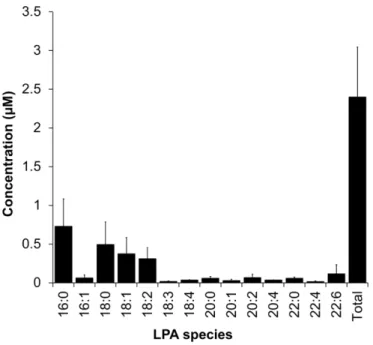
20:0, 20:1, 20:2, 20:4, 22:0, 22:4, and 22:6, were present in low (0.01–0.1 μM) level 310 (Supplementary Fig. 2). 311 4. Discussion 312
Research in medicinal herbs has identified many active components that exert anti-ulcer effects
313
with diverse mechanisms of action. These include compounds belonging to flavonoids, alkaloids,
314
tannins, and saponins [28, 29]. Dietary phospholipids, such as soy-derived PC, are also shown as
315
reducing agents for NSAIDs-induced gastric ulcer [30]. In this study, we found that LPA in
316
medicinal herbs is a potential component for prevention of gastric mucosal injury. This notion is
317
based on several observations. Firstly, synthetic LPA and herbal LPA showed ameliorative
318
activity against cytotoxic effect of indomethacin. Secondly, LPAs and LPA-rich lipid enhanced
319
PGE2 production, an important cytoprotective factor in GI mucosa. Thirdly, peony root lipid, an 320
LPA-rich herbal lipid identified here, significantly ameliorated indomethacin-induced gastric
321
lesions in mice. Lastly, there were many LPA-rich herbs in Chinese traditional medicines used
322
for the treatment of GI disorders. We discuss on mechanisms of these effects in detail.
323
A well-known mechanism of NSAID-induced gastric mucosal lesions is the inhibition of
324
COX-1 and COX-2 enzymes and a resulting decrease in gastroprotective PGE2. The importance 325
of PGE2 in the integrity of stomach mucosa is evident from the fact that common anti-ulcer drugs, 326
such as rebamipide and geranylgeranylacetone, up-regulate COX-2, leading to the enhancement
327
of PGE2 production [31, 32]. Consistent with our previous study [16], LPA from medicinal herb 328
was found to enhance PGE2 production in human gastric cancer cell line, MKN74 cells. We also 329
showed that peony root lipid, which has concentrated LPA, enhanced PGE2 production with Gi-330
mediated manner. Surprisingly, the efficacy of PGE2 production of peony root lipid is higher 331
than that expected from the LPA content in the lipid extract. At present, we do not know other
332
components in peony root lipid that enhance PGE2 production along with LPA. Considering that 333
PGE2-enhancement was completely abolished by PTX, there may be components that increase 334
LPA action in the lipid. Further study is needed for clarification of this point.
335
NSAIDs have been reported to induce apoptosis in gastric mucous cells [25, 26]. The
336
mechanism of the cytotoxicity is uncoupling of mitochondrial oxidative phosphorylation and
337
inhibition of the electron transport chain, leading to depletion of intracellular ATP, cellular Ca2+
338
toxicity, and generation of reactive oxygen species [13]. It is also reported that NSAIDs
339
chemically interact with cell membrane phospholipids, disrupt membrane permeability, and form
340
membrane pores [13]. In this study, we found that pretreatment of MKN74 cells with LPA
341
prevents indomethacin-induced cell shape change, LDH leakage and cell death. The protective
342
effect of LPAs against indomethacin-induced cell death was completely abolished by PTX,
343
indicating the involvement of Gi-coupled receptor.
344
It has been reported that LPA stimulates proliferation of diverse types of cells, including
345
gastric cancer cells [33]. In this study, we found that LPA stimulated proliferation of MKN74
346
cells. The proliferative effect of LPA was partially but significantly abolished by PTX. The
347
partial inhibition of PTX in LPA-induced proliferation of MKN74 indicates the involvement of
348
Gi as well as G12/13 and Gq/11/14 in the LPA response. Our result is consistent with previous
349
reports showing that LPA stimulates proliferation of NIH3T3 in both PTX sensitive and
-350
insensitive manners [34].
351
We observed membrane budding-like structure in LPA-treated MKN74 cells (Supplementary
352
Fig. 3). They form almost vesicles, and their size is considerably smaller than those of
induced membrane blebs reported by Valentine et al. [35]. At present, we do not know the
354
biological significance of this phenomenon induced by LPA. Miyake et al. reported that MKN28
355
cells secrete mucin when the plasma membrane of the cells is injured in the presence of calcium
356
[36]. They also found microvilli on the plasma membrane of MKN28 cells. If the phenomenon
357
observed here is one of the steps of mucin secretion from gastric cells, the physiological function
358
of the LPA-induced vesicle secretion is to strengthen the mucus gel layer, a protective barrier of
359
the stomach wall.
360
We found that LPA2 and LPA5 are predominant LPAR in MKN74 cells. This is good 361
agreement with the expression profile of LPARs in human stomach tissue
362
[http://www.proteinatlas.org/]. Here, we showed that LPA-induced PGE2 production, anti-363
apoptosis, and proliferation in MKN74 cells were all Gi-mediated responses. LPA2 has been 364
known to couple with Gi in many cells [8, 37, 38]. On the other hand, LPA5 seems to be coupled 365
with Gq rather than Gi in many cells [39, 40]. Considering these facts, it is reasonable to assume
366
that LPA2 is involved in these observations and that LPA2 expressed on the apical membrane of 367
gastric mucous cells [16] plays important role in gastric mucosal integrity.
368
In this study, for the first time, we determined the LPA concentration in stomach fluid. We
369
found that LPA concentration in stomach fluid was 2.4 µM. The LPA concentration in stomach
370
fluid is three times higher than that in saliva (0.9 µM) [41] and similar or relatively higher level
371
to that in human plasma or serum LPA (0.1–2.4 μM) [42-44]. The presence of LPA in stomach
372
fluid is reasonable because LPA receptors are expressed in the apical side of gastric mucosal
373
cells [14, 16]. We found that the level of LPA in the stomach fluid is in a range that induces a
374
diverse response in gastric cells in vitro (Fig. 4, 5, 6). We also found that the abundant LPA
375
species in the stomach fluid were 16:0, 18:0, 18:1, and 18:2 LPA. These LPA species are potent
agonists for LPA receptors [45] and showed potent gastro protective effects in vivo and in vitro
377
(Fig. 3, 5 ). These results suggested that endogenous LPAs have a possibility to play vital roles in
378
gastric epithelial cells, and that ingested LPAs contribute to mucosal integrity by augmentation
379
of LPA in stomach fluid.
380
5. Conclusion
381
The present study revealed the abundant existence of LPA in medicinal herbs that are used for
382
treatment of GI disorders. An LPA-rich herb, peony root had a significant gastro protective effect
383
on NSAID-induced gastric ulcer. We also revealed that in addition of PGE2 enhancement, LPA 384
protects against NSAID-induced acute cell toxicity and stimulates the proliferation of gastric
385
cells. LPA2 in gastric mucosal cells are considered to be involved in these LPA actions. 386 Conflicts of interest 387 No conflicts of interest 388 Author contributions 389
S. A. is primary author of manuscript, conducted most of experiments and data analysis. S. W.,
390
A. T., K. K. and T. T. designed this study. A. Y. contributed to analysis of medicinal herbs. K. F.,
391
M. M. R., T. F., T. S. and T. I. contributed to cultured cell experiments. K. M. conducted mass
392
spectrometric analysis of LPA in stomach fluid and receptor expression analysis. E. K. and K. T.
393
conducted microscopic observation including electron microscope. All authors of the manuscript
394
have approved this manuscript.
395 396
Funding sources
This research was supported by a Grant-in-Aid for the Cooperative Research Project from the
398
Joint Usage/Research Center, (Joint Usage/Research Center for Science-Based Natural
399
Medicine) Institute of Natural Medicine, University of Toyama in 2015, the Tokyo Biochemical
400
Research Foundation (Tokyo, Japan), Grants-in-aid from the Ministry of Education, Science,
401
Sports, and Culture of Japan (15K07430), the research program for Development of an
402
Intelligent Tokushima Artificial Exosome (iTEX), and Sugiyama Sangyou Kagaku Research
403
Foundation.
404
Acknowledgements
405
We thank Teru Ikoma for her technical assistance for animal experiment.
406 407
References
408
[1] W. H. Moolenaar, L. A. Van Meeteren, B. N. Giepmans, The ins and outs of
409
lysophosphatidic acid signaling, Bioessays, 26 (2004), pp. 870–881.
410
[2] Y. C. Yung, N. C. Stoddard, J. Chun, LPA receptor signaling: pharmacology, physiology,
411
and pathophysiology, J. Lipid Res., 55 (2014), pp. 1192–1214.
412
[3] A. Tokumura, Physiological significance of lysophospholipids that act on the lumen side of
413
mammalian lower digestive tracts, J. Health Sci., 57 (2011), pp. 115–128.
414
[4] C. C. Yun, A. Kumar, Diverse roles of LPA signaling in the intestinal epithelium, Exp. Cell
415
Res., 333 (2015), pp. 201–207.
[5] C. Li, K. S. Dandridge, A. Di, K. L. Marrs, E. L. Harris, K. Roy, J. S. Jackson, N. V.
417
Makarova, Y. Fujiwara, P. L. Farrar, D. J. Nelson, G. J. Tigyi, A. P. Naren,
418
Lysophosphatidic acid inhibits cholera toxin-induced secretory diarrhea through CFTR-dependent
419
protein interactions, J. Exp. Med., 202 (2005), pp. 975–986.
420
[6] A. Singla, A. Dwivedi, S. Saksena, R. K. Gill, W. A. Alrefai, K. Ramaswamy, P. K. Dudeja,
421
Mechanisms of lysophosphatidic acid (LPA) mediated stimulation of intestinal apical Cl
-422
/OH- exchange, Am. J. Physiol. Gastrointest. Liver Physiol., 298 (2010), pp. G182–189.
423 424
[7] W. Deng, L. Balazs, D. A. Wang, L. Van Middlesworth, G. Tigyi, L. R. Johnson,
425
Lysophosphatidic acid protects and rescues intestinal epithelial cells from radiation- and
426
chemotherapy-induced apoptosis, Gastroenterol., 123 (2002), pp. 206–216.
427 428
[8] W. Deng, E. Shuyu, R. Tsukahara, W. J. Valentine, G. Durgam, V. Gududuru, L. Balazs, V.
429
Manickam, M. Arsura, L. Van Middlesworth, L. R. Johnson, A. L. Parrill, D. D. Miller, G.
430
Tigyi, The lysophosphatidic acid type 2 receptor is required for protection against
radiation-431
induced intestinal injury, Gastroenterol., 132 (2007), pp. 1834–1851.
432 433
[9] S. J. Lee, G. Leoni, P. A. Neumann, J. Chun, A. Nusrat, C. C. Yun, Distinct phospholipase
C-434
β isozymes mediate lysophosphatidic acid receptor 1 effects on intestinal epithelial
435
homeostasis and wound closure, Mol. Cell Biol., 33 (2013), pp. 2016–2028.
436 437
[10] J. D. Kaunitz, Y. Akiba, Gastroduodenal mucosal defense: role of endogenous
438
mediators, Curr. Opin. Gastroenterol., 20 (2004), pp. 526–532.
[11] L. Laine, K. Takeuchi, A. Tarnawski, Gastric mucosal defense, and cytoprotection: bench to
440
bedside. Gastroenterol., 135 (2008), pp. 41–60.
441
[12] P. B. Halverson, Nonsteroidal anti-inflammatory drugs: benefits, risks, and COX-2
442
selectivity, Orthop. Nurs., 18 (1999), pp. 21–26.
443
[13] C. Musumba, D. M. Pritchard, M. Pirmohamed, Review article: cellular and molecular
444
mechanisms of NSAID-induced peptic ulcers, Aliment. Pharmacol. Ther., 30 (2009), pp.
445
517–531.
446
[14] T. Tanaka, K. Morito, M. Kinoshita, M. Ohmoto, M. Urikura, K. Satouchi, A. Tokumura, Orally
447
administrated phosphatidic acid and lysophosphatidic acids ameliorate aspirin-induced stomach
448
mucosal injury in mice, Dig. Dis. Sci., 58 (2013), pp. 950–958.
449
[15] S. Afroz, T. Ikoma, A.Yagi, K. Kogure, A. Tokumura, T. Tanaka, Concentrated phosphatidic acid
450
in cereal brans as potential protective agents against indomethacin-induced stomach ulcer, J. Agric.
451
Food Chem., 64 (2016), pp. 6950–6957.
452
[16] T. Tanaka, M. Ohmoto, K. Morito, H. Kondo, M. Urikura, K. Satouchi, A. Tokumura, Type 2
453
lysophosphatidic acid receptor in gastric surface mucous cells: possible implication of
454
prostaglandin E2 production, Biofactors, 40 (2014), pp. 355–361. 455
[17] W. P. Bi, H. B. Man, M. Q. Man, Efficacy and safety of herbal medicines in treating gastric
456
ulcer: a review. World J. Gastroenterol., 20 (2014), pp. 17020–17028.
[18] M. Adachi, G. Horiuchi, N. Ikematsu, T. Tanaka, J. Terao, K. Satouchi, A. Tokumura,
458
Intragastrically administrated lysophosphatidic acid protect against gastric ulcer in rats under
water-459
immersion restraint stress, Dig. Dis. Sci., 56 (2011), pp. 2252–2261.
460
[19] T. Tanaka, G. Horiuchi, M. Matsuoka, K. Hirano, A. Tokumura, T. Koike, K. Satouchi, Formation
461
of lysophosphatidic acid, a wound-healing lipid, during digestion of cabbage leaves, Biosci.
462
Biotechnol. Biochem., 73 (2009), pp. 1293–1300.
463
[20] E. G. Bligh, W. J. Dyer, A rapid method of total lipid extraction and purification, Can. J.
464
Biochem. Physiol., 37 (1959), pp. 911–917.
465 466
[21] A. Chalvardjian, E. Rudnicki, Determination of lipid phosphorus in the nanomolar range,
467
Anal. Biochem., 36 (1970), pp. 225–226.
468 469
[22] J. L. Weihrauch, Y. S. Son, Phospholipid content of foods, J. Am. Oil Chem. Soc., 60
470
(1983), pp. 1971–1978.
471 472
[23] K. Tsuboi, Y. Okamoto, N. Ikematsu, M. Inoue, Y. Shimizu, T. Uyama, J. Wang, D. G. Deutsch, M.
473
P. Burns, N. M. Ulloa, A. Tokumura, N. Ueda, Enzymatic formation of N-acylethanolamines from
474
N-acylethanolamine plasmalogen through N-acylphosphatidylethanolamine-hydrolyzing
475
phospholipase D-dependent and -independent pathways, Biochim. Biophys. Acta, 1811 (2011),
476
pp.565–577.
[24] T. Tanaka, A. Kassai, M. Ohmoto, K. Morito, Y. Kashiwada, Y. Takaishi, M. Urikura, J.
478
Morishige, K. Satouchi, A. Tokumura, Quantification of phosphatidic acid in foodstuffs using a
479
thin-layer-chromatography-imaging technique. J. Agric. Food Chem., 60 (2012), pp. 4156–4161.
480
[25] G. H. Zhu, B. C. Wong, M. C. Eggo, C. K. Ching, S. T. Yuen, E. Y. Chan, K. C. Lai, S. K. Lam,
481
Non-steroidal anti-inflammatory drug-induced apoptosis in gastric cancer cells is blocked by
482
protein kinase C activation through inhibition of c-myc, Br. J. Cancer, 79 (1999), pp. 393–400.
483
[26] W. Tomisato, S. Tsutsumi, K. Rokutan, T. Tsuchiya, T. Mizushima, NSAIDs induce both necrosis
484
and apoptosis in guinea pig gastric mucosal cells in primary culture, Am. J. Physiol. Gastrointest.
485
Liver Physiol., 281 (2001), pp. G1092–1100.
486
[27] M. Bickel, J. G. L. Kauffman, Gastric gel mucus thickness: effect of distention, 16, 16-dimethyl
487
prostaglandin E2, and carbenoxolone, Gastroenterol., 80 (1981), pp. 770–775. 488
[28] L. C. K. Junior, J. R. Santin, R. Niero, S. F. Andrade, V. C. Filho, The therapeutical lead potential
489
of metabolites obtained from natural sources for the treatment of peptic ulcer, Phytochem. Reviews,
490
11 (2012), pp. 567–616.
491
[29] N. Z. Jesus, H. Falcao, I. F. Gomes, T. J. Laite, G. R. Lima, J. M. Filho, J. F. Tavares, M. S. Silva,
492
P. F. Filho, L. M. Batista, Tannins, peptic ulcers and related mechanisms, Int. J. Mol. Sci., 13
493
(2012), pp. 3203–3228.
494
[30] L. M. Lichtenberger, Role of phospholipids in protection of the GI mucosa, Dig. Dis. Sci., 58
495
(2013), pp. 891–893.
[31] W. H. Sun, S. Tsuji, M. Tsujii, E. S. Gunawan, N. Kawai, A. Kimura, Y. Kakiuchi, M. Yasumaru,
497
H. Iijima, Y. Okuda, Y. Sasaki, M. Hori, S. Kawano, Induction of cyclooxygenase-2 in rat gastric
498
mucosa by rebamipide, a mucoprotective agent, J. Pharmacol. Exp. Ther., 295 (2000), pp. 447–452.
499
[32] T. Nishida, Y. Yabe, H. Y. Fu, Y. Hayashi, K. Asahi, H. Eguchi, S. Tsuji, M. Tsujii, N. Hayashi, S.
500
Kawano, Geranylgeranylacetone induces cyclooxygenase-2 expression in cultured rat gastric
501
epithelial cells through NF-kappaB, Dig. Dis. Sci., 52 (2007), pp. 1890–1896.
502
[33] S. Ramachandran, D. Shida, M. Nagahashi, X. Fang, S. Milstien, K. Takabe, S. Spiegel,
503
Lysophosphatidic acid stimulates gastric cancer cell proliferation via ERK1-dependent
504
upregulation of sphingosine kinase 1 transcription, FEBS Lett., 584 (2010), pp. 4077– 4082.
505
[34] V. Radhika, J. H. Ha, M. Jayaraman, S. T. Tsim, D. N. Dhanasekaran, Mitogenic signaling by
506
lysophosphatidic acid (LPA) involves G alpha 12, Oncogene, 24 (2005), pp. 4597– 4603.
507
[35] W. Valentine, Y. Fujiwara, R. Tsukahara, G. Tigyi, Lysophospholipid signaling: Beyond the EDGs,
508
Biochem. Biophys. Acta, 1780 (2008), pp.597– 605.
509
[36] K. Miyake, T. Tanaka, P. L. McNeil, Disruption-induced mucus secretion: repair and protection,
510
PLoS Biol., 4 (2006), pp. 1525–1531.
511
[37] K. J. Jeong, S. Y. Park, J. H. Seo, K. B. Lee, W. S. Choi, J. W. Han, J. K. Kang, C. G. Park, Y. K.
512
Kim, H. Y. Lee, Lysophosphatidic acid receptor 2 and Gi/Src pathway mediate cell motility
513
through cyclooxygenase 2 expression in CAOV-3 ovarian cancer cells. Exp. Mol. Med., 40 (2008),
514
pp. 607–616.
[38] C. C. Yun, H. Sun, D. Wang, R. Rusovici, A. Castleberry, R. A. Hall, H. Shim, LPA2 receptor 516
mediates mitogenic signals in human colon cancer cells, Am. J. Physiol. Cell Physiol., 289 (2005),
517
pp. C2–C11.
518
[39] C. W. Lee, R. Rivera, S. Gardell, A. E. Dubin, J. Chun, GPR92 as a new G12/13- and Gq-coupled
519
lysophosphatidic acid receptor that increases cAMP, J. Biol. Chem., 281 (2006), pp. 23589–23597.
520
[40] K. Kotarsky, A. Boketoft, J. BristulfJ, N. ENilsson, A. Norberg, S. Hansson, C. Owman, R. Sillard,
521
L. M Leeb-Lundberg, B. Olde, Lysophosphatidic acid binds to and activates GPR92, a G
protein-522
coupled receptor highly expressed in gastrointestinal lymphocytes, J. Pharmacol. Exp. Ther., 318
523
(2006), 619–628.
524
[41] T. Sugiura, S. Nakane, S. Kishimoto, K. Waku, Y. Yoshioka, A. Tokumura, Lysophosphatidic acid,
525
a growth factor-like lipid, in the saliva. J. Lipid Res., 43 (2002), pp. 2049–2055.
526
[42] M. Murph, T. Tanaka, J. Pang, E. Felix, S. Liu, R. Trost, A. K. Godwin, R. Newman, G. Mills,
527
Liquid chromatography mass spectrometry for quantifying plasma lysophospholipids: potential
528
biomarkers for cancer diagnosis, Methods Enzymol., 433 (2007), pp. 1–25
529
[43] S. Hosogaya, Y. Yatomi, K. Nakamura, R. Ohkawa, S. Okubo, H. Yokota, M. Ohta, H. Yamazaki,
530
T. Koike, Y. Ozaki, Measurement of plasma lysophosphatidic acid concentration in healthy
531
subjects: strong correlation with lysophospholipase D activity, Ann. Clin. Biochem., 45 (2008), pp.
532
364–368.
533
[44] J. Aoki, A. Taira, Y. Takanezawa, Y. Kishi, K. Hama, T. Kishimoto, K. Mizuno, K. Saku, R.
534
Taguchi, H. Arai, Serum lysophosphatidic acid is produced through diverse phospholipase
535
pathways, J. Biol. Chem., 277 (2002), pp. 48737–48744.
[45] K. Bandoh, J. Aoki, A. Taira, M. Tsujimoto, H. Arai, K. Inoue, Lysophosphatidic acid (LPA)
537
receptors of the EDG family are differentially activated by LPA species. Structure-activity
538
relationship of cloned LPA receptors, FEBS Lett., 478 (2000), pp. 159–165.
539
540 541
542
Fig. 1. Abundance of LPA in medicinal herbs
543
Amounts of (A) LPA and (B) PA isolated from herbs were determined by measurement of their
544
lipid phosphorus. Data represent means ± SD of three independent experiments. Herbs and foods
545
except for cabbage leaves were dry weight. ND: Not detectable (less than 2 µg/g).
546 547
548
Fig. 2. MALDI TOF mass spectra of PA and LPA isolated from peony root
549
(A) PA and (B) LPA from peony root were analyzed by MALDI-TOF MS as their Phos-tag
550
complexes. Chemical structure shown is a complex of a phosphate monoester compound with
551
Phos-tag.
553
Fig. 3. Anti-ulcer effect of peony root lipid and powder
554
(A, B) Peony root lipid was suspended in 3% CMC. Aliquots of the suspension (0.2 ml) were
555
orally administered to mice. After 30 min, 0.2 ml of indomethacin (22.9 mg/kg) in 3% CMC was
556
intragastrically administered. The total lengths of lesions on the stomach wall were measured at
557
5 h after injection of indomethacin. (B) Indicated concentrations are based on the amounts of
558
total phospholipid. For example, “1 mM PL” indicates administration of peony lipid containing
559
0.2 µmol of phospholipid in 0.2 ml of the suspension. Values in the parenthesis indicate
560
concentrations of LPA in peony root lipid suspensions. This is deduced from Table1. The
561
numbers of mice of each group were 15 for control (0 mM) and 5–15 for others. ***P<0.005
562
versus control. (C) Mice were intragastrically administered 0.2 ml water or 0.2 ml peony root
563
powder suspended in water. Peony root powder in a dose of 2 g/kg body weight contains 1 mM
564
phospholipids. Mice were sacrificed 3 h after aspirin administration (300 mg/kg). The numbers
565
of mice of each group were 5–10. ***P<0.005 versus control. Synthetic LPA (16:0 LPA) at 1
566
mM (5.7 µmol/kg body weight) was used as positive control.Error bar represents SD.
568 569 570 571 572 573 574 575 576 577 578 579 580
Fig. 4. Enhancement of PGE2 production by LPA and peony root lipid.
581
(A) mRNA profile of LPARs (LPA1-6) of MKN74 cells. (B) MKN74 cells repleted with 582
arachidonic acid were preincubated with the indicated concentration of LPA or peony root lipid
583
for 5 h before stimulation with 1 µM A23187 for 10 min. PGE2 released into the culture media 584
was measured using an ELISA kit. Indicated values in parenthesis are the concentration of LPA
585
in the peony root lipid. Each value shown is mean ± SD. (C) MKN74 cells were incubated with
586
3 µM synthetic LPA (18:1 LPA) or 3 µM peony root lipid in the absence or presence of 100
587
ng/ml of PTX for 5 h before stimulation with 1 µM A23187 for 10 min. PGE2 released into the 588
culture media was measured using an ELISA kit.Each value shown is the mean ± SD. *P<0.05,
589
**P<0.01, ***P<0.005 versus control and ## P<0.01.
591
Fig. 5. LPA protect indomethacin-induced cell injury in MKN74 cells.
592
(A) Serum-starved MKN74 cells were incubated with 0.8 mM indomethacin with or without 10
593
μM 18:1 LPA. After 30 h, dead cells are stained with propidium iodide (PI). The image shown is
594
phase-contrast microscopic photo merged with a fluorescent microscopic photo for indication of
595
PI-positive cells. (B, C) Serum-starved MKN74 cells were incubated with 0.8 mM indomethacin
596
in the absence or presence of increasing concentration of LPAs. (B) The percentage of dead cells
was calculated by counting PI-positive cells in several randomized subfields in each dish from
598
three different experiments. (C) LDH leakage in the culture media was determined to know the
599
extent of the cellular damage. (D) MKN74 cells were incubated with 0.8 mM indomethacin in
600
the absence or presence of 10 μM 18:1 LPA or 16:0 LPC with or without PTX (100 ng/ml). Flow
601
cytometry was performed 30 h after indomethacin treatment. Values in the flow cytometry chart
602
indicate the % of dead cells. Data represent means ± SD of three independent experiments.
603
*P<0.05, **P<0.01, ***P<0.005 versus control and ## P<0.01. 604
606
607
Fig. 6. LPA stimulated proliferation of MKN74 cells
608
(A, B) MKN74 cells were serum starved for 24 h, and then, treated with different concentrations
609
of LPA. After 24 h, the extent of proliferation was determined by direct counting or BrdU
610
incorporation. (C) The proliferation assay was conducted in the presence or absence of 100
611
ng/ml of PTX or with 16:0 LPC (10 μM) instead of 18:1 LPA. Data represent means ± SD of
612
three independent experiments. *P<0.05, **P<0.01 versus control and #P<0.05. 613
615
Fig. 7. LPA concentration in a mouse stomach fluid
616
Stomachs of fasted mice were isolated and cut along the greater curvature. The stomach mucosal
617
surface was washed with a small amount of PBS. Lipids were extracted from the stomach
618
washing solution and subjected to LC/MS/MS by using 17:0 LPA as an internal standard. Data
619
represent means ± SD of three independent experiments.
620 621
Table 1
622
LPA is highly concentrated in the lipid fraction of peony root.
623
Cabbage Soybean Peony root
µg/g ( %) µg/g (%) µg/g (%) Total phospholipid 2300 ± 160 - 13000 ± 2700 - 2200 ± 140 - PA 540 ± 110 (24) 450 ± 20 (3.0) 460 ± 20 (21) LPA 9 ± 1 (0.4) 5 ± 2 (0.03) 240 ± 20 (11)
Value in cabbage is wet weight. Values in soybean and peony root are dry weight. Values in
624
parentheses are percentage in total phospholipid.
Supplementary Fig. 1
Supplementary Fig. 1. Two-dimensional TLC of peony root lipid
Peony root lipid was separated by two-dimensional TLC. The solvent systems for the first and second chromatography were chloroform/methanol/28% ammonia (60:35:8, v/v/v) and chloroform/acetone/methanol/acetic acid/water (50:20:10:13:5, v/v/v/v/v), respectively. PA, phosphatidic acid; LPA, lysophosphatidic acid; PC, phosphatidylcholine; PE, phosphatidylethanolamine; PI, phosphatidylinositol. Dit, Dittmer-positive spot indicating phospholipid.
Supplementary Fig. 2
Supplementary Fig. 2. Determination of LPA molecular species in a mouse stomach fluid by LC/MS/MS.
A stomach of fasted mouse was isolated, and cut along the greater curvature. The stomach inner surface was washed with a small amount of PBS. Lipid was extracted from the washing solution, and subjected to LC/MS/MS for determination of LPA using 17:0 LPA as an internal standard.
Supplementary Fig. 3
Supplementary Fig. 3. LPA-induced structural change in plasma membrane of MKN74 cells.
MKN74 cells were treated with 10 μM 16:0 LPA for 3 h and fixed with 3% glutaraldehyde. After treatment with 1% osmium, cells were stained with 2% uranyl acetate. Serial section of the cells in each 70–80 nm in thickness, were cut with an ultramicrotome and examined with an electron microscope. Large protrusions with budded vesicles were observed in plasma membrane of the cells.
Supplementary Table 1.
Primer sequences and PCR conditions used in this study
Name Sequence Denaturation Annealing Extension LPA1 Forward: 5’-GAGGAATCGGGACACCATGAT-3’
Reverse: 5’-ACATCCAGCAATAACAAGACCAATC-3’
94°C/30 sec 67°C/30 sec 72°C/30 sec
LPA2 Forward: 5’-CATCATGCTTCCCGAGAACG-3’
Reverse: 5’-GGGCTTACCAAGGATACGCAG-3’
94°C/30 sec 67°C/30 sec 72°C/30 sec
LPA3 Forward: 5’-GCTCCCATGAAGCTAATGAAGACA-3’
Reverse: 5’-AGGCCGTCCAGCAGCAGA-3’
94°C/30 sec 67°C/30 sec 72°C/30 sec
LPA4 Forward: 5’-CAGTGCCTCCCTGTTTGTCTTC-3’
Reverse: 5’-GAGAGGGCCAGGTTGGTGAT-3’
94°C/30 sec 67°C/30 sec 72°C/30 sec
LPA5 Forward: 5’-AGCAACACGGAGCACAGGTC-3’
Reverse: 5’-CCAAAACAAGCAGAGGGAGGT-3’
94°C/30 sec 67°C/30 sec 72°C/30 sec
LPA6 Forward: 5’-CCGCCGTTTTTGTTCAGTC-3’
Reverse: 5’-GAGATATGTTTTCCATGTGGCTTC-3’
Supplementary Table 2. Molecular species composition of PA in medicinal herbs *34:3 34:2 34:1 36:5 36:4 36:3 36:2 Peony root 9 53 − 10 28 − − Cimicifuga rhizome − 75 − − 25 − − Panax rhizome − 60 16 − 24 − − Atractylodes rhizome − 67 − − 33 − −
Atractylodes lancea rhizome − 66 − − 34 − −
Phellodendron bark − − − − − − −
Ginger rhizome (steamed) − − − − 100 − −
Corydalis tuber − − − − − − −
Licorice root − 62 − − 38 − −
Ginger rhizome (dry) 16 75 − − 9 − −
Moutan cortex − 55 − − 45 − − Fennel fruit − 32 16 − 16 22 14 Pinellia tuber − − − − − − − Bupleurum root − 65 − − 35 − − Schisandra fruit − 25 − − 45 28 − Poria sclerotium − − − − − − − Amomum seed 15 40 45 − − − − Zedoary rhizome − − − − − − − Sophora root − 53 − − 47 − − Coptis rhizome − 47 − 9 30 14 −
The possible assignable PA species are 16:0/18:3 (34:3), 16:0/18:2 (34:2), 16:0/18:1 (34:1), 18:2/18:3 (36:5), 18:1/18:3 or 18:2/18:2 (36:4), 18:1/18:2 (36:3), and 18:1/18:1 (36: 2). *Total carbon number and number of double bonds in the fatty acid residues. The horizontal dashes lines indicate "not detectable". Our detection limit of PA in MALDI-TOF MS method is around 3 nmol/g herbs.
Supplementary Table 3. Molecular species composition of LPA in medicinal herbs
The fatty acyl moieties of LPA are designated in terms of the number of carbon atoms and double bonds. The horizontal dashes lines indicate "not detectable". Our detection limit of LPA in MALDI-TOF MS method is around 3 nmol/g herb.
16:0 18:1 18:2 18:3 Peony root 29 − 71 − Cimicifuga rhizome 42 8 41 9 Platycodon root 12 − 83 4 Panax rhizome 42 − 58 − Atractylodes rhizome 33 − 67 −
Atractylodes lancea rhizome 41 − 58 −
Phellodendron bark 36 − 56 8
Ginger rhizome (steamed) 23 41 36 −
Corydalis tuber 35 − 59 6
Licorice root 17 14 58 11
Ginger rhizome (dry) 44 − 46 −
Moutan cortex 21 14 57 8 Fennel fruit 41 27 32 − Pinellia tuber 41 − 58 − Bupleurum root 100 − − − Schisandra fruit − − − − Poria sclerotium − − − − Amomum seed − − − − Zedoary rhizome − − − − Sophora root − − − − Coptis rhizome − − − −
Highlights
• The concentration of LPA in mouse stomach fluid was determined to be 2.4 μM.
• Peony root powder, a medicinal herb used for the treatment of gastrointestinal disorders, contained highly concentrated LPA.
• The lipid of peony root showed an ameliorative effect against indomethacin-induced gastric ulcer in mice.
• LPA and LPA-rich herbal lipid enhanced PGE2 production and reduce cytotoxicity of