A
s there are many individuals in Japan with developmental dysplasia of the hip (DDH), there are various types of femoral marrow cavities [1,2]. Conse
quently, careful attention is needed when selecting a cementless stem. As a variety of cementless stems have been developed, clinically and radiographicallyfavor
able outcomes have been reported [36]. Cementless stem fixation has also been performed numerous times in Japan [7].
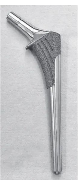
The PerFix910 (Kyocera, Osaka, Japan) stem is a doublewedge metaphyseal filling straight stem (Fig.1),
that is made of Ti6Al4V with a hydroxyapatite (HA) processed porous surface. Khanuja et al. [8] classified all of the currently available uncemented stems into six types, based on morphology and fixation. The PerFix910 is classified as type 2 in Khanuja’s classification [8], and it is designed to be secured by proximal fixation in two planes, i.e., the anteroposterior (AP) and mediolateral (ML) planes.
When a cementless stem is inserted, a pressfit of the porous coating into the cortical bone and filling is important. Once the initial fixation is achieved, the promotion of osseointegration into the porous coating
CopyrightⒸ 2020 by Okayama University Medical School.
http ://escholarship.lib.okayama-u.ac.jp/amo/
Original Article
Ten-Year Outcomes of Total Hip Arthroplasty Using Fit-and-Fill Type Cementless Collared Straight Stem Implants: Relationship between
the Initial Contact Status and Stress Shielding
Tomoaki Sanki, Hirosuke Endo*, Tomonori Tetsunaga, Takayuki Furumatsu, Kazuki Yamada, and Toshifumi Ozaki
Department of Orthopaedic Surgery, Okayama University Graduate School of Medicine, Dentistry and Pharmaceutical Sciences, Okayama 700-8558, Japan
We investigated the relationship between the initial contact status and stress shielding in total hip arthroplasty (THA) using fit-and-fill type straight-stem implants. In addition we evaluated the clinical and radiographic outcomes. Subjects were 100 hips of 94 patients who underwent THA and were followed-up for ≥10 years.
Contact areas with the femoral cortical bone were investigated according to the zonal distribution of Gruen using postoperative CT images. Depending on the number of contact areas, the patients were classified into high contact [HC], medium contact [MC], and low contact [LC] groups. Radiographic and clinical outcomes were evaluated. In the HC group (20 hips), severe stress shielding was observed in 12 hips, which was statisti- cally significant (p=0.008). In the LC group (29 hips), mild stress shielding was observed in 27 hips which was statistically significant (p<0.001). No significant differences were observed among the 3 groups in clinical out- comes, Harris hip score (p=0.719) or Japanese Orthopedic Association (JOA) score (p=0.301). In insertion of cementless collared fit-and-fill type straight-stem implants, severe late stress shielding of the femoral bone may occur if high contact of the femoral component is achieved. However, the degree of stress shielding does not result in adverse clinical outcomes.
Key words: fitandfill, stress shielding, cementless straight stem, total hip arthroplasty
Received March 27, 2019 ; accepted August 8, 2019.
*Corresponding author. Phone : +81862357273; Fax : +81862239727
Email : [email protected] (H. Endo) Conflict of Interest Disclosures: No potential conflict of interest relevant to this article was reported.
contributes to longterm stability. In the case of a straight stem, a fitandfill concept is needed to obtain good initial fixation and favorable clinical outcomes [911].
We have routinely inserted cementless straight stem implants to achieve a fitandfill with the aim of metaphyseal filling. However, we have observed severe stress shielding in cases with filling in the distal part of the straight stem. We thus hypothesized that cases in which there are wide contact areas between the implant and cortical bone might experience severe stress shield
ing over the longterm. The purpose of this study was to investigate the relationship between the initial bone contact status of cementless collared straightstem implants after surgery and the patients’ radiographic and clinical outcomes.
Patients and Methods
Patients. Between April 2005 and May 2008, a total of 276 patients underwent a primary THA at our institution, and collared straightstem implants were used in 100 hips in 94 of these patients. Cemented stems and modular type stems were used in other patients if anteversion of the femoral bone was ≥40 degrees as measured by 3D template software. When the anteversion was <40 degrees, various cementless stems were used depending on the surgeon’s preference.
We included these 94 patients in the present retrospec
tive cohort analysis. This study was approved by our
institution’s ethics committee (approval no. 180920) and was conducted in accordance with the ethical stan
dards of the 1964 Declaration of Helsinki as revised in 1983 and 2000. The patients’ mean age at the time of their THA was 60.2 years (range 3187 years), and the preoperative diagnoses were osteoarthritis (78 hips, 78%), avascular necrosis (11 hips, 11%), and rheuma
toid arthritis (11 hips, 11%). The incidence of DDH was 63% (63 of 100 hips). All of the patients were fol
lowedup for >10 years, and the mean followup dura
tion was 10.4 years (range 1013 years). Six patients underwent staged bilateral THA. The patients’ mean height was 152 cm (range 130175 cm); their mean body weight was 54.5 kg (range 31.075.9 kg), and their mean body mass index (BMI) was 23.3 kg/m2 (range 15.633.2 kg/m2). The mean canal flare index (CFI) was 4.2 (range 2.86.3). We used the Dorr classification of preoperative hip joint plain radiographs to assess and classify the femoral bone quality as type A (champagne flute canal), type B (normal canal), or type C (stove
pipe canal) [12]. With this system, 24 hips were classi
fied as type A, 75 hips as type B, and one hip as type C.
Surgical procedure. The THA surgeries were per
formed by 3 orthopedic surgeons (S.M., H.E., and K.F.) each with experience of >500 THAs. All patients were placed in the lateral decubitus position for surgery. The surgical approaches were selected by the surgeons depending on the degree of each patient’s pelvic defor
mity. Cementless titanium hemisphere cups (AMS HA;
Kyocera Medical, Osaka, Japan) were used with cementless titanium and doublewedge or metaphyseal
filling designed stems (PerFix910 HA; Kyocera Medical).
Collared stems were used in all cases. The femur was scraped with a handpowered reamer and a broach, and the stem was inserted using the pressfit technique. We used 3D template software for the preoperative plan
ning and the decision regarding the stem size. We per
formed hand rasping toward the planned stem size.
When rotational stability was observed and the collar could prevent subsidence, we approved an undersized stem to avoid intraoperative fracture. The patients were allowed full weight bearing as tolerated with a walker from the day after surgery.
Radiographic evaluation. For the postoperative evaluations, CT images from the pelvis to the knee joint were taken 1 week after surgery by a multislice CT scanner (Discovery CT 750 HD; GE Medical Systems, Milwaukee, WI, USA). The image settings were as fol
Fig. 1 PerFix910 HA collard stem (Kyocera, Osaka, Japan).
lows: tube voltage 120 kV; tube current 150 mA; slice thickness 2 mm; and slice pitch 2 mm. The CT image data saved in DICOM format were transferred into 3D templating software ver. 03.08.05 (Kyocera Medical).
Images were captured in the coronal and sagittal planes centered on the longitudinal axis of the stem.
We counted the contact areas between the implant and cortical bone based on Gruen zones [13] (Fig.2), and we assessed the state of initial contact. We then
classified the hips into 3 groups according to the num
ber of contact areas on the coronal and sagittal planes: the HC (high contact; ≥7 contact areas), MC (medium contact; ≥4 and <6 contact areas), and LC (low contact; ≤3 contact areas) groups (Fig.3).
We assessed the stress shielding, spot welds, cortical hypertrophy, and pedestal to evaluate bone remodeling around the implant using the latest followup AP radio
graph in each group. Zero to 2nd degree stress shield
ing was defined as mild, while 3rd and 4th degree stress shielding was defined as severe based on Engh’s classifications [10]. Spot welds were recorded according to Gruen’s zonal distribution [13]. Stem subsidence was measured based on the criteria of Johnston and Loudon [14,15]. Stem alignment in the sagittal plane was clas
sified as anterior tilt (≥3 degrees), or neutral and poste
rior tilt (≥3 degrees) based on the criteria of Müller [16].
Clinical evaluation. Followups were conducted at 2,4,6 months, and 12 months and every 12 months thereafter at the patient’s surgeon’s office. The clinical evaluation of function and pain was performed by the operative surgeon (E.H. or T.T) with the use of the Harris Hip Score (HHS) [17] and the Japanese Orthopedic Association (JOA) hip score [18]. We investigated whether reimplantation was performed.
Statistical analyses. We performed a oneway analysis of variance (ANOVA) and the nonparametric KruskalWallis test to compare the patients’ demo
graphic data on age, height, body weight, BMI, CFI, HHS and JOA score. The chisquared test and a resid
L M A P
1
2
3 4 5
6
7 8
9
10 11 12
13 14
B A
Fig. 2 CT images centered on the longitudinal axis of the stem.
Numbers indicate the Gruen zones. A, Coronal plane; B, Sagittal plane.
A B C
Fig. 3 CT images centered on the longitudinal axis of the stem. We classified the patients into three groups according to the numbers of contact areas on the coronal and sagittal planes. A, HC group; B, MC group; C, LC group.
ual analysis were used to compare sex, diagnosis, Dorr classification, rate of DDH, stress shielding, frequency of spot welds, cortical hypertrophy, pedestal, subsid
ence, and stem alignment. We carried out the statistical analyses using the Statistical Package for the Social Sciences (SPSS), ver. 20.0 (IBM SPSS Statistics for Windows, Ver. 19.0. Armonk, NY, USA). Values of p<0.05 were considered significant.
Results
We counted the contact areas between the implant stem and cortical bone using the postoperative CT images, and we classified the hips into 3 groups: the HC group (20 hips), MC group (51 hips), and LC group (29 hips) (Table 1). The patients’ demographic data did not differ significantly among the 3 groups. In the HC group, severe stress shielding was observed in 12 hips (60.0%) showing a statistically significant differ
ence among the 3 groups (p=0.008) (Fig.4). In the MC
B C
A D
Fig. 4 Radiographs and CT images of a 66-year-old woman with osteonecrosis (HC group). A, Preoperative radiograph; B, Post- operative radiograph; C, CT images at 1 week after surgery. Contacts between the implant and cortical bone in zones 2, 3, 5, 6, 9, 10, 12, and 13 were identified; D, 10 years after surgery. Severe stress shielding (grade 4) was observed.
Table 1 The patient demographic data
HC Group (20hips) MC Group (51hips) LC Group (29hips) p Value
Age (years) 62.0±13.4 (31-63) 61.3±11.6 (33-87) 62.3±10.6 (41-78) 0.937a
Gender : female/male 19/1 42/9 25/4 0.384a
Diagnosis 0.918b
Osteoarthritis 15 39 24
DDH 14/15 (93.3%) 28/39 (71.8%) 21/24 (87.5%) 0.119b
Osteonecrosis 3 6 2
Rheumatoid Arthrisis 2 6 3
Height (m) 1.49±0.08 (1.33-1.64) 1.52±0.08 (1.30-1.75) 1.54±0.08 (1.37-1.75) 0.154a
Weight (kg) 52.0±10.1 (31.0-71.1) 55.0±10.5 (31.7-75.9) 55.4±10.3 (31.1-74.7) 0.476a
BMI (kg/m2) 23.1±3.8 (15.6-30.7) 23.5±3.9 (15.9-33.2) 23.1±4.0 (15.6-33.1) 0.851a
CFI 4.1±0.8 (3.3-6.3) 4.3±0.6 (2.8-5.6) 4.0±0.5 (2.8-5.3) 0.178a
Dorr type A/B/C 4/16/0 16/35/0 4/24/1 0.233b
DDH, developmental dysplasia of the hip; BMI, body mass index; CFI, canal flare index.
Age, Height, Weight, BMI, and CFI are expressed as mean ± standard deviation, and range.
aOne-way analysis of valiance. bChi-square test.
group, severe stress shielding was observed in 20 hips (39.2%) and mild stress shielding was observed in 31 hips (60.8%) not showing a statistically differences among the 3 groups (p=0.368). In the LC group, mild stress shielding was observed in 27 hips (93.1%) with a statistically significant difference among the 3 groups (p<0.001) (Fig.5). Spot welds were observed in 71 hips (71.0%), and these were in zone 2 in 37 hips (37.0%), and zone 6 in 65 hips (65.0%). There was no significant difference in spot welds among the 3 groups (p=0.086).
Cortical hypertrophy was observed in 8 hips (8.0%), in zone 3 in 5 hips (5.0%), and in zone 5 in 3 hips (3.0%).
There was no significant difference in cortical hypertro
phy among the 3 groups (p=0.562). Pedestals were observed in 13 hips (13.0%) with no significant differ
ence among the 3 groups (p=0.131). Stem subsidence of ≥2 mm was not observed. In 33 of the 47 hips (70.2%) in which the stem collar was not in contact with the medial cortex of the femoral neck at 1 week after surgery, the collar was in contact within 2 mm subsid
ence at the final followup. Regarding stem alignment with the sagittal plane, the neutral position was observed in 73 hips (73.0%) and anterior tilt in 27 hips (27.0%); no posterior tilt was observed. In the HC group, the neutral position was observed in all 20 hips, which was statistically significant (p=0.009). The HHS values showed a statistically significant improvement from 34.8 (range 760) preoperation to 80.1 (range 5195) at the final followup. The JOA score showed a
statistically significant improvement from 35.2 (range 567) preoperation to 80.9 (range 50100) at the final followup. Significant clinical improvements were observed in each of the 3 contact groups, and there were no significant differences in the clinical improve
ment among the groups. No reimplantation or other reoperations were performed for any reason. The sur
vival rate was 100% (Table 2).
Discussion
A variety of cementless femoral stems have been associated with favorable clinical and radiographic out
comes [36]. The fitandfill type straight stem is widely used worldwide, with good longterm results [3,19
21].This study investigated the relationship between the initial contact status of the PerFix910HA collared stem and stress shielding at a mean of 10 years after surgery, in 100 hips of 94 patients who underwent a primary THA. Severe stress shielding was observed in the patients with 7 or more initial contact areas. In con
trast, severe stress shielding was not observed in any patients with 3 or fewer initial contact areas. Spot welds were observed particularly in zones 6 and 9, where osseointegration was observed on the distal end of the porous coating. Cortical hypertrophy and pedestals were observed in 8 and 13 patients respectively, with no significant differences among the 3 groups. No reim
B C
A D
Fig. 5 Radiographs and CT images of a 62-year-old woman with osteoarthritis (LC group). A, Preoperative radiograph; B, Posto- perative radiograph; C, CT images at 1 week after surgery. Contacts between the implant and cortical bone in zones 8, 9, and 12 were identified; D, 10 years after surgery. Mild stress shielding (grade 2) was observed.
plantation or other reoperation was performed. The survival rate of the PerFix910 stem was 100%.
Regarding clinical outcomes, the HHS and JOA scores were improved in all three groups, and there was no significant difference among the groups. Radiographic findings did not affect clinical outcomes.
The PerFix910 stem is a type 2 stem according to Khanuja’s classification [8]. The type 2 stem is typically a doublewedge metaphysealfilling stem designed to contact the cortical bone in the AP and ML planes.
Compared with the singlewedge type 1 stem, the type 2 stem has a wider AP plane with a distally tapering or rounding form for the purpose of filling the canal.
Nakashima et al. [22] reported the >10year outcomes of 131 hips surgically treated using the PerFix910 col
lared stem. In their study stem with revision as an end
point, the survival rate >12 years was 99.2%. In addi
tion, the radiographic evaluations revealed bone ingrowth fixation of the stem without radiolucency or stem subsidence around the coating areas in all cases, in parallel with the favorable clinical outcomes. Epinette and Manley et al. [21] reported the >15year outcomes
of 571 hips in 504 patients who were treated by THA using the Omnifit stem (Stryker Osteonics, Allendale, NJ, USA) as longterm outcomes of the same type 2 stem. The stem revision rate of 0.7% and the survival rate of 99.2% after 17 years represented satisfactory results. However, unfavorable outcomes with a similar type of stem have also been reported. Nishino et al. [23]
investigated the correlation between distal bone remod
eling and stress shielding with a >10years followup in 50 hips (41 patients) who underwent THA using the Synergy stem (Smith & Nephew Orthopaedics, Memphis, TN, USA). They reported that severe stress shielding was observed in almost half of the hips (23 cases/50 hips [46%]), and spot welds and cortical hypertrophy were found in the distal end (zones 3 and 5) of the stem. They considered that these adverse effects could be attributed to a mismatch between the femur morphology of the Japanese patients and the stem size.
When a cementless straight stem is inserted, ade
quate metaphyseal filling contributes to the initial fixa
tion and stability of the stem, secondarily promoting
Table 2
HC Group (20hips) MC Group (51hips) LC Group (29hips) p Value
Stress shielding
Grade 1/2/3/4 0/8/6/6 7/24/17/3 4/23/2/0 <0.001a Grade ≧3 12 (60.0%) 20 (39.2%) 2 (6.9%)
Spot welds
Anterior-posterior 12 (60.0%) 34 (66.7%) 25 (86.2%) 0.086a Zone 2 9 (45.0%) 15 (29.4%) 13 (44.8%)
Zone 6 8 (40.0%) 32 (62.7%) 25 (86.2%)
Medial-lateral 8 (40.0%) 25 (49.0%) 16 (55.2%) 0.579a Zone 9 6 (30.0%) 17 (33.3%) 15 (51.7%)
Zone 13 4 (20.0%) 15 (29.4%) 9 (31.0%)
Cortical hypertrophy 2 5 1 0.562a
Pedestal 1 10 2 0.131a
Subsidence 0 0 0
Sagittal stem alignment 0.009a
anterior tilt 0 17 10
neutral 20 34 19
posterior tilt 0 0 0
JOA hip score
preoperative 38.8±13.8 (15-67) 34.7±12.0 (5-61) 33.7±13.5 (10-61) 0.422b postoperative 80.6±7.7 (64-93) 83.2±6.9 (62-100) 81.2±6.9 (61-93) 0.301b Harris Hip Score
preoperative 36.0±12.7 (15-60) 34.8±11.2 (7-54) 34.0±12.3 (13-54) 0.868b postoperative 80.9±7.5 (64-94) 82.3±6.1 (64-95) 81.6±5.5 (61-91) 0.719b JOA hip score, The Japanese Orthopaedic Associatoin evaluation standard of hip joint function.
JOA hip score and Harris Hip Score are expressed as mean ± standard deviation, and range.
aChi-square test. bOne-way analysis of valiance.
osseointegration into the porous coating, and ulti
mately leading to good clinical results [911]. Insufficient filling is likely to cause thigh pain and/or stem loosen
ing, resulting in poor clinical outcomes [11,13,24].
Gosens et al. [25] stated that femoral fitandfill pre
dicted radiologic changes, but not clinical results. They concluded that there was no correlation between clini
cal parameters and the radiologic phenomena. Laine et al. [11] reported bone remodeling at 5 years after sur
gery using the fitandfill stem; they concluded that satisfactory metaphyseal filling provided stability of the stem in the location and promoted ingrowth and bone remodeling, while stress shielding advanced in the proximal metaphysis. In the same sense, dependence on stem filling is likely to cause distal fixation, rather than proximal fixation, and to induce severe stress shielding [10,23,26].
In this study, we used 3D template software to match the CT image to the long axis of the stem at 1 week postoperatively, and we evaluated the contact areas between the stem and cortical bone in both the coronal and sagittal planes. Evaluations based on the CT image software have been reported to be more accu
rate than radiographic evaluations using frontal and lateral views of the contact areas and the filling status [10,11]. In the HC group, many contacts with cortical bone from the metaphysis to the distal part of the stem such as zones 2 to 6 and zones 9 to 13 were observed. In these cases, severe stress shielding was observed due to distal fixation. In the MC group, many contacts with cortical bone mainly at the metaphysis were observed.
The degrees of stress shielding differed depending on the part of fixation. In the LC group, there were signifi
cantly less contacts with cortical bone from the metaph
ysis to the distal part of the stem, but many contacts in zones 8 and 9 in the lateral view were observed. In these cases, severe stress shielding was not observed due to proximal fixation. Although favorable filling was achieved in the cases that had many contact areas between the stem and cortical bone, significant and severe stress shielding was observed. These results sug
gested that strong initial fixation was achieved in the distal part of the stem, but not in the proximal coating part.
By contrast, in the cases that had only a few areas of contact and insufficient filling, severe stress shielding was not observed. These results indicated that even insufficient filling did not induce implant loosening or
unfavorable radiographic findings. As insufficient canal filling may not ensure the initial stability of the cement
less stem, stem subsidence may occur. However, sub
sidence of ≥2 mm did not occur in any patient in this study. Two reasons for this result are considered:
The first reason is the use of the collared system in all patients. Some studies have reported that contact between the medial cortex of the femoral neck and the collar of the stem prevents stem subsidence [2729]. In the present study, in 33 of the 47 hips (70.2%) in which the stem collar was not in contact with the medial cor
tex of the femoral neck at 1 week after surgery, the col
lar was in contact within 2 mm subsidence at the final followup. Some studies reported that the presence or absence of the collar was unrelated to the prevention of stem subsidence [3032]. We used 3D template software for preoperative planning and for selecting the stem size. We performed hand rasping toward the planned stem size. We approved undersized stem to avoid intraoperative fracture if rotational stability was obtained and the collar could prevent subsidence. Thus, even if the stem size was small, the collar at the medial cortex of the femoral neck might have prevented subsidence of ≥2 mm. The stem would then be stuck at the proximal part of the femoral bone, and this might have caused osseointegration at the coating area.
The second reason is stem insertion in anterior tilt alignment in the lateral view, which we observed in cases using onesizesmaller stems. Kim and Mulliken et al. [24,33] mentioned that filling of the straight stem is inclined to be achieved in the lateral view. However, stem insertion in anterior tilt alignment may have allowed insufficient filling and a lack of distal fixation.
This suggests that even if stem size was small in the frontal view, stem insertion in anterior tilt alignment caused proximal fixation as the result of contact in zones 8 and 9.
Although this logic may be contradictory, the use of a onesize smaller collared stem may be useful to avoid severe stress shielding due to sufficient filling. In the insertion of cementless collared fitandfill type straightstem implants, severe late stress shielding may occur if high contact of the femoral component is achieved.
This study has several limitations as follows: first, use of collared stems in all patients, which were not compared with collarless stems; secondly, this study was retrospective. The use of onesize smaller stems and
stem insertion in anterior tilt alignment was not intended. Thus our concept, that we should “not aim to achieve sufficient filling” requires further prospective study.
In conclusion, we investigated the relevance of the early postoperative contact status between the implant and cortical bone with the mean 10year outcome of stress shielding in 100 hips of 94 patients, who under
went THA using the PerFix910 HA collared stem, which is a doublewedge metaphyseal filling cementless straight stem. In cases that had many contact areas with the cortical bone, significantly severe stress shielding was observed. In contrast, in the cases that had fewer contact areas, severe stress shielding was not observed.
When a cementless collared fitandfill type straight
stem implant is inserted, severe late stress shielding may occur if there is a large amount of contact between the femoral component and cortical bone from the metaphysis to the distal part of the stem. However, the degree of stress shielding does not affect the rate of adverse clinical outcomes.
Acknowledgments. We thank Dr Shigeru Mitani and Dr Kazuo Fujiwara for their professional surgery.
References
1. Sugano N, Ohzono K, Nishii T, Haraguchi K, Sakai T and Ochi T:
Computed-tomography-based computer preoperative planning for total hip arthroplasty. Comput Aided Surg (1998) 3:320-324.
2. Sugano N, Noble PC, Kamaric E, Salama JK, Ochi T and Tullos HS: The morphology of the femur in developmental dysplasia of the hip. J Bone Joint Surg Br (1998) 80:711-719.
3. Capello WN, DʼAntonio JA, Jaffe WL, Geesink RG, Manley MT and Feinberg JR: Hydroxyapatite-coated femoral components:
15-year minimum follow up. Clin Orthop Relat Res (2006) 453:75- 4. Engh CA Jr, Mohan V, Nagowski JP, Sychterz Terefenko CJ and 80.
Engh CA Sr: Influence of stem size on clinical outcome of primary total hip arthroplasty with cementless extensively porous-coated femoral components. J Arthroplasty (2009) 24: 554-559.
5. Bojescul JA, Xenos JS, Callaghan JJ and Savory CG: Results of porous-coated anatomic total hip arthroplasty without cement at fif- teen years: a concise follow-up of a previous report. J Bone Joint Surg Am (2003) 85:1079-1083.
6. Kim YH: Long-term results of the cementless porous-coated ana- tomic total hip prosthesis. J Bone Joint Surg Br (2005) 87: 623- 7. Akiyama H, Hoshino A, Iida H, Shindo H, Takakura Y, Miura H, 627.
Yamamoto K, Yoshiya S, Hasegawa Y, Shimamura T, Kurosaka M, Otsuka H, Kawanabe K, Kawate K, Harada Y and Nakamura T: A pilot project for the Japan arthroplasty register. J Orthop Sci (2012) 17: 358-369.
8. Khanuja HS, Vakil JJ, Goddard MS and Mont MA: Cementless
femoral fixation in total hip arthroplasty. J Bone Joint Surg Am (2011) 93: 500-509.
9. Huiskes R: The various stress patterns of press-fit, ingrown, and cemented femoral stems. Clin Orthop Relat Res (1990) 261: 27- 10. Engh CA, Bobyn JD and Glassman AH: Porous-coated hip 38.
replacement. The factors governing bone ingrowth, stress shield- ing, and clinical results. J Bone Joint Surg Br (1987) 69:45-55.
11. Laine HJ, Puolakka TJ, Moilanen T, Pajamäki KJ, Wirta J and Lehto MU: The effects of cementless femoral stem shape and proximal surface texture on ʻfit and fillʼ characteristics and bone remodeling. Int Orthop (2000) 24: 184-190.
12. Dorr LD, Faugere MC, Mackel AM, Gruen TA, Bognar B and Malluche HH: Structural and cellular assessment of bone quality of proximal femur. Bone (1993) 14: 231-242.
13. Gruen TA, McNeice GM and Amstutz HC: “Modes of failure” of cemented stem-type femoral components: a radiographic analysis of loosening. Clin Orthop Relat Res (1979) 141:17-27.
14. Johnston RC, Fitzgerald RH Jr, Harris WH, Poss R, Müller ME and Sledge CB: Clinical and radiographic evaluation of total hip replacement: a standard system of terminology for reporting results.
J Bone Joint Surg Am (1990) 72:161-168.
15. Loudon JR: Femoral prosthetic subsidence after low-friction arthro- plasty. Clin Orthop Relat Res (1986) 211:134-139.
16. Müller M, Duda G, Perka C and Tohtz S: The sagittal stem align- ment and the stem version clearly influence the impingement-free range of motion in total hip arthroplasty: a computer model-based analysis. Int Orthop (2016) 40:473-480.
17. Harris WH: Traumatic arthritis of the hip after dislocation and ace- tabular fractures: treatment by mold arthroplasty. An endresult study using a new method of result evaluation. J Bone Joint Surg Am (1969) 51: 737-755.
18. Imura S: The Japanese Orthopaedic Association: evaluation chart of hip joint functions. J Jpn Orthop Assoc (1995) 69: 864-867.
19. García-Cimbrelo E, Bru-Pomer A, García-Benítez B, Hernández- Blanco M and Vaquero J: Multicentric and prospective study of the Summit cementless stem. Hip Int (2010) 20: 63-69.
20. Meding JB, Ritter MA, Keating EM and Berend ME: Twenty-year followup of an uncemented stem in primary THA. Clin Orthop Relat Res (2015) 473: 543-548.
21. Epinette JA and Manley MT: Uncemented stems in hip replace- ment--hydroxyapatite or plain porous: does it matter? Based on a prospective study of HA Omnifit stems at 15-years minimum fol- low-up. Hip Int (2008) 18:69-74.
22. Nakashima Y, Sato T, Yamamoto T, Motomura G, Ohishi M, Hamai S, Akiyama M, Hirata M, Hara D and Iwamoto Y: Results at a minimum of 10 years of follow-up for AMS and PerFix910 HA-coated cementless total hip arthroplasty: impact of cross- linked polyethylene on implant longevity. J Orthop Sci (2013) 18:
962-968.
23. Nishino T, Mishima H, Kawamura H, Shimizu Y, Miyakawa S and Ochiai N: Follow-up results of 10-12 years after total hip arthro- plasty using cementless tapered stem -- frequency of severe stress shielding with synergy stem in Japanese patients. J Arthroplasty (2013) 28: 1736-1740.
24. Mulliken BD, Nayak N, Bourne RB, Rorabeck CH and Bullas R: Early radiographic results comparing cemented and cementless total hip arthroplasty. J Arthroplasty (1996) 11: 24-33.
25. Gosens T, Sluimer JC, Kester AD and van Langelaan EJ: Femoral fit predicts radiologic changes, but not clinical results, in Mallory- head total hip arthroplasties. Clin Orthop Relat Res (2005) 432:
138-147.
26. Issa K, Stroh AD, Mont MA and Bonutti PM: Effect of bone type on clinical and radiographic outcomes of a proximally-coated cementless stem in primary total hip arthroplasties. J Orthop Res (2014) 32:1214-1220.
27. Flecher X, Blanc G, Sainsous B, Parratte S and Argenson JN: A customized collared polished stem may reduce the complication rate of impaction grafting in revision hip surgery: a 12-year fol- low-up study. J Bone Joint Surg Br (2012) 94: 609-614.
28. Campbell D, Mercer G, Nilsson KG, Wells V, Field JR and Callary SA: Early migration characteristics of a hydroxyapa- tite-coated femoral stem: an RSA study. Int Orthop (2011) 35:
483-488.
29. Simpson DJ, Kendrick BJ, Hughes M, Glyn-Jones S, Gill HS, Rushforth GF and Murray DW: The migration patterns of two ver- sions of the Furlong cementless femoral stem: a randomised, con-
trolled trial using radiostereometric analysis. J Bone Joint Surg Br (2010) 92: 1356-1362.
30. Al-Najjim M, Khattak U, Sim J and Chambers I: Differences in subsidence rate between alternative designs of a commonly used uncemented femoral stem. J Orthop (2016) 13: 322-326.
31. Meding JB, Ritter MA, Keating EM and Faris PM: Comparison of collared and collarless femoral components in primary uncemented total hip arthroplasty. J Arthroplasty (1997) 12: 273-280.
32. Weber E, Sundberg M and Flivik G: Design modifications of the uncemented Furlong hip stem result in minor early subsidence but do not affect further stability: a randomized controlled RSA study with 5-year follow-up. Acta Orthop (2014) 85: 556-561.
33. Kim YH and Kim VE: Uncemented porous-coated anatomic total hip replacement. Results at six years in a consecutive series. J Bone Joint Surg Br (1993) 75:6-13.