Mutational Studies on Catalytic Domain of D -stereospecific Amidohydrolase from
Streptomyces sp. 82F2
D
YASMEEN YOUSIF AHMED ELYAS
2018
DEDICATION
This thesis work is dedicated to:
To my husband, Ayman, who has been a constant source of support and encouragement during the challenges of graduate school life, to Menatallah my sweet daughter and also to Ahmed my lovely son.
This work also dedicated to my parents, Yousif Elyas and Mhasien AbduElgader.
Yasmeen,
ACKNOWLEDGMENT
My profound gratitude goes to Almighty Allah, the omnipotent and omniscient, all the praise and glory are to Him alone for giving me the strengths, patience, knowledge, health, time, resources, opportunity and blessing to complete this study.
First, I would like to express my sincere gratitude to my advisor Prof. Jiro Arima, Department of Agricultural, Biological, and Environmental Sciences, Faculty of Agriculture, Tottori University, for his continuous support, valuable guidance, scholarly inputs and consistent encouragement I received throughout the research work. This feat was possible only because of the unconditional support provided by prof Arima, person with an amicable and positive disposition, prof Arima has always made himself available to clarify my doubts despite his busy schedules and I consider it as a great opportunity to do my doctoral programme under his guidance and to learn from his research expertise. Thank you, Prof Arima, for all your help and support.
My sincere gratitude goes to my Ph.D. co-supervisor Professor Hiroyuki Azakami, Department of Biological Chemistry, Faculty of Agriculture, Yamaguchi University, for extended discussions and valuable suggestions which have contributed greatly to the progress of this research.
Also, I would like to thank my former supervisor Dr. Nuha Elkhatim for her inspiration and advice during the first stage of my academic life.
My deepest thankfulness and appreciation offers to Dr. Isam Ali Mohamed Ahmed, Associate Professor Department of Food and Agricultural Sciences, King Saud University, who recommended and encouraged me to study in ]apan, for his remarkable advice and help during the application process.
I am forever thankful to my colleagues at the Laboratory of Bio-Functional Chemistry, a former student and current for their friendship and support, and for creating a cordial working environment, especially, Mr. Kazusa Miyatani, Mr. Yoshitaka Isoda, Mr. Taro Amano.
I gratefully acknowledge the contributions of the Ministry of Education, Science, Sports and Culture of Japan for providing the financial support which removed financial concerns from my decision to embark on this journey.
My thanks and appreciation goes to the staff members of the International Affairs Division Center, United Graduate School of Agricultural Sciences, Tottori University for their unlimited help and support through my study and personal life.
I would like to thank my colleagues, the academic, technical, and administrative staff of Khartoum University for allowing me to complete my study in Japan.
My thanks and appreciation goes to Dr. Yasir Serag Alnor and Dr.
Nasreen Mohamed Kamal, for their generous support and encouragement. I received a lot of help from them both in my research and personal life.
My deepest love and appreciation goes to Ikuko Sekino, Japanese language teacher for her patient, generosity, and emotional support, she has always been there for me in spite of her busy life.
My deep and sincere gratitude to my family for their continuous and unparalleled love, help and support. I am grateful to my sisters Yousra and Yathrib and my brother Ahmed for help and support. I am forever indebted to my parents, my mother Mahasin Abdulgadir and my father Yousif Elyas for their unconditional love, Patience, prayers and for giving me the opportunities and experiences that have made me who I am. They selflessly encouraged me to explore new directions in life and seek my own destiny. This journey would not have been possible if not for them, and I dedicate this milestone to them.
Special thanks and love also goes to my second family, my father-in-law Mr. MohamedZain AbduElrahim and my mother-in-law Afaf Khidir For their endless understanding, support, love, and prayers.
Finally, I owe thanks to a very special person, my husband, Aiman for his continued and unfailing love, support and understanding during my pursuit of a Ph.D. degree that made the completion of thesis possible. You were always around at times I thought that it is impossible to continue, you helped me to keep things in perspective. I greatly value his contribution and deeply appreciate his belief in me. I appreciate my kids, my little girl Menaallah and my little boy Ahmed for abiding my ignorance and the patience they showed during my study time. Words would never say how grateful I am to both of you.
I consider myself the luckiest in the world to have such a lovely and caring family, standing beside me with their love and unconditional support.
Yasmeen Yousif Elyas 2018
I
TABLE OF CONTENTS
TABLE OF CONTENT ... I LIST OF FIGURES ... V LIST OF TABLES ... VII LIST OF ABBREVIATIONS ... VIII
CHAPTER ONE: GENERAL INTRODUCTION
1.1 Introduction ... 1
1.2 Serine protease ... 2
1.2.1 Classification of Serine Peptidase ... 3
1.2.2 The catalytic mechanism of serine peptidase ... 5
1.3 D-stereospecific Amidohydrolase (DAH): ... 10
1.3.1 Structure of the DAH:... 13
1.3.2 Mechanism of catalytic reaction of DAH ... 15
1.4 RESEARCH OBJECTIVES ... 17
CHAPTER TWO: Active site pocket of Streptomyces D-stereospecific amidohydrolase has functional roles in aminolysis activity 2.1 INTRODUCTION ... 19
2.2 MATERIALS AND METHODS ... 23
2.2.1 Materials, bacterial strains, and plasmids ... 23
2.2.2 Enzyme assay ... 24
2.2.3 Mutagenesis ... 24
2.2.4 Expression and purification of WT and mutant enzymes ... 24
2.2.5 Assay of aminolysis activity ... 25
2.2.6 Measurement of the weight of the aminolysis product ... 27
2.2.7 MS analysis... 27
2.2.8 Methanol release assay ... 27
2.2.9 Thermal stability test ... 28
2.2.10 Structural model of mutant DAHs ... 28
2.3 RESULTS... 30
2.3.1 Effect of the mutation of residues of the pocket on pNA-releasing activity ... 30
2.3.2 Effects of the mutation of residues in the pocket on aminolysis activity ... 31
2.3.3 Properties of I338A DAH from an aminolysis point of view ... 36
2.3.4 Enzyme kinetics ... 38
2.3.5 Effects of the Ile338 mutation on enzyme activity and thermal stability ... 40
2.3.6 Acyl acceptor preferences of WT and mutant DAHs ... 43
2.4 DISCUSSION ... 45
2.5 Conclusion ... 47
CHAPTER THREE: Effect of active site pocket structure modification of D-stereospecific amidohydrolase on the recognition of stereospecific and hydrophobic substrates 3.1 Introduction ... 49
3.2 Materials and Methods ... 51
3.2.1 Materials, bacterial strains, and plasmids ... 51
3.2.2 Structural model of mutant DAHs ... 53
3.2.3 Mutagenesis ... 53
3.2.4 Expression and purification of WT and mutant DAHs ... 53
3.2.5 Methanol release assay ... 54
3.2.6Assay of aminolysis and hydrolysis activity ... 55
3.2.7 MS analysis... 55
3.3. Results ... 58
III
3.3.1 Effect of Phe-substitution on acyl-enzyme intermediate formation activity ... 58
3.3.2 Effect of the Ala267 mutation on acyl-enzyme intermediate formation activity ... 60
3.3.3 Kinetic analysis of the WT and mutant DAHs ... 62
3.3.4 Effect of Mutations on Aminolysis Activity ... 63
3.4 Discussion ... 66
3.5 Conclusion ... 69
CHAPTER FOUR: Enhancement of aminolysis activity by space filling of active site pocket of D-stereospecific amidonhydrolase 4.1. INTRODUCTION ... 70
4.2Materials and Methods ... 72
4.2.1 Materials, bacterial strains, and plasmids ... 72
4.2.2 Structural model of mutant DAHs ... 73
4.2.3 Mutagenesis ... 73
4.2.4 Expression and purification of WT and mutant DAHs ... 73
4.2.5 Enzyme reaction ... 74
4.2.6 Methanol release assay ... 75
4.2.7 MS analysis... 75
4.2.8 c(DP-LR) synthesis ... 75
4.3 Results ... 76
4.3.1 Effect of space filling of active site pocket on aminolysis activity ... 76
4.3.2 Effect of space filling of active site pocket on c(DP-LR) synthetic activity ... 81
4.3.3Time dependence on c(DP-LR) synthesis ... 81
4.4 Discussion ... 84
5.5 Conclusion ... 86 CHAPTER FIVE
Summury for the study... 88 ... 92 References: ... 95
V
LIST OF FIGURES
Fig. 1.1 Scheme of the hydrolysis and aminolysis reactions catalyzed by serine peptidases... 3
Fig. 1.2 The catalytic triad of chymotrypsin (PDB code 4CHA) ... 8
Fig. 1.3 The generally accepted mechanism for serine proteases. ... 9
Fig. 1.4 Overall structure around the active site Ser of DAH... 11
Fig. 1.5 The local structure of the active site pocket of DAH ... 12
Fig. 1.6 Comparison of the cavity and pockets shapes of (A) DAH ... 14
Fig. 1.7 Proposed mechanism for cyclo(D-Pro L-Arg) [c(D-Pro L-Arg)] production by a D- stereospecific amidohydrolase. ... 16
Fig. 2.1 Cavity shape and residues in the active site pocket... 23
Fig. 2.2 SDS-PAGE of partially purified wild-type (WT) and mutant D-stereospecific amidohydrolases (DAHs)... 29
Fig. 2.3 Effect of mutation on p-nitroanilide (pNA) release activity from D-Phe-pNA. ... 31
Fig. 2.4 Effects of mutation on methanol release, hydrolysis, and aminolysis. ... 33
Fig. 2.5 Precipitates in the reaction mixtures of WT and mutant DAHs representing aminolysis reactions. ... 34
Fig. 2.6 Time dependence of the reaction products by catalysis with the WT and I338A DAHs.35 Fig. 2.7 Effect of 1,8-DAO on methanol release (A), hydrolysis (B), and aminolysis (B) of WT and I338A DAHs. ... 37
Fig. 2.8 Effects of Ile338 mutations on methanol release, hydrolysis, and aminolysis by DAH. 41 Fig. 2.9 Thermal stabilities of WT and mutant DAHs. ... 42
Fig. 2.10 Changes in the ratio of aminolysis products via the mutations. ... 44
Fig. 3.1 Cavity and pocket in DAH. (A) Cross-sectional view of the molecular surface of DAH. ... 52
Fig. 3.2 SDS-PAGE of partially purified wild-type (WT) and mutant D-stereospecific amidohydrolases (DAHs). ... 56
Fig. 3.3 Comparison of the local structures of DAH and DAA (A) and pocket shapes of WT DAH and the predicted structures of the mutants (B G). ... 57
Fig. 3.4 Reaction scheme of the DAH reaction toward aminoacyl derivatives. ... 61
Fig. 3.5 Effect of mutation on methanol release. ... 61
Fig. 3.6 Effect of mutation on methanol release, hydrolysis, and aminolysis activities in the presence of 50 mM 1,8-DAO as acyl acceptor. ... 64
Fig. 3.7 Local structures (upper panel) and predicted pocket shapes (lower panel) of WT ... 67
Fig. 4.1 Scheme of the hydrolysis and aminolysis reactions catalyzed by serine peptidases... 72
Fig. 4.2 SDS-PAGE of partially purified wild-type (WT) and mutant D-stereospecific amidohydrolases (DAHs)... 76
Fig. 4.3 Structure of active site pocket of DAH. ... 79
Fig. 4.3 Effect of mutation on Ac-Phe-Trp production by aminolysis function of DAH. ... 80
Fig. 4.4 Effect of mutation on c(DP-LR) synthesis by aminolysis function of DAH. ... 83
VII
LIST OF TABLES
Table 1: Amino acid and their abbreviations ... IX
Table 1.1.: Known diversity of serine peptidase structure and catalytic mechanism. ... 6
Table 2.1: Sequences of primers used for site-directed mutagenesis ... 26
Table 2.2: Enzyme kinetics for hydrolysis and aminolysis by the WT and I338A DAHs. ... 39
Table 3.1: Sequences of mutagenesis primers used for site-directed mutagenesis ... 56
Table 3.2: Enzyme kinetics for acyl-enzyme intermediate formation (methanol release). ... 65
Table 4.1: Sequences of mutagenesis primers used for site-directed mutagenesis ... 76
LIST OF ABBREVIATIONS 1,8-DAO 1,8-diaminooctane
Amp Ampicillin BSA bovine serum albumin D.W. Distilled water
DAH D-stereospecific amidohydrolase
DMSO dimethyl sulfoxide
LB Luria-Bertani
LC liquid chromatography
MS mass spectrometry
OEt ethyl ester
OMe methyl ester
PAGE polyacrylamide gel electrophoresis PCR polymerase chain reaction
pNA p-nitroaniline
SDS sodium dodecyl sulfate
TEMED N,N,N,N-tetramethylethylenediamine Tris Tris (hydroxymethyl) amino methane
IX
Table 1: Amino acid and their abbreviations
Amino acid 3 letters 1 letter Amino acid 3 letters 1 letter
Alanine Ala A Leucine Leu L
Arginine Arg R Lysine Lys K
Asparagine Asn N Methionine Met M
Aspartate Asp D Phenylalanine Phe F
Aspartate or Asparagine
Asx B Proline Pro P
Cysteine Cys C Serine Ser S
Glutamate Glu E Threonine Thr T
Glutamine Gln Q Tryptophan Trp W
Glutamate or Glutamine
Glx Z Tyrosine Tyr Y
Glycine Gly G Valine Val V
Histidine His H
Isoleucine Ile I
CHAPTER ONE
GENERAL INTRODUCTION
1.1 INTRODUCTION
Proteases likely arose at the earliest stages of protein evolution as simple destructive enzymes necessary for protein catabolism and the generation of amino acids in primitive organisms. For many years, studies on proteases focused on their original roles as blunt aggressors associated with protein demolition. However, the realization that, beyond these nonspecific degradative functions, proteases act as sharp scissors and catalyze highly specific reactions of proteolytic processing, producing new protein products, inaugurated a new era in protease research. Depends on their catalytic mechanisms, proteases are classified into six main classes: aspartic, threonine, metallo, cysteine, serine, and glutamic proteases, although glutamic proteases have not been found in mammals so far. Human cells produce at least 569 proteases including 194 metallo, 176 serine, 150 cysteine, 28 threonine and 21 aspartic proteases(López-otín and Matrisian 2007). Proteases cleave proteins from either the N- terminus or C-terminus (exopeptidases), or in the middle of a protein (endopeptidases). Much primary knowledge about the function of proteases stems from investigations of the digestive system and intracellular protein turnover. However, proteases are also considered as extremely important signaling molecules influenced by various biological events, such as apoptosis, blood coagulation, extracellular tissue remodeling and DNA replication(Rawlings and Barrett 1994; López-otín and Matrisian 2007).
Proteases have great medical and pharmaceutical importance due to their key role in biological processes and in the life-cycle of many pathogens. They are extensively applied
2
enzymes in several sectors of industry and biotechnology, furthermore, numerous research applications require their use, including production of Klenow fragments, peptide synthesis, digestion of unwanted proteins during nucleic acid purification, cell culturing and tissue dissociation, preparation of recombinant antibody fragments for research, diagnostics and therapy, exploration of the structure-function relationships by structural studies, removal of affinity tags from fusion proteins in recombinant protein techniques, peptide sequencing and proteolytic digestion of proteins in proteomics (Mótyán János András 2013).
Based on the catalytic mechanism and the presence of amino acid residue(s) at the active site the proteases can be grouped as aspartic proteases, cysteine proteases, glutamic proteases, metalloproteases, asparagine proteases, serine proteases, threonine proteases, and proteases with mixed or unknown catalytic mechanism. The current classification system further classifies the proteases into families based on sequence similarities, furthermore, homologous families are grouped into clans using a structure-based classification (Rawlings and Barrett 1993; Rawlings 2016). Classification and nomenclature of proteolytic enzymes, as well as a detailed description of individual proteases, is available in the MEROPS database (Rawlings et al. 2018).
1.2 SERINE PROTEASE
Almost one-third of all proteases can be classified as serine proteases, named for the nucleophilic Ser residue at the active site (Rawlings et al. 2016). Serine peptidases are the most widely studied group in biology, they can be found in eukaryotes, prokaryotes, archaea and viruses, interest in this family due to their widespread and distinctive role in a host of potential physiological and pathological process including digestion, hemostasis, apoptosis, signal transduction, and immune response (Hedstrom 2002a).
The catalytic action of serine peptidases depends on the interplay of a nucleophile, a general base, and an acid. In the first step of reaction a nucleophilic attack by the catalytic serine residue (active site Ser) on the carbonyl atom of the substrate resulting in a covalent acyl- enzyme intermediate and a new peptide amino terminus. a second nucleophilic attack, by a water molecule, leads to hydrolysis of the acyl-enzyme, releasing the new carboxyl group and restoring the catalytic Ser residue to its initial state. (Zakharova et al. 2009). In the presence of high concentrations of primary amines, which can not be achieved under physiological condition an amide bond is formed through nucleophilic attack by an amino group instead of a water molecule forms an amide bond (Fig. 1.1) (Bratovanova and Petkov 1987; John J. Perona and Charles S. Craik 1995). In this reaction, termed aminolysis the primary amine acts as an acyl acceptor. Because amino acids can act as acyl acceptors, the unnatural aminolysis reaction of serine peptidases has attracted attention as a method for synthesizing various peptides that cannot be produced under natural conditions. (Arima et al.
2016)
Fig. 1.1 Scheme of the hydrolysis and aminolysis reactions catalyzed by serine peptidases(Arima et al. 2016)
1.2.1 Classification of Serine Peptidase
4
The MEROPS protease classification system counts 16 super-families (as of 2013) each containing many families. Each superfamily uses the catalytic triad or dyad in a different protein fold and represents the convergent evolution of the catalytic mechanism.
Families are grouped in a clan if there are indications, principally from tertiary structure comparisons, that they arise from a common ancestor. For every family and clan, there is an identifier that shows the catalytic type of the peptidases contained in the group. The
identific metallo
catalyt (Polgár 2005).Each clan, family, and the inhibitor is assigned to an identifier. For a clan, the identifier consists of two letters. The first indicates the catalytic
itors that are proteins
sequentially as each clan is identified. An example of a clan identifier is CA, which includes cysteine peptidases with a papain-like fold. For a family, the identifier consists of an initial letter, again corresponding to the catalytic type and a number. An example is C1, the family of papain-like cysteine peptidases (Rawlings et al. 2018).
Based on Barrett and Rawlings classification, the peptidases are divided into clans on the base of the catalytic mechanism and to families based on common ancestry (Rawlings 2009;
Rawlings et al. 2016). Over 908300 peptidases protein sequences classified into 62 clans and 268 families (MEROPS 2018) (Rawlings et al. 2018). Serine peptidases represent almost one of third of all known proteolytic. More than 26000 serine peptidases have been classified into 13 clans and 82 families. The family name stems from the nucleophilic Ser in the enzyme
active site, which attacks the carbonyl moiety of the substrate peptide bond to form an acyl- enzyme intermediate. Nucleophilicity of the catalytic Ser is typically dependent on a catalytic triad of Asp, His, and Ser residues, commonly referred to as the charge relay system.
1.2.2 The catalytic mechanism of serine peptidase
The hallmark of serine peptidases is that they contain the so-
Ser/His/Asp triad. Although the classical serine proteases are the most widespread in nature,
are observed. Such variations include the triads Ser/His/Glu, Ser/ His/His, and Ser/Glu/Asp, and include the dyads Ser/Lys and Ser/His. Other variations are seen with certain serine and threonine peptidases of the Ntn hydrolase superfamily that carry out catalysis with a single active site residue (Ekici et al. 2008). A summary of catalytic units in all serine peptidase families, primary specificity and the fold that harbors them are provided in Table 1.1.
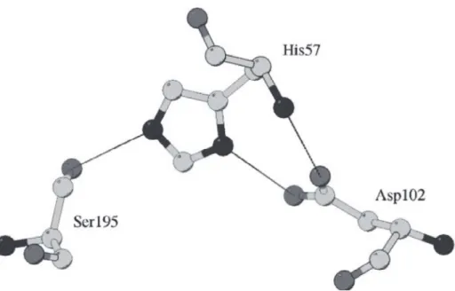
The first well-characterized mechanism of action of serine peptidases was established primarily by the kinetic studies of chymotrypsin by Bender and his co-workers in the 1960s (Matthews et al. 1977). The X-ray data showed that the serine and histidine are the potent residues to catalysis and they are in proper position to function in the catalytic mechanism.
As shown in figure (1.2) the OG of Ser195 and the NE2 of His57 are within hydrogen bonding distance. The other nitrogen atom (ND1) of the imidazole ring is hydrogen bonded to the carboxyl group of Asp102 so that the hydrogen bond is shielded from water by several amino acid residues. The geometric relation of Asp102, His57, and Ser195 led to the postulation that His57 serves for transferring the proton from Ser195 to Asp102 in a charge relay mechanism. However, a proton relay from the highly basic serine OH group to the acidic aspartate is chemically unlikely (Polgár and Bender 1969). It is a more reliable assumption that Asp102 may be involved in the stabilization of the ion-pair generated between the imidazolium ion and the negatively charged-tetrahedral intermediate, and that Asp102 may participate in the orientation of the correct tautomer of His57 relative to Ser195.
Nuclear magnetic resonance (NMR) and neutron diffraction studies have then confirmed that it is the imidazole and not the aspartate that is protonated (Polgár 2005).
8
Fig. 1.2 The catalytic triad of chymotrypsin (PDB code 4CHA) (Polgár 2005)
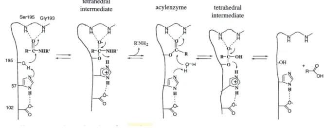
All proteases must overcome three obstacles to hydrolyze a peptide bond: (a) amide bonds are very stable due to electron donation from the amide nitrogen to the carbonyl. For comparison, a simple bond, while a p-nitrophenyl ester is 300000× more alkyl ester is 3000× more reactive than an amide reactive. Proteases usually activate an amide bond via the interaction of the carbonyl oxygen with a general acid, and may also distort the peptide bond to disrupt resonance stabilization; (b) water is a poor nucleophile; proteases always activate water, usually via a general base; and (c) amines are poor leaving groups; proteases protonate the amine prior to expulsion. Serine proteases perform these tasks very efficiently: the rates of peptide hydrolysis by serine proteases are 1010-fold greater than the uncatalyzed reactions.
Obviously, these mechanisms of catalysis are not confined to peptide hydrolysis; serine proteases also readily hydrolyze other acyl compounds, including amides, anilides, esters, and thioesters. Figure 1.3 displays the generally accepted mechanism for chymotrypsin-like serine proteases. In the acylation half of the reaction, Ser195 attacks the carbonyl of the
peptide substrate, assisted by His57 acting as a general base, to yield a tetrahedral intermediate. The resulting His57-H+ is stabilized by the hydrogen bond to Asp102. The oxyanion of the tetrahedral intermediate is stabilized by interaction with the main chain NHs of the oxyanion hole. The tetrahedral intermediate collapses with the expulsion of leaving the group, assisted by His57-H+ acting as a general acid, to yield the acyl-enzyme intermediate.
The deacylation half of the reaction essentially repeats the above sequence: water attacks the acyl-enzyme, assisted by His57, yielding a second tetrahedral intermediate. This intermediate collapses, expelling Ser195 and carboxylic acid product. The transition states of the acylation and deacylation reactions will resemble the high energy tetrahedral intermediates, and the
literature. It is worth noting that a web of hydrogen bonding interactions links the substrate binding sites to the catalytic triad. As the reaction proceeds, changes in bonding and charge at the scissile bond will propagate to more remote enzyme-substrate interactions, and vice versa. (Hedstrom 2002a)
Fig. 1.3 The generally accepted mechanism for serine proteases. (Hedstrom 2002a)
10
1.3 D-STEREOSPECIFIC AMIDOHYDROLASE (DAH):
By Screening of 2000 soil isolates, D-stereospecific amidohydrolase (DAH) has been identified from the culture supernatant of Streptomyces sp. 82F2. The enzyme belongs to the S12 peptidase family with catalytic Ser / Lys dyad (Ser86 and Lys 89). DAH recognizes D- aminoacyl derivative as a substrate and amide bond is formed by aminolysis. The enzyme preferentially uses the D-aminoacyl derivative as the acyl donor and uses the L-amino acid and its derivative as the acyl acceptor to produce a dipeptide with the DL- configuration(Arima et al. 2011a, b). Therefore, DAH has been used to synthesize biologically active dipeptide such as cyclic dipeptide family 18 chitinase inhibitor, cyclo (D- Pro-L-Arg) in a one-step/one-pot reaction(Arima et al. 2011b).
Numbers of enzymes in the MEROPS peptidase database belonging to the S12 family, which have a catalytic Ser / Lys dyad, exhibit high aminolysis activity for various types of substrates such as amides esters and peptides (Kato et al. 1989, 1990; Pratt and Frère 2013). Up to date, five crystal structures of S12 family peptidases (including DAH) have been resolved: D-Ala-D- Ala carboxypeptidase B D, D-peptidase), class C b-lactamase, D-amino acid amidase (DAA) and D-stereospecific aminopeptidase D, D-peptidase, and D-stereospecific aminopeptidase catalyze peptide bond formation by their aminolysis activity (Lobkovskya et al. 1994; Kelly and Kuzin 1995; Bompard-Gilles et al. 2000; Okazaki et al. 2007; Arima et al. 2016). Based on these crystal structures, the structural factors responsible for the substrate specificities have been identified (Delmarcelle et al. 2005b).
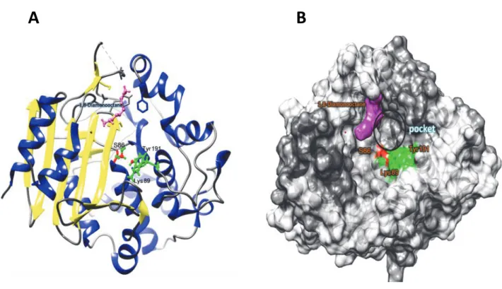
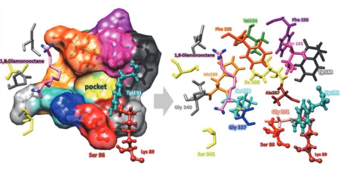
Fig. 1.4 Overall structure around the active site Ser86 of DAH. (A) The overall structure of DAH that interacts with 1,8-diaminooctane. a-Helices and b sheets are shown in red and yellow, respectively. (A) The overall structure of DAH that bounded with 1,8-diaminooctane. -Helices and sheets are shown in blue and yellow, respectively. The bound 1,8-diaminooctane and active site residues (Ser86, Lys89, and Tyr191) are shown as a purple and green ball and stick models, respectively. (B) Surface model of DAH. The active site Ser86 bound 1,8-
diaminooctane and active site residues ( Lys89 and Tyr191) are shown as red, purple and green, respectively.
A B
12
Fig. 1.5 The local structure of the active site pocket of DAH, and the residues composing the active site pocket. The active site residues (Ser86 and Lys89 and Tyr191) are shown as a ball and stick model according to the atom type. Other residues are shown as sticks colored according to the atom type.
1.3.1 Structure of the DAH:
Comparative analysis of DAH with homologous enzymes in same family S12 and crystallographic study provide insight into structural factors that affect the substrate specificity, stereoselectivity, and aminolysis activity. DAH can recognizes 1,8-diaminooctane and other amino acid compounds as acyl acceptor in aminolysis reaction, therefore, the crystal structure of DAH that binds 1,8-diaminooctane was determined at a resolution of 1.49 Å(Arima et al. 2016)
Figure (1.4) shows the overall structure of DAH, the enzyme consists of -rich and ß-rich -rich region is formed by residues 38 86 and 224 379, and consists of six main antiparallel b- strands flanked by four a-helices. The a-helix rich region (residues 87 223) includes a -helices Figure (1.4). The two motifs, Ser-x-x-Lys and Tyr-x-Asn, located at the N-terminal (Ser86 Lys89) and the internal region (Tyr191 Asn193) of DAH, respectively. Crystallographic analysis of the enzyme revealed that DAH possesses a large cavity that leads to the catalytic center. The side chains of Ser86 and Lys89 that create a catalytic Ser/Lys dyad in S12 family peptidases (Kelly and Kuzin 1995) were positioned at the center of the large cavity (Fig.1.4 A). hr191, which is close to the Ser/Lys dyad, acts as the general base to activate Ser as a nucleophile for the acyl donor during the acylation step and the water molecule during hydrolysis. In addition, there is a small pocket close to the catalytic center of Ser86, Ly89, and Tyr191 (Fig 1.4 B, and Fig 1.5). Structures comparison of family S12 enzymes, D, D-peptidase (PDB: 3PTE), D-amino acid amidase (PDB: 2EFX).
Have revealed that DAH possess the largest cavity leading to catalytic center and large active site pocket beside DAA, therefore the large cavity and active site pocket allow the
14
substrate to enter, and accommodate the large side chain if acyl donor substrate (Fig 1.6) (Arima et al. 2016).
Fig. 1.6 Comparison of the cavity and pockets shapes of (A) DAH, (B) D, D-peptidase (Protein Data Bank code: 3PTE) and (C) DAA (Protein Data Bank code: 2EFX). The hydrophobic surfaces are also shown (left). Active site Ser residues of the enzymes are shown as ball and stick models colored according to the atom type. Corresponding cross- sectional views are shown on the right. The structures of respective enzymes are superimposed on the model of DAH, and then the cross-sectional view of respective enzymes with the same angle is created. The enzyme molecules are shown as hydrophobic surfaces.
(Arima et al. 2016).
A
B
C
1.3.2 Mechanism of catalytic reaction of DAH
Several stereospecific peptidases that recognize an amide bond involving D-amino acid residues have been reported (Asano et al. 1989; Arima et al. 2010b). They are regarded as being associated mainly with the biosynthesis and remodeling of peptidoglycan. Among them, peptidases such as D-Ala D-Ala carboxypeptidase B from Streptomyces sp. R61 (Kumar and Pratt 2005), D-aminopeptidase from Ochrobactrum anthropi (Kato et al. 1990), and D-peptidase from Bacillus cereus(Komeda and Asano 1999) have the function of an aminolysis reaction. The enzymes belonging to clan SE, the enzymes of the S11, S12, and S13 peptidase families, which are specialized for roles in bacterial cell wall metabolism.
The synthesis of diverse DL-configuration dipeptides in a one-pot reaction was demonstrated by using a function of the aminolysis reaction of a D-stereospecific amidohydrolase from Streptomyces sp.,(Arima et al. 2011b) a clan SE, S12 family peptidase categorized as a peptidase with D-stereospecificity. The enzyme was able to use various aminoacyl derivatives, including L-aminoacyl derivatives, as acyl donors and acceptors. Investigations of the specificity of the peptide synthetic activity revealed that the enzyme preferentially used D- aminoacyl derivatives as acyl donors. In contrast, L-amino acids and their derivatives were preferentially used as acyl acceptors. Consequently, the synthesized dipeptides had a DL- configuration when D- and L-aminoacyl derivatives were mixed in a one-pot reaction(Arima et al. 2011b).
DAH is also applicable for to synthesize biologically active dipeptides, such as cyclo(D-Pro L-Arg), with the conversion rate of D- Pro-OBzl and L-Arg-OMe to cyclo(D-Pro L-Arg) being greater than 65%. The cyclization of D-Pro L-Arg-OMe occurred non-enzymatically (Fig.
1.7)(Arima et al. 2011b).
1.4 RESEARCH OBJECTIVES
According to the previously mentioned literature review, Serine proteases comprise nearly one-third of all known proteases identified to date and play crucial roles in a wide variety of cellular as well as extracellular functions, including the process of blood clotting, protein digestion, cell signaling, inflammation, fibrinolysis, fertilization, complement activation during immune responses and protein processing. (Rawlings and Barrett 1994; Page and Di Cera 2008) Owing to the expanding roles for serine proteases, there has been increasing interest in the identification, structural, and functional characterization of all members of the serine protease family of enzymes in humans and other organisms. Therefore, understanding the mechanism and structural factors affecting substrate recognition is important for the rational modification of enzymes and aimed at developing a convenient biocatalyst for peptide synthesis.
The assumption and concepts of the current study were to understand the structural implications of the D-Stereospecific amidohydrolase residues in and around the active site pocket and to investigate the effects of mutations of residues constituting the pocket on the catalysis and substrate specificity activity of DAH.
The Specific aims of the study were:
1- Mutational study on residues DAH active site pocket residues which are thought to involve in substrates recognition. We used site-directed mutagenesis to analyze the potential effects of alteration of eight residues to alanine aiming to expand the size of DAH pocket and investigated any changes in substrate recognition especially in the term of aminolysis reaction.
2- Assessing the effect of size/shape changes to the pocket on its substrate stereoselectivity and hydrophobic substrates recognition.
18
3- To assess the effect of the space filling of the active site pocket of DAH on catalytic activity to increase the aminolysis activity of the enzyme.
CHAPTER TWO
ACTIVE SITE POCKET OF STREPTOMYCES D-STEREOSPECIFIC AMIDOHYDROLASE HAS FUNCTIONAL ROLES IN AMINOLYSIS ACTIVITY
2.1 INTRODUCTION
Serine peptidases are widespread and abundant and play key roles in all living organisms.
The enzymes are classified into 53 families (S1 S81 in the MEROPS peptidase database [http://merops.sanger.ac.uk/] (Rawlings et al. 2016; Rawlings 2016), and account for over 30%
of all known proteolytic enzymes. The catalytic mechanism of all known serine peptidases involves a nucleophilic Ser residue that attacks the carbonyl moiety of the substrate peptide bond to form an acyl-enzyme intermediate. The substrate is an acyl donor. The intermediate is then hydrolyzed through nucleophilic attack by an activated water molecule. In the presence of high concentrations of primary amines, an amide bond is formed through nucleophilic attack by an amino group instead of a water molecule (Bratovanova and Petkov 1987; Arima et al. 2014). The primary amine acts as an acyl acceptor. This enzymatic aminolysis reaction has been used for the syntheses of various biologically active peptides, such as Kyotorphin (Arima et al. 2010a) infusion material (Yokozeki and Hara 2005) and carnosine (Heck et al. 2007; Arima et al.
2010b)
The nucleophilicity of the catalytic Ser of most serine peptidases is dependent on a catalytic triad of Asp, His, and Ser residues, commonly referred to as the charge relay system(Dodson and Wlodawer 1998; Delmarcelle et al. 2005a; Polgár 2005). In contrast, variations in the architecture of the active site, such as the Ser/Ser/Lys triad and Ser/Lys dyad, have also been reported (Paetzel and Dalbey 1997; Hedstrom 2002b; Shin et al. 2003; Paetzel
20
and J. Strynadka 2008). Recently, we found a new serine peptidase, D-stereospecific amidohydrolase (DAH) from Streptomyces sp. 82F2 (Arima et al. 2011a). The enzyme belongs to the S12 peptidase family, which has a catalytic Ser/Lys dyad (Ser86 and Lys89). The most characteristic feature of DAH is the recognition of D-amino acyl derivatives as substrates and the formation of amide bonds by aminolysis. In DAH aminolysis reactions, the enzyme preferentially uses D-aminoacyl derivatives as acyl donors and L-amino acids and their derivatives as acyl acceptors and produces dipeptides with a DL-configuration(Arima et al.
2011b). In fact, DAH has been used to synthesize a cyclic dipeptide as the lead compound of the pesticide cyclo-(D-Pro-L-Arg), which inhibits family 18 chitinases (Houston et al. 2002), in a one-step/one-pot reaction (Arima et al. 2011b).
Several of the enzymes of the S12 family exhibit high aminolysis activity toward various types of substrates including amides, esters, and peptides (Kato et al. 1989, 1990; Pratt and Frère 2013). The functions are thought to be mainly associated with the biosynthesis or remodeling of peptidoglycan. Five crystal structures of S12 family peptidases, including DAH (PDB: 3WWX), have been reported to date: D-aminopeptidase (PDB: 1EI5), D, D-peptidase (PDB: 3PTE), class C -lactamase (PDB: 1BLS), D-amino acid amidase (PDB: 2EFX) (Lobkovskya et al. 1994; Kelly and Kuzin 1995; Bompard-Gilles et al. 2000; Okazaki et al. 2007; Arima et al. 2016). Among them, D, D-peptidase and D-stereospecific aminopeptidase, as well as DAH, catalyze the formation of peptide bonds by aminolysis(Kato et al. 1990; Bompard-Gilles et al. 2000). Their substrate specificities vary, and the structural factors responsible for the substrate specificity of hydrolysis have been identified (Delmarcelle et al. 2005b; Sauvage et al. 2008) However, the mechanisms of substrate (both acyl donors and acceptors) recognition for aminolysis remain
unclear but represent an important issue for understanding the characteristics of this class of enzymes and their detailed function in biosynthesis or remodeling of peptidoglycan.
Crystallographic analysis of DAH revealed that the enzyme possesses a large cavity. The side chains of Ser86 and Lys89 that create the catalytic Ser/Lys dyad are positioned at the center bottom of the cavity (Arima et al. 2016). In addition, there is a pocket close to this catalytic center (Fig.3.1). The overall structures of S12 enzyme family members are similar, although there are significant differences in terms of the shapes and sizes of the cavities and active site pockets among them. DAH recognizes L-aminoacyl derivatives specifically as acyl acceptors.
However, the exact positions that recognize L-amino acids in the cavity of DAH have not been found. Okazaki et al. reported that the structure of the D-amino acid amidase pocket fits L-Phe and D-Phe (Okazaki et al. 2008a). In this structure, unlike the bound D-Phe, the bound L-Phe does not form an acyl-enzyme intermediate. Thus, we assumed that the pocket functions to recognize acyl acceptors for aminolysis. In addition to L-aminoacyl derivatives, DAH recognizes 1,8-diaminooctane (1,8-DAO) as an acyl acceptor in aminolysis (Arima et al. 2016), and the crystal structure of DAH has been refined in the presence of 1,8-DAO. Although the bound 1,8- DAO in the DAH crystal structure is positioned at the side of the cavity (Fig. 2.1), the distance between Ser86 and 1,8-DAO is 7.3 Å, which is too great for nucleophilic attack. Therefore, the 1,8-DAO binding form is thought not to reflect the location of the nucleophilic attack on the acyl acceptor (Arima et al. 2016). For that reason, an investigation into the effects of mutations of residues constituting the pocket on aminolysis could provide new insights. The aim of this study was to evaluate the role of the residues constituting the DAH pocket via mutations. We used site- directed mutagenesis to analyze the potential effects of altered residues in the DAH pocket, which is thought to bind substrates and investigated any changes in substrate recognition,
22
especially in aminolysis. The data indicated that Ile338 is an important residue for substrate recognition among the DAH pocket residues.
Fig. 2.1 Cavity shape and residues in the active site pocket. The pictures show the overall structure (left), a cross-sectional view (center), and a close-up view of D-stereospecific amidohydrolase (DAH). The bound 1,8-diaminooctane (1,8-DAO) molecule is shown as a stick or ball and stick. The active site Ser86 residue is shown in red, the residues composing the active site are shown in green (left and center) or blue, and the region associated with 1,8-DAO binding is shown in yellow.
2.2 MATERIALS AND METHODS
2.2.1 Materials, bacterial strains, and plasmids
Peptides and aminoacyl ester derivatives were purchased from Bachem AG, Aldrich Chemical Co. Inc., Sigma Chemical Co., Novabiochem Corp., and Wako Pure Chemical Industries Ltd.
Escherichia coli JM109 was used as a host strain for general cloning procedures and E. coli Rosetta (DE3) was used as a host strain for gene expression. Plasmid pET-82F2DAP (with the DAH gene inserted into the MscI-NcoI gap of pET-22b (Arima et al. 2011b) was used for the expression of wild-type DAH (WT) and as a template for mutagenesis.
24
2.2.2 Enzyme assay
For routine assays, enzyme activity was determined via a continuous spectrophotometric assay with D-Phe-pNA as the substrate. Substrate solution (10 L; 10 mM) was added to 90 L of a mixture containing 200 mM Tris-maleate (pH 6.5) and 5 g·mL-1 of the enzyme at 25 °C.
The increased absorption at 405 nm caused by the release of pNA was monitored continuously with a microtiter plate reader (680; Bio-Rad Laboratories Inc.). The initial activity rate was determined from the linear part of the optical density profile of pNA measured using the same instrument.
2.2.3 Mutagenesis
Site-directed mutagenesis for the construction of mutant enzymes was conducted by inverse PCR using two pairs of primers containing a point mutation (Table 2.1). The PCR program consisted of 18 cycles for 1 min at 95 °C, 1 min at 65 °C, and 8 min at 68 °C. The PCR product was treated with Dpn I at 37 °C overnight. Thereafter, it was transfected into competent E. coli JM109 cells according to the manufac
confirmed by sequencing.
2.2.4 Expression and purification of WT and mutant enzymes
E. coli Rosetta (DE3) harboring pET-82F2DAH or the expression vector for mutant DAH production was cultivated at 25 °C for 48 h in 50 mL of Overnight Expression Instant TB medium (Novagen Inc.). Since DAH is an extracellular enzyme (Arima et al. 2011a), the culture was centrifuged to remove the cells and the recombinant enzyme was purified using the
following procedures. The culture supernatant was dialyzed against 20 mM sodium citrate (pH 5.5). The dialysate was loaded onto a Vivapure-S spin column (Sartorius AG) equilibrated with 20 mM sodium citrate (pH 5.5). After washing with the same buffer containing 0.1 M NaCl, the bound protein was eluted with the same buffer, this time containing 0.4 M NaCl. The homogeneity of the purified proteins was confirmed by 12% SDS-PAGE under denaturing conditions (Laemmli 1970).
2.2.5 Assay of aminolysis activity
Aminolysis and hydrolysis reactions are competitive and hydrolysis tends to occur at high temperatures; thus, the reaction was conducted at 4 °C to suppress hydrolysis. In addition, pH 8.5 was adopted for the aminolysis reaction to avoid the non-enzymatic degradation of ester substrates. The aminolysis activity of DAH was assayed as follows. First, 5 µL (or 50 µL) of 0.1 mg·mL-1 enzyme solution was added to 40 µL (or 400 µL) of the mixture containing 35 µL (or 350 µL) of 0.5 M Tris-HCl (pH 8.5) and 5 µL (or 50 µL) of 0.5 M 1,8-DAO. The reaction was initiated by adding 5 µL (or 50 µL) of an acyl donor substrate (aminoacyl derivative solutions dissolved in dimethyl sulfoxide at an appropriate concentration [ca. 0.5 M]). The reaction was then continued at 4 °C for 2 30 min. The reaction was terminated by adding 50 µL (or 500 µL) 0.5 M HCl to the mixture. The reaction mixture was then analyzed by measuring the released methanol and the weight of the reaction product, and by using mass spectrometry (MS).
26
Table 2.1 Sequences of primers used for site-directed mutagenesis
Mutations Primer name
Y144A
DAH Y144A_F DAH Y144A_R
CATCTACAGCGCGACCGAGGACCCCGCCTT AAGGCGGGGTCCTCGGTCGCGCTGTAGATG
T145A
DAH T145A_F DAH T145A_R
ATCTACAGCTACGCCGAGGACCCC GGGGTCCTCGGCGTAGCTGTAGAT
F150A DAH F150A_F DAH F150A_R
AGGACCCCGCCGCGCAGGCCAAGGTCTT AAGACCTTGGCCTGCGCGGCGGGGTCCT
V154A
DAH V154A_F DAHV154A_R
TTCCAGGCCAAGGCGTTCGGCCCCGGCTT AAGCCGGGGCCGAACGCCTTGGCCTGGAA
F155A
DAH F155A_F DAHF155A_R
GCCAAGGTCGCCGGCCCCGGCTTC GAAGCCGGGGCCGGCGACCTTGGC
I266A
DAH I266A_F DAH I266A_R
CTCAACCCGAGCGCCGCGGGCGCG CGCGCCCGCGGCGCTCGGGTTGAG
I338A
DAH I338A_F DAHI338A_R
CACGGCGGCGGCGCCCACGGCTCG CGAGCCGTGGGCGCCGCCGCCGTG
H339A
DAH H339A_F DAH H339A_R
GGCGGCGGCATCGCCGGCTCGTCC GGACGAGCCGGCGATGCCGCCGCC
I338G
DAH I338G_F DAH I338G_R
ACGGCGGCGGCGGCCACGGCTCGT ACGAGCCGTGGCCGCCGCCGCCGT
I338S
DAH I338S_F DAH I338S_R
ACGGCGGCGGCAGCCACGGCTCGT ACGAGCCGTGGCTGCCGCCGCCGT
I338D
DAH I338D_F DAH I338D_R
ACGGCGGCGGCGACCACGGCTCGT ACGAGCCGTGGTCGCCGCCGCCGT
I338F
DAH I338F_F DAH I338F_R
ACGGCGGCGGCTTCCACGGCTCGT ACGAGCCGTGGAAGCCGCCGCCGT
2.2.6 Measurement of the weight of the aminolysis product
DAH reaction mixtures for aminolysis sometimes appear as white precipitates. Before the reaction, the weights of the reaction tubes were measured and used as the basis weight of the reaction container. After the reaction, the precipitates were collected by centrifugation (13000 × g, 10 min) and dried at 60 °C for around 1 day. The weights of the dried products were measured by using an analytical balance (ATX84; Shimadzu).
2.2.7 MS analysis
The molecular masses of the products of DAH catalytic activity were determined via matrix- assisted laser desorption ionization time-of-flight (MALDI-TOF) MS and electrospray ionization (ESI TOF) MS. For MALDI-TOF MS analysis, the precipitates were collected by centrifugation (13000 × g, 10 min) and washed twice with distilled water. They were then suspended in 5 L of MALDI matrix solution (150 mM 2, 5-dihydroxybenzoic acid in 50%
acetonitrile). A small amount of each sample (ca. 1 L) was dropped onto the target frame and dried, and the samples were analyzed using Autoflex TOF (Bruker Daltonics Inc.).
For ESI-TOF MS analysis, the reaction mixture was diluted with a 200-fold volume of 0.1%
formic acid. After the solution was filtered, 5 µL from each sample was analyzed by using an ESI-TOF MS system (LCT Premier XE or Quattro Micro API; Waters Corp.). The data were processed using a computer program (MassLynx; Waters Corp.).
2.2.8 Methanol release assay
28
Liberated methanol, which is equivalent to the formation of the acyl-enzyme intermediate in the first step of the catalytic reaction, was measured with the 4-aminoantipyrine phenol method (Allain et al. 1974) coupled with an alcohol oxidase reaction. The DAH reaction mixture (10 µL) was added to 90 µL of the mixture containing 140 mM Tris-HCl (pH 8.0), 0.5 mM 4- aminoantipyrine, 2 mM phenol, 0.1 mg·mL-1 horseradish peroxidase, and 0.1 mg·mL-1 alcohol oxidase. Absorbance was determined at 490 nm using a microtiter plate reader (680; Bio-Rad Laboratories Inc.) after incubation for 30 min at room temperature. The concentration of the liberated methanol was measured from the linear part of the optical density profile of standard methanol using the same instrument.
2.2.9 Thermal stability test
The thermal stability of wild-type and mutant DAHs was investigated as follows: the purified enzyme solution (0.1 mg/mL) was incubated for 30 min at temperatures of 30 60oC.
Subsequently, the samples were assayed for residual activity using the methanol release activity assay described above.
2.2.10 Structural model of mutant DAHs
The sequences of the primary structures of the mutant DAHs were aligned with the sequences of the WT (Protein Data Bank accession no. 3WWX). The alignment data were submitted to SWISS-MODEL (Arnold et al. 2006a; Biasini et al. 2014a) to generate a homology model of mutant DAHs based on the template structure of WT. The structural models obtained were analyzed using UCSF Chimera software (Arnold et al. 2006a).
Fig. 2.2 SDS-PAGE of partially purified wild-type (WT) and mutant D-stereospecific amidohydrolases (DAHs).
30
2.3 RESULTS
2.3.1 Effect of the mutation of residues of the pocket on pNA-releasing activity
DAH possesses a small pocket close to the catalytic center of Ser86 (Arima et al. 2016) (Fig. 2.1). The space, shape, and electrostatic environment of the pocket are believed to confer the substrate specificity of DAH. To elucidate the way that the pocket relates to the substrate specificity, especially during the aminolysis reaction of DAH, we performed mutational analysis focusing on the residues in the pocket. Based on the crystal structure of DAH, eight residues (Tyr144, Thr145, Phe150, Val154, Phe155, Ile266, Ile338, and His339) that constitute the pocket (Fig. 2.1) were selected and substituted with alanine. As judged by SDS-PAGE, all mutants could be purified as described in the Materials and Methods section (Fig. 2.2). The release of pNA is accompanied by the formation of an acyl-enzyme intermediate (Fig. 2.3A). We first assessed the effect of the mutation on pNA-releasing activity with D-Phe-pNA. Mutants release of pNA by D-Phe-pNA at pH 8.5 tended to be higher or at the same level as that at pH 6.5, whereas that by the WT showed optimum activity at pH 6.5 (Fig. 2.3B). The results indicated that the optimum pH for the formation of the acyl-enzyme intermediate was shifted to alkaline especially in the mutants of (Y144, T145, F150, I266, and I338).
Fig. 2.3 Effect of mutation on p-nitroanilide (pNA) release activity from D-Phe-pNA. (A) Reaction scheme of the pNA release via DAH. (B) pNA release activity of the wild-type (WT) and mutant DAHs. p-Nitroaniline is a product of the formation of the acyl-enzyme intermediate when D-Phe-pNA is used as a substrate. The reaction was performed in a solution containing 1 mM D-Phe-pNA at pH 6.5 (gray bars) or 8.5 (black bars) at 25 °C. Each value represents the mean ± standard deviation (SD) of values from four independent experiments.
2.3.2 Effects of the mutation of residues in the pocket on aminolysis activity
DAH possesses a small pocket close to the catalytic center of Ser86 (Arima et al. 2016) (Fig.2.1).
The space, shape, and electrostatic environment of the pocket are believed to confer the substrate specificity of DAH. To elucidate the way that the enzyme pocket relates to substrate specificity, especially during aminolysis reactions of DAH, we performed mutational analysis focusing on the residues in the pocket. Based on the crystal structure of DAH, eight residues (Tyr144, Thr145,
32
Phe150, Val154, Phe155, Ile266, Ile338, and His339) that constitute the pocket (Fig. 2.1) were selected and substituted with alanine. SDS-PAGE confirmed the purity of all mutants as described in the Materials and Methods section (Fig. 2.2). To investigate the possible effects of mutations on aminolysis, we examined the products of the enzyme reaction by using Ac-D-Phe methyl ester (Ac-D-Phe-OMe) in combination with 1,8-DAO as the acyl-donor and acyl-acceptor substrates, respectively, under optimum conditions for aminolysis (i.e., at 4 °C and pH 8.5) (Arima et al. 2011a).
In the reaction mixtures of each enzyme, precipitates appeared after several minutes (Fig.2.5).
MS analysis showed that these precipitates were composed of the condensation products of Ac- D-Phe-OMe and 1,8-DAO, Ac-D-Phe-1,8-DAO, and Ac-D-Phe-1,8-DAO-Ac-D-Phe (Fig.2.5).
Therefore, we assumed that precipitation represented the catalytic activity for aminolysis (Fig.
2.4A). As portrayed in Fig. 2.4B 2.4D, the same level of methanol release (acyl-enzyme intermediate formation) but a lower level of Ac-D-Phe formation (hydrolysis) and a higher level of precipitation formation (aminolysis) were observed in a 15-min reaction with the I338A mutant. The results indicated that DAH aminolysis was enhanced by substitution of Ile338 with Ala.
34
of hydrolysis reaction products after 15 min with the WT and mutant DAHs. (D) Precipitation formation activity of the WT and mutant DAHs. Precipitates were assumed to be a product of aminolysis. The values were calculated from the weights of the precipitates after 15-min enzyme reactions. In all graphs, the reactions were performed in solutions containing 20 mM Ac-D-Phe- OMe and 50 mM 1,8-DAO at pH 8.5 and 4 °C for 15 min. Each value represents the mean ± SD of values from four independent experiments.
Fig. 2.5 Precipitates in the reaction mixtures of WT and mutant DAHs representing aminolysis reactions.
reaction started. The reaction was performed in a solution containing 20 mM Ac-D-Phe methyl ester (Ac-D-Phe -OMe) and 50 mM 1,8-diaminooctane (1,8-DAO) at pH 8.5 and 4 °C for 15 min.
Photographs were taken after centrifugation to obtain the precipitates.
Fig. 2.6 Time dependence of the reaction products by catalysis with the WT and I338A DAHs.
(A) Methanol release and substrate consumption. Open circles show the concentrations of methanol; closed circles show the concentrations of Ac-D-Phe-OMe. (B) Concentrations of hydrolysis products after the respective reaction times. (C) Weights of the precipitates after the respective reaction times. All graphs represent reactions performed in solutions containing 20 mM Ac-D-Phe-OMe and 50 mM 1,8-DAO at pH 8.5 and 4 °C. Each value represents the mean ± SD of values from four independent experiments.
36
2.3.3 Properties of I338A DAH from an aminolysis point of view
We focused on I338A DAH, which showed the strongest mutational effect on aminolysis, and investigated the properties of this mutant in detail. Figure 2.6 shows the time courses of Ac-D- Phe-OMe consumption, methanol release (acyl-enzyme intermediate formation), hydrolysis product release, and aminolysis product release when Ac-D-Phe-OMe and 1,8-DAO were used as substrates. The release of methanol and consumption of the substrate with I338A were higher than those with the WT (Fig. 2. 6A). In contrast, the production of Ac-D-Phe (hydrolysis product) was lower with I338A than with WT, as portrayed in Fig. 2.6B. The lower productivity of Ac-D-Phe in the I338A reaction was presumed to be caused by a lower susceptibility to nucleophilic attack by water or an increase in susceptibility to nucleophilic attack by 1,8-DAO on the acyl-enzyme intermediate via the mutation. In addition, the rate of precipitation with I338A was higher than that with the WT and quickly reached saturation (10 min, Fig. 2.6C). The results indicated that modification of the 338 Ile side-chain structure led to enhanced aminolysis activity.
The effects of 1,8-DAO concentration on methanol release, hydrolysis, and aminolysis activity for WT and I338A were assessed. The results showed lower hydrolysis activity for I338A compared with WT (Fig.2.7). In contrast, methanol release, which was thought to reflect an increase in the reaction rate for Ac-D-Phe-1,8-DAO production, was much higher for I338A than WT.
Fig. 2.7 Effect of 1,8-DAO on methanol release (A), hydrolysis (B), and aminolysis (B) of WT and I338A DAHs. (B) Open circles, the reaction rate for Ac-D-Phe production; closed circles, reaction rates for Ac-D-Phe-1,8-DAO production. Reactions were performed in a solution containing 20 mM Ac-D-Phe-OMe at pH 8.5 and 4°C for 5 min. Each value represents the mean ± standard deviation of values from eight independent experiments.
38
2.3.4 Enzyme kinetics
We further examined the enzyme kinetics of the aminolysis and hydrolysis reactions of I338A and the WT. As summarized in Table 2.2, Thekcat for methanol release was related to only Ac- D-Phe-OMe; that is, the theoretical max value for the nucleophilic attack of DAH on Ac-D-Phe- OMe. In contrast, the kcat values for hydrolysis and aminolysis are the theoretical max values for the nucleophilic attack of water and 1,8-DAO, respectively. Therefore, the rate-limiting step in this enzyme reaction is the formation of the acyl-enzyme intermediate Table 2.2: the kcat of I338A for methanol release from Ac-D-Phe-OMe increased on addition of 1,8-DAO (from 80.1 s-
1 to 304 s-1) and the Km value of I338A for methanol release from Ac-D-Phe-OMe also changed (from 3.78 mM to 19.5 mM). In contrast, the kcat and Km of the WT for methanol release from Ac-D-Phe-OMe under the respective conditions remained almost the same (kcat: 96.4 95.2 s-1, Km: 3.30 4.95 mM).
In terms of Ac-D-Phe-OMe hydrolysis, the changes in kcat of both enzymes on the addition of 50 mM 1,8-DAO were almost identical (WT changed from 96.4 s-1 to 46.7 s-1, and I338A from 80.1 s-1 to 43.7 s-1). However, the apparent Km of I338A increased on addition of 1,8-DAO (from 3.78 mM to 7.07 mM), while that of WT decreased (from 3.30 mM to 2.01 mM).
Thus, the kinetics of DAH aminolysis were dramatically affected by the mutation. The kcat value of I338A under the respective fixed conditions (50 mM 1,8-DAO or 50 mM Ac-D-Phe-OMe) was higher than that of the WT.
40
2.3.5 Effects of the Ile338 mutation on enzyme activity and thermal stability
The effects of the structural modification of Ile338 on hydrolysis and aminolysis were examined by constructing several mutants (I338G, I338S, I338D, and I338F) and investigating the ability of each to form the acyl-enzyme intermediate and mediate aminolysis and hydrolysis. The purity of the mutants was assessed via SDS-PAGE (Fig. 2.2).
A different effect was observed in the investigation into methanol release, hydrolysis, and aminolysis of Ac-D-Phe-OMe in the presence of 1,8-DAO. Aminolysis via DAH was significantly enhanced by substitutions with Ala and Ser. That is, the mutants exhibited higher levels of methanol release (acyl-enzyme intermediate formation), lower hydrolysis activity, and a five-fold greater formation of precipitates than the WT (Fig. 2.8). Substitutions with Gly and Phe also moderately enhanced aminolysis. However, the acyl-enzyme intermediate formation of 1338F was lower than that of the WT. In contrast, substitution with Asp decreased the ability of DAH to form the acyl-enzyme intermediate and mediate hydrolysis and aminolysis
.
This indicates that the introduction of a negative charge at position 338 of DAH negatively affects DAH activity in the presence of Ac-D-Phe-OMe.We next examined the thermal stability of wild-type and mutant DAHs. As shown in Fig.
2.9, they exhibited slightly lower stability with respect to temperature (up to 45 oC) than the WT.
Based on the results, the mutation of Ile338 might lead to a change in the local flexibility of the active site pocket.
Fig. 2.8 Effects of Ile338 mutations on methanol release, hydrolysis, and aminolysis by DAH.
(A) Methanol release via WT and mutant DAHs to Ac-D-Phe-OMe. The values were calculated from the concentrations of methanol. (B) Comparison of the concentrations of the hydrolysis products (Ac-D-Phe) of the WT and mutant DAHs. (C) WT and mutant DAHs precipitate formation activity. The values were calculated from the weights of the precipitates. All graphs represent comparisons of the products following reactions performed in solutions containing 20 mM Ac-D-Phe-OMe and 50 mM 1,8-DAO at pH 8.5 and 4 °C for 5 min (the length of the time that precipitation takes to occur without completion of the reactions). Each value represents the mean ± SD of values from four independent experiments.* p < 0.05; ** p < 0.02; *** p < 0.01
t test.
42
Fig. 2.9 Thermal stabilities of WT and mutant DAHs. Symbols in each panel: gray circle WT;
filled diamond mutant enzyme. Each value represents the mean ± SD of values from four independent experiments.
2.3.6 Acyl acceptor preferences of WT and mutant DAHs
Finally, we examined the aminolysis ability of the WT and mutant DAHs using Ac-D-Phe-OMe as an acyl donor and several amino acids and peptides as acyl acceptors to investigate the changes in acyl acceptor specificities of the mutants. In this experiment, Gly, Gly-OMe, Gly-Gly ((Gly)2), Gly-Gly-Gly ((Gly)3), L-Leu, L-Leu ethyl ester (-OEt), L-Leu-L-Leu ((Leu)2), L-Leu-L- Leu-L-Leu (Leu)3) were used as the acyl acceptor substrates.
Aminolysis was dramatically enhanced with mutations of Ile338 to Ala, Ser, and Phe when using L-Leu as an acyl acceptor (Fig. 2.10 B). In the reaction using Gly and glycyl peptides as acyl acceptors, several mutants (I338A, I338S, and I338F) exhibited enhanced aminolysis compared with the WT (approximately two-fold); the trends in the effects of the mutations were similar to the reaction using L-Leu and 1,8-DAO (Fig. 2.10 A). In contrast, the effects of mutations on aminolysis using Gly-OMe, leucyl peptides, and L-Leu-OEt differed from those using 1,8-DAO, amino acids (Gly and L-Leu), and glycyl peptides as acyl acceptors. The activity levels of all mutants were almost the same as that of the WT when using Gly-OMe as an acyl acceptor (Fig. 2.10 A). In the reaction using leucyl peptides as acyl acceptors, only I338F exhibited enhanced aminolysis compared with the WT (Fig. 2.10 A). In contrast, mutants other than I338F exhibited enhanced aminolysis with L-Leu-OEt as an acyl acceptor, which was in contrast to the reaction with leucyl peptides.
2.4 DISCUSSION
Among the overall structures of the S12 enzyme family members, significant differences are observed in the shapes and sizes of the cavities and pockets. In particular, the enzymes, including DAH, that catalyzes aminolysis, possess large cavities. Crystallographic analysis of DAH with 1,8-DAO indicates that 1,8-DAO binds to the middle of the cavity and the pocket and side chain of Ser86 in DAH are accessible to acyl donor substrates (Arima et al. 2016). Therefore, a large cavity is considered necessary to confer binding to the acyl acceptor without covering the active site Ser and its pocket.
DAH prefers L-amino acids and their derivatives as acyl acceptors and they cannot utilize D-amino acids (Arima et al. 2011b). This indicates that DAH recognizesonly L-amino acids as acyl acceptors and bypasses D-amino acids. As described in the Introduction section, the pocket structure of DAA, which is homologous to that of DAH, fits L-Phe without the formation of an acyl-enzyme intermediate. Thus, we assumed that the pocket functions to recognize acyl acceptors for aminolysis. In this study, we found that recognition of the acyl acceptor in aminolysis is changed by substituting Ile338 with another residue.
Our results clearly demonstrated that substitution with Ala and Ser specifically led to a decrease in hydrolytic activity and an increase in aminolysis when 1,8-DAO was used as the acyl acceptor (Figs. 2.6 and 3.8). The kcat and Km values of WT for methanol release from Ac-D-Phe- OMe in the absence of 1,8-DAO were almost identical to those of I338A (Table 2.2). These results indicate that the DAH potentials for substrate affinity and the formation of an acyl- enzyme intermediate for Ac- -Phe-OMe are barely affected by the mutation. The Km for methanol release increased after adding 1,8-DAO. This result is consistent with our previous reports (Arima et al. 2011b, 2016); that is, the increase in Km implies that the binding of 1,8- DAO interferes with Ac-D-Phe-OMe binding to the enzyme. The Km for methanol release of
46
I338A with Ac-D-Phe-OMe was dramatically increased via the addition of 1,8-DAO; however, that of the WT changed only slightly (Table 2.2). Thus the mutant is considered to have a high inclination to bind 1,8-DAO. The kcat for methanol release of I338A was also dramatically increased by the addition of 50 mM 1,8-DAO; therefore, we presumed that the enhanced 1,8- DAO binding via mutation led to the increase in aminolysis activity.
In addition to the results using 1,8-DAO, a different effect on aminolysis activity by mutation was observed when leucyl peptides were used as acyl acceptor substrates. That is, the activities for leucyl peptides were apparently reduced by mutations of Ile338 to Gly, Ala, and Ser (Fig. 2.10). Among the eight residues that constitute the pocket, Ile338 exists at the position closest to the active center Ser (5.05 Å). Thus, mutations of Ile338 to Gly, Ala, or Ser presumably expand the space in the active center of the pocket. In addition, a thermal stability test indicated that the mutation might affect the local flexibility of the pocket near the active center Ser. According to the report of Fields (Fields 2001), molecular flexibility and rigidity are important characteristics in the activity and stability of proteins. In particular, the high structural flexibility of psychrophilic enzymes may allow better interactions with substrates and could explain their higher catalytic rate, lower thermostability, and lower activation energy requirements compared with those of mesophilic and thermophilic enzymes. Therefore, changes in local flexibility are considered to allow an increase in the ability to accommodate the acyl acceptor substrate (1,8-DAO), which leads to changes in aminolysis activity. Changes in the local environment have different effects among Ile338 mutants; thus, the effects on aminolysis mediated by Ile338 mutations also differ. In the I338F mutant especially, leucyl derivatives are accepted as preferable acyl acceptor substrates, whereas the steric space of the pocket seems to be narrower than that of the other mutants. Although we could not find a reason for the low