Arg tyrosine kinase is involved in homologous recombinational DNA repair
著者 Li Yingzhu, Shimizu Hiroko, Xiang Shuang‑Lin, Maru Yoshiro, Takao Noriaki, Yamamoto Ken‑ichi journal or
publication title
Biochemical and biophysical research communications
volume 299
number 5
page range 697‑702
year 2002‑12‑01
URL http://hdl.handle.net/2297/1650
Arg tyrosine kinase is involved in homologous recombinational DNA repair
Yingzhu Li, Hiroko Shimizu, Shuang-Lin Xiang, #Yoshiro Maru, Noriaki Takao, and Ken-ichi Yamamoto§
Department of Molecular Pathology, Cancer Research Institute, Kanazawa University, Kanazawa, Ishikawa 920-0934, and the #Department of Pharmacology, Tokyo Women s Medical University, 8-1 Kawada-cho, Shinjuku-ku,, Tokyo 162-8666, Japan.
§To whom requests for reprints should be addressed, at Department of Molecular Pathology, Cancer Research Institute, Kanazawa University, 13-1 Takaramachi, Kanazawa, Ishikawa 920-0934, Japan. Phone, 81-76-265- 2755; Fax, 81-76-234-4516; E-mail, [email protected];
The Abbreviations used are: A-T, ataxia telangiectasia; ATM, ataxia telangiectasia mutated; DSB, double
stranded break; HR, homologous recombination; IR, ionizing radiation; PI, phosphatidylinositol
Abstract
c-Abl plays important roles in cellular response to DNA damage. However, possible roles for Arg (Abl-related gene) in DNA damage response are unknown. Here we show that ionizing radiation (IR)-induced Rad51 focus formation is reduced in Arg-deficient cells generated from a chicken B cell line by targeted disruption. This is consistent with the findings that Arg-deficient cells display hypersensitivity to IR, elevated frequencies of IR-induced chromosomal aberrations, and reduced targeted integration frequencies. All of these abnormalities in DNA damage repair are also observed in ATM-deficient cells but not in c-Abl-deficient cells. Finally, we show that Arg interacts with and phosphorylates Rad51 in 293T cells. These results suggest that Arg plays a role in homologous recombinational (HR) DNA repair by phosphorylating Rad51.
Key Words:, c-Abl family, tyrosine phosphorylation, double strand break, homologous
recombinational DNA repair, Rad51 focus
Introduction
Ataxia telangiectasia (A-T) is an autosomal recessive disease characterised by radiosensitivity and chromosomal instability. The 350 kDa product of ATM, the gene responsible for A-T, is related to a family of large phosphatidylinositol 3 (PI3)-kinase domain-containing proteins involved in cell cycle control and/ or DNA repair. The other members of this family include ATR and DNA-dependent protein kinase. Recent work has shown ATM to act on a number of important effector proteins involved in the cellular reaction to DNA damage, including c-Abl(1). c-Abl is an ubiquitously expressed nonreceptor-type tyrosine kinase(2) and is activated by DNA damage in an ATM-dependent manner(3, 4). It plays important roles in growth arrest(5, 6) and apoptosis(7-10), and may also function in DNA repair through the phosphorylation of Rad51(11, 12), a key molecule in homologous recombinational (HR) DNA repair(13, 14).
Arg (Abl-related gene), the only other known member of the c-Abl family, shares considerable structural and sequence homology with c-Abl in the N-terminal SH3, SH2, and tyrosine kinase domains(15), and abnormal variants of Arg are implicated in some human lymphoid malignancies(16, 17). However, the roles played by Arg in the cellular response to DNA damage are unknown. In the present study, we show that Arg-deficient cells generated from the chicken B cell line by targeted disruption display various abnormalities in HR DNA repair. We further show that Arg associates with and phosphorylates Rad51 in vivo. Our study thus provides evidence for an Arg-Rad51 link in DNA repair.
Materials and Methods
Expression plasmids. The full-length human Arg cDNA was constructed from partial and overlapping cDNA clones, which were isolated by library screening and RT-PCR based on the published human cDNA sequences(15). The full-length Arg, c-Abl(5), and Rad51(18) cDNA expression vectors with Flag- or HA-tags (Flag-Arg-wt, Flag-c-Abl-wt, and HA-Rad51) were prepared by inserting the entire Arg, c-Abl, or Rad51 coding sequences in the correct orientation into Flag- or HA-tagged eukaryotic expression vectors driven by a human elongation factor-1 promoter (the tag sequences were located upstream of the coding sequences). The conserved lysine residues in the Arg (amino acid 337) and c-Abl (amino acid 290) tyrosine kinase domains were mutated to arginine using a PCR strategy. These kinase-dead c-Abl and Arg mutant cDNAs were then inserted into the expression vector (Flag-Arg-kd and Flag-c-Abl-kd).
Gene targeting. A partial chicken Arg cDNA was obtained by degenerative RT-PCR methods
from mRNA extracted from DT40 cells. Using this cDNA fragment as a probe, chicken Arg
genomic clones were isolated by screening an EMBL3 SP6/T7 library of genomic DNA from the
liver of adult male Leghorn chicken (Clontech, Palo Alto, CA) by standard procedures. As shown
in Figure 1A, about 1000 bp of the genomic sequence encoding the N-terminal portion of the
chicken Arg kinase domain was replaced with selection marker gene cassettes under the control
of the b-actin promoter. To generate the Arg
-/-mutant clones, cells were transfected with a
targeting vector that carried the neomycin gene, and selected in medium containing 2 mg/ml
neomycin (Sigma) after the first transfection. The cells were then transfected with a histidinol-
resistance cassette, and selected with both 1 mg/ml histidinol (Sigma) and 0.5 mg/ml neomycin
after the second transfection. RT-PCR analysis of chicken Arg mRNA expression was performed
using the following primers: 5’-AATCTGGTGCAGTTATTAGGTGTGTGTACC-3’ and 5’-
AATGCCTGGGTATGGTGACATCCCATAGGT-3’.
Immunoprecipitation and immunoblotting. 293T cells were grown in Dulbecco's Modified Eagle's Medium (DMEM, Gibco-BRL, Life Technologies) supplemented with 10% heat- inactivated fetal calf serum (FCS, Gibco-BRL), 50 units/ml penicillin G, and 50 mg/ml streptomycin sulfate (Gibco-BRL) in a 5% CO
2humidified incubator. The cells were transiently transfected with 0.5 mg of the various expression vectors as indicated in the Figures, using the calcium precipitation method. At 48 hours after transfection, whole-cell lysates were prepared and were immunoprecipitated with mouse anti-Flag (M5, Sigma) or rat high-affinity anti-HA (Roche) antibodies. Mouse anti-Flag, rat anti-HA, or mouse monoclonal anti-phosphotyrosine (4G10, Upstate) antibodies were used as the primary antibodies for immunoblotting; horse anti- mouse IgG HRP-linked (New England Biolabs) or goat anti-rat IgG HRP-linked (Funakoshi) antibodies were used as the secondary antibodies.
Immunofluorescent visualization of Rad51 foci. Rad51 foci were visualized using confocal microscopy (LSM510, Carl Zeiss Co., Ltd.) of cells stained with an anti-Rad51 antibody, as described previously(19, 20). Following microscopy and image processing with Adobe Photoshop v5.0, color-inverted images were printed and distinct Rad51 foci were counted.
Colony survival assay, karyotype analysis, and measurements of targeted integration
frequencies. The measurement of surviving colonies and chromosomal aberrations following X-
ray irradiation were carried out as described previously(21). Targeting with various constructs as
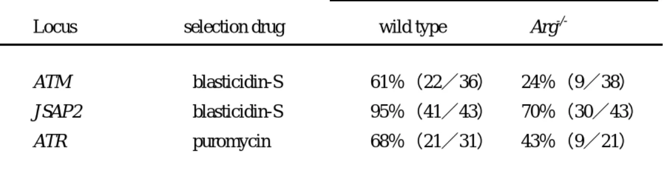
shown in Table 2 and the subsequent analysis of drug-resistant clones were done as described
previously(21). Targeting vectors for the chicken Atr and JSAP2 loci will be described elsewhere.
Results and Discussion
To study possible roles for Arg in the cellular response to IR, we generated Arg
-/-cells from a chicken B cell line (DT40) by targeted disruption. The Arg locus was disrupted by sequential transfection of the cells with two targeting vectors (Figure 1A; see Methods). The successful targeted integration was confirmed by Southern blot analysis as the appearance of mutant 8-kbp and 9-kbp KpnI genomic fragments (Figure 1B), and the disruption of the Arg gene was verified by RT-PCR analysis (Figure 1C).
Previous studies showed that c-Abl interacts with and phosphorylates Rad51 on tyrosine
in response to DNA damage. However, the biological significance of these findings in HR DNA
repair is not yet clear, as c-Abl-mediated phosphorylation negatively affected Rad51's activity in
one set of experiments, but enhanced its association with Rad52 in another(11, 12). More
recently, the BCR/ABL oncogenic tyrosine kinase has been shown both to enhance Rad51
expression and to phosphorylate it, resulting in drug resistance(22). On the other hand, we have
recently shown that Rad51 focus formation and DSB repair capacity in c-Abl
-/-DT40 cells is not
grossly impaired(19), although various DSB repair defects have been well documented in ATM
-/-DT40 cells(19-21). These results indicate that, while ATM are indispensable in DSB repair in
eukaryotic cells, there must be redundant functions for c-Abl in DSB repair, at least in chicken
DT40 cells and mouse fibroblasts(23). One possibility is that Arg substitutes for c-Abl in the
genome maintenance of the c-Abl
-/-cells. To clarify this issue, we analyzed the appearance of
Rad51 foci in Arg
-/-, c-Abl
-/-, and ATM
-/-DT40 cells following IR. Rad51 foci are subnuclear
aggregates, believed to represent intermediate structures formed during the recombination
required to repair radiation-induced or replication-associated DNA damage(24). As shown in
Figure 2, the disruption of Arg as well as of ATM led to impairment in the formation of Rad51 foci, although Rad51 focus formation was normal in c-Abl
-/-DT40 cells.
To analyze further the DSB DNA repair capacity of Arg
-/-DT40 cells, wild-type and mutant clones that were plated on methylcellulose were irradiated with 4 or 8 Gy of X-ray, and the percentage of surviving clones was determined relative to the numbers of colonies arising on untreated plates. As shown in Table 1, ATM
-/-DT40 cells were extremely X-ray sensitive, as we reported previously(19-21), and Arg
-/-DT40 cells were moderately sensitive; c-Abl
-/-DT40 cells did not exhibit hypersensitivity to X-rays, in agreement with our recent results(19). To confirm the results of this X-ray sensitivity assay, we examined chromosomal aberrations in Arg
-/-DT40 cells following IR treatment. As we reasoned and reported previously, an increase in induced chromosomal aberrations observed within 3 hours after IR (i.e., cells irradiated during the S-G
2phase of the cell cycle) would reflect a defect in the HR repair pathway, and ATM
-/-DT40 cells displayed highly increased levels of chromosomal aberrations within 3 hours of X-ray irradiation(19-21). Interestingly, disruption of Arg but not c-Abl resulted in a significant increase in chromosomal aberration frequencies (Figure 3). We further observed a slight but significant reduction in targeted integration frequencies in Arg
-/-DT40 cells (Table 2), as we reported previously for ATM
-/-DT40 cells(21). Consistent with these defects in DNA repair, enhanced IR- induced apoptosis was observed in Arg
-/-DT40 cells (data not shown), as in ATM
-/-DT40 cells(19). However, unlike in ATM
-/-DT40 cells(21), IR-induced mitotic delay was normal in Arg
-/-