MRI‑based assessment of acute effect of head‑down tilt position on intracranial hemodynamics and hydrodynamics
著者 石田 翔太
著者別表示 Ishida Shota journal or
publication title
博士論文本文Full 学位授与番号 13301甲第4908号
学位名 博士(保健学)
学位授与年月日 2019‑03‑22
URL http://hdl.handle.net/2297/00056978
doi: https://doi.org/10.1002/jmri.25781
Creative Commons : 表示 ‑ 非営利 ‑ 改変禁止 http://creativecommons.org/licenses/by‑nc‑nd/3.0/deed.ja
MRI-Based Assessment of Acute Effect of Head-Down Tilt Position on Intracranial
Hemodynamics and Hydrodynamics
Shota Ishida, MS,1,2 Tosiaki Miyati, PhD, DMSc,1* Naoki Ohno, PhD,1 Shinnosuke Hiratsuka, MS,3 Noam Alperin, PhD,4 Mitsuhito Mase, MD, PhD,5 and
Toshifumi Gabata, MD, PhD6
Purpose: To quantify the acute effect of the head-down tilt (HDT) posture on intracranial hemodynamics and hydrodynamics.
Materials and Methods: We evaluated the intracranial physiological parameters, blood flow-related parameters, and brain morphology in the HDT (–68 and –128) and the horizontal supine (HS) positions. Seven and 15 healthy subjects were scanned for each position using 3.0 T magnetic resonance imaging system. The peak-to-peak intracranial volume change, the peak-to-peak cerebrospinal fluid (CSF) pressure gradient (PGp-p), and the intracranial compliance index were calculated from the blood and CSF flow determined using a cine phase-contrast technique. The brain volumetry was conducted using SPM12. The measurements were compared using the Wilcoxon signed-rank test or a pairedt-test.
Results:No measurements changed in the –68HDT. ThePGp-pand venous outflow of the internal jugular veins (IJVs) in the –128 HDT were significantly increased compared to the HS (P < 0.001 and P 5 0.025, respectively). The cross- sectional areas of the IJVs were significantly larger (P < 0.001), and the maximum, minimum, and mean blood flow velocity of the IJVs were significantly decreased (P5 0.003,< 0.001, and 5 0.001, respectively) in the –128 HDT. The mean blood flow velocities of the internal carotid arteries were decreased (P 5 0.023). Neither position affected the brain volume.
Conclusion: Pressure gradient and venous outflow were increased in accordance with the elevation of the intracranial pressure as an acute effect of the HDT. However, the CSF was not constantly shifted from the spinal canal to the cranium.
Level of Evidence:2 Technical Efficacy:Stage 1
J. MAGN. RESON. IMAGING 2018;47:565–571.
Invasive procedures are typically used for monitoring the intracranial physiology despite the risk of complications and morbidity.1,2 Magnetic resonance imaging (MRI)-based techniques can noninvasively provide physiological informa- tion on the brain, such as cerebral blood flow (CBF).3Flow measurements using phase-contrast (PC) MRI may quantita- tively assess intracranial physiological parameters, such as the intracranial volume change (ICVC), the cerebrospinal fluid (CSF) pressure gradient (PG), and the intracranial compliance index (ICCI). Previous reports using PC cine
MRI showed that the intracranial biomechanical properties were altered in patients with idiopathic normal pressure hydrocephalus,4–6 Chiari malformation,7 and idiopathic intracranial hypertension.8
Intracranial hydrodynamics and cerebral hemodynam- ics are strongly affected by body posture due to gravitational hydrostatic pressure changes.9,10 Notably, most neuroradio- logical examinations are conducted in patients in a horizon- tal supine position (HS). However, cephalad fluid shifts are driven by a loss of the hydrostatic pressure in the head-
View this article online at wileyonlinelibrary.com. DOI: 10.1002/jmri.25781 Received Feb 14, 2017, Accepted for publication May 18, 2017.
*Address reprint requests to: T.M., Division of Health Sciences, Graduate School of Medical Sciences, Kanazawa University, 5-11-8, Kodatsuno, Kanazawa, Ishikawa, 920-0942, Japan. E-mail: [email protected]
From the1Division of Health Sciences, Graduate School of Medical Sciences, Kanazawa University, Ishikawa, Japan;2Radiological center, University of Fukui Hospital, Fukui, Japan;3Department of Radiology, Shiga University of Medical Science Hospital, Shiga, Japan;4Department of Radiology, University of Miami, Miami, Florida, USA;5Department of Neurosurgery and Restorative Neuroscience, Graduate School of Medical Sciences, Nagoya City University,
Aichi, Japan; and6Department of Radiology, Kanazawa University Hospital, Ishikawa, Japan.
down tilt position (HDT).11An HDT, in which the physio- logical responses are similar to those in microgravity envi- ronments, is exploited for the ground-based evaluation of intracranial conditions in space.11 A previous report using sonography showed that the cross-sectional area (CSA) in the right internal jugular vein (IJV) was significantly increased in the HDT position.12 Sun et al reported that CBF velocity was decreased in the HDT.13 A recent study reported that cerebral hemodynamics changed during an HDT using PC MRI.14 MRI-based measurements enabled us to noninvasively observe the posture-related changes in the intracranial physiology. Furthermore, the HDT is easily implemented inside of the bore of a general MRI scanner.
While cerebral hemodynamics, intracranial hydrodynamics, brain tissue volumes, and brain morphology are linked to one another,15these parameters have been separately studied and reported. CSF flow dynamics in the HDT have not been previously investigated. We hypothesized that a com- prehensive MRI-based assessment of intracranial physiology associated with the HDT will provide more detailed infor- mation on the biodynamical faculties of the brain.
Materials and Methods Subjects and Data Acquisition
This prospective study was approved by our Institutional Review Board. All data acquisitions and analyses were performed in healthy male volunteers with no known history of neurological dis- ease. The purpose and procedures of our investigation were fully explained to all subjects, and the study was performed only after we obtained informed consent from each volunteer.
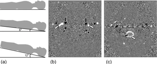
We used three different tilt angle positions, ie, HS of 08, a slight HDT (sHDT) of –68, and a moderate HDT (mHDT) of –128(Fig. 1a). The subjects were scanned using the following pulse
sequences immediately after a posture change to the sHDT or mHDT from the HS.
On a 3.0 T MRI (Signa HDxt, GE Healthcare, Milwaukee, WI), the retrospective electrocardiogram-synchronized PC cine MRI was used to obtain transcranial blood flow, CSF flow, and spinal cord displacement in each tilt angle position (seven men in the sHDT study, 23 6 1 years; 15 men in the mHDT study, 2361 years). A transverse imaging plane was set perpendicular to the flow direction at the mid C2 level. PC MRI was performed using the following parameters: repetition time (TR) 11 msec, echo time (TE) 4 msec, slice thickness 5 mm, field of view (FOV) 140 mm, matrix size 256, flip angle (FA) 208, and number of sig- nal averaged (NSA) 1. The velocity encoding (VENC) was set at 90 cm/s for blood flow and 7–10 cm/s for CSF flow. Subsequently, 3D fast spoiled gradient-echo (3D-FSPGR) was performed with a TR 6.8–7.0 msec, TE 2.5 msec, slice thickness 1 mm, FOV 230–
256 mm, matrix size 256, FA 128, and NSA 1 (seven men in the sHDT study, 236 1 years; eight men in the mHDT study, 236 1 years).
Calculation of Intracranial Physiological Parameters
We used pulsatility-based segmentation16to automatically delineate the lumen boundaries of the internal carotid arteries (ICAs), verte- bral arteries (VAs), and IJVs on velocity-mapped phase images (Fig. 1b,c). TheICVCwas calculated using Eqs. (1) and (2):
ICVCðtÞ5½QAðtÞ2QVðtÞ2QCSFðtÞDt (1) ICVCðTÞ5 X
Cardiac cycle
½QAðtÞ2QVðtÞ2QCSFðtÞDt50 (2)
whereQA(t) is the arterial volumetric flow rate,QV(t) is the venous volumetric flow rate,QCSF(t) is the CSF oscillatory flow, and Tis the time period of one cardiac cycle.
The peak-to-peak ICVC during the cardiac cycle (ICVCp-p) was calculated from Eq. (3):
FIGURE 1: Schematic diagrams of the three different tilt angle positions and velocity-mapped phase images:(a)horizontal supine position (top), slight head-down tilt position (middle), and moderate head-down tilt position (bottom),(b)velocity-mapped phase image with a VENC of 90 cm/s for blood flow, and(c) VENC of 7 to 10 cm/s for CSF flow measurements. (b) ICA 5 internal carotid artery (black arrow), VA5vertebral artery (black arrowhead), IJV5internal jugular vein (white arrow); and (c) CSF (white arrow), cord (black arrow).
Journal of Magnetic Resonance Imaging
566 Volume 47, No. 2
ICVCp2p5ICVCmax2ICVCmin (3)
where theICVCmaxandICVCminare the maximum and minimum ICVCduring the cardiac cycle, respectively.
Next, we assumed that CSF is a Newtonian fluid, and calcu- lated the craniospinal CSFPGduring the cardiac cycle from a sim- plified Navier-Stokes equation17:
PG52q @V
@t 1V rV
1l r2V (4)
whereqis the fluid density (1.0007 g/cm3),lis the fluid viscosity (1.1 cP), and V is the velocity vector. To correct for the loss of pressure derived from the difference in cross-sectional flow area, PG was normalized by multiplying by the CSF flow area. The peak-to-peakPG(PGp-p) was calculated from Eq. (5):
PGp2p5PGmax2PGmin (5)
where thePGmaxand PGminare the maximum and minimumPG during the cardiac cycle, respectively.
Finally, theICCIwas calculated from Eq. (6):
ICCI5ICVCp2p=PGp2p (6)
In addition to these parameters, we also evaluated the CSA and blood flow velocity of the aforementioned vessels.
All statistical analyses were performed using SPSS for Win- dows, v. 23.0 (Chicago, IL). The Wilcoxon signed-rank test was used for statistical comparisons in the sHDT study. In the mHDT study, the intracranial physiological and blood flow parameters were assessed using a pairedt-test. AP-value < 0.05 was defined as statistically significant.
Data Analysis of Voxel-Based Morphometry
We used SPM12 software for voxel based morphometry (VBM) analysis.18 First, a bias correction was computed for the 3D- FSPGR images. The corrected images were segmented into gray matter (GM), white matter (WM), and CSF. A study-specific tem- plate was created using the Diffeomorphic Anatomical Registration using Exponentiated Lie Algebra algorithm.19 The images were transformed to the Montreal Neurological Institute space, and smoothing was performed using an 8 mm full-width at half- maximum (FWHM) Gaussian kernel. The spatially normalized images were modulated by multiplying the relative volumes derived from the Jacobian determinant. After modulation, the GM, WM, and CSF volumes were computed. We calculated the total intracra- nial volume (TIV) by summing each brain compartment volume.
Whole-tissue volumes of each brain compartment were compared by the Wilcoxon signed-rank test. Region-wise volumetric compari- sons between the groups were statistically performed using a paired t-test.P<0.05 was considered statistically significant.
Results
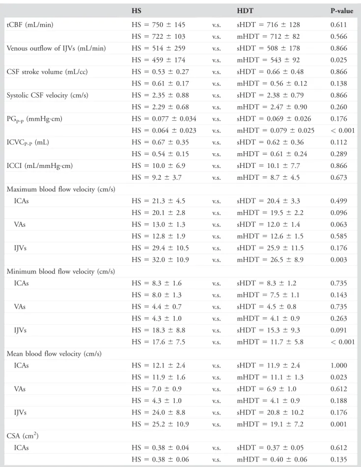
None of the intracranial physiological and blood flow parameters showed significant differences between the sHDT and the HS (n 5 7; Table 1). All nonsignificant P- values were as follows: P5 0.611 for tCBF; P5 0.866 for
venous outflow of IJVs; P 5 0.866 for CSF stroke volume;
P 5 0.866 for systolic CSF velocity; P 5 0.176 for PGp-p; P 5 0.112 for ICVCp-p;P 5 0.866 for ICCI; P 5 0.499, 5 0.063,5 0.176 for maximum blood flow velocity of the ICAs, VAs, and IJVs, respectively; P 5 0.735,5 0.735, 5 0.091 for minimum blood flow velocity of the ICAs, VAs, and IJVs, respectively; P 5 1.000, 5 0.612, 5 0.176 for mean blood flow velocity of the ICAs, VAs, and IJVs, respectively; and P 5 0.612, 5 0.176, 5 0.128 for the CSA of the ICAs, VAs, and IJVs, respectively.
In contrast, the PGp-p in the mHDT (7.9 6 2.5 3 1022 mmHgcm) was significantly higher when compared to the HS (6.4 6 2.3 3 1022 mmHgcm; P < 0.001).
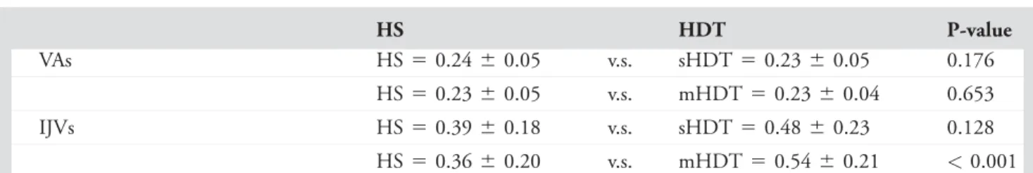
The venous outflow of the IJVs was significantly increased in the mHDT (543 6 92 mL/min) compared with the HS (459 6 174 mL/min; P 5 0.025). The mean blood flow velocity of the ICAs in the mHDT (11.1 6 1.3 cm/s) was significantly decreased when compared to the HS (11.9 6 1.6 cm/s; P 5 0.023). The maximum, minimum, and mean blood flow velocity of the IJVs were significantly decreased in the mHDT (P 5 0.003, < 0.001, 5 0.001, respectively), whereas the CSA of the IJVs was significantly larger in the mHDT (0.54 6 0.21 cm2) compared to the HS (0.36 6 0.20 cm2; P < 0.001). None of the other parameters significantly varied between the mHDT and the HS (n 5 15; Table 1). All nonsignificant P-values were as follows: P 5 0.566 for tCBF; P 5 0.138 for CSF stroke volume;P50.260 for systolic CSF velocity; P50.289 for ICVCp-p; P 5 0.673 for ICCI; P 5 0.096, 5 0.585 for maximum blood flow velocity of the ICAs and VAs, respec- tively;P 50.143,5 0.263 for minimum blood flow veloc- ity of the ICAs and VAs, respectively; P 5 0.188 for mean blood flow velocity of the VAs;P 50.135,50.653 for the CSA of the ICAs and VAs, respectively.
For both datasets, there were no significant differences in any of the whole-tissue volumes of each brain compart- ment or TIV (Table 2). P-values for the sHDT study (n5 7) were as follows:P 5 0.735 for GM volume, P 5 0.612 for WM volume, P 5 0.499 for CSF volume, and P 5 0.499 for TIV. The results of the statistical tests for the mHDT (n5 8) study were as follows:P 5 0.889 for GM volume, P 5 0.889 for WM volume, P 5 0.674 for CSF volume, andP5 0.674 for TIV.
There were no statistical differences between the region-wise measured tissue volumes for both datasets.
Discussion
None of the intracranial physiological or blood flow param- eters were significantly changed in the sHDT condition. We assessed the intracranial physiological parameters immedi- ately after a position change. The loading induced by lower- ing the head did not exceed the intracranial compensatory faculties, due to the low tilt angle and extremely short
TABLE 1. Intracranial Physiological and Blood Flow Parameters Obtained in the Horizontal Supine Position (HS) and the Head-Down Tilt (HDT) Positions
HS HDT P-value
tCBF (mL/min) HS5 7506 145 v.s. sHDT 57166128 0.611
HS5 7226 103 v.s. mHDT 5712682 0.566
Venous outflow of IJVs (mL/min) HS5 5146 259 v.s. sHDT 55086178 0.866
HS5 4596 174 v.s. mHDT 5543692 0.025
CSF stroke volume (mL/cc) HS5 0.5360.27 v.s. sHDT 50.666 0.48 0.866 HS5 0.6160.17 v.s. mHDT 50.566 0.12 0.138 Systolic CSF velocity (cm/s) HS5 2.3560.88 v.s. sHDT 52.386 0.79 0.866 HS5 2.2960.68 v.s. mHDT 52.476 0.90 0.260
PGp-p(mmHgcm) HS5 0.0776 0.034 v.s. sHDT 50.06960.026 0.176
HS5 0.0646 0.023 v.s. mHDT 50.07960.025 <0.001
ICVCP-P(mL) HS5 0.6760.35 v.s. sHDT 50.626 0.36 0.112
HS5 0.5460.15 v.s. mHDT 50.616 0.24 0.289
ICCI (mL/mmHgcm) HS5 10.066.9 v.s. sHDT 510.16 7.7 0.866
HS5 9.26 3.7 v.s. mHDT 58.764.5 0.673
Maximum blood flow velocity (cm/s)
ICAs HS5 21.364.5 v.s. sHDT 520.46 3.3 0.499
HS5 20.162.8 v.s. mHDT 519.56 2.2 0.096
VAs HS5 13.061.3 v.s. sHDT 512.06 1.4 0.063
HS5 12.861.9 v.s. mHDT 512.66 1.5 0.585
IJVs HS5 29.4610.5 v.s. sHDT 525.96 11.5 0.176
HS5 32.0610.9 v.s. mHDT 526.56 8.9 0.003 Minimum blood flow velocity (cm/s)
ICAs HS5 8.36 1.6 v.s. sHDT 58.361.2 0.735
HS5 8.06 1.3 v.s. mHDT 57.561.1 0.143
VAs HS5 4.46 0.7 v.s. sHDT 54.560.8 0.735
HS5 4.36 1.0 v.s. mHDT 54.160.9 0.263
IJVs HS5 18.368.8 v.s. sHDT 515.36 9.3 0.091
HS5 17.667.5 v.s. mHDT 511.76 5.8 <0.001 Mean blood flow velocity (cm/s)
ICAs HS5 12.162.4 v.s. sHDT 511.96 2.4 1.000
HS5 11.961.6 v.s. mHDT 511.16 1.3 0.023
VAs HS5 7.06 0.9 v.s. sHDT 56.961.0 0.612
HS5 4.36 1.0 v.s. mHDT 54.160.9 0.188
IJVs HS5 24.068.8 v.s. sHDT 520.86 10.2 0.176
HS5 25.2610.9 v.s. mHDT 519.16 7.2 0.001 CSA (cm2)
ICAs HS5 0.3860.04 v.s. sHDT 50.376 0.05 0.612
HS5 0.3860.06 v.s. mHDT 50.406 0.06 0.135
Journal of Magnetic Resonance Imaging
568 Volume 47, No. 2
duration exposure. In contrast to our results, a recent report documented that the CBF and the venous outflow of the IJVs were decreased after 4.5 hours in a –68 HDT.14 According to these results, the duration of the posture may be a profound factor when investigating the effects of a low- angle HDT on the intracranial condition.
The PGp-p in the mHDT was significantly larger than that in the HS, as lowering the head altered the gravitational hydrostatic pressure gradient. We deduced that the increase in PG in the mHDT indicated an elevation of intracranial pressure in accordance with the law of fluid mechanics and not derived from a cephalad fluid shift.20,21 The PG is the most sensitive to postural changes of the intracranial physio- logical parameters. Therefore, the CSF dynamics associated with posture is the most important factor to evaluate the acute effects of a HDT. Our brain volume measurement of each tissue compartment results also support this inference, since the whole-tissue volumes of each brain compartment and the TIV were not significantly different between the positions. Due to the stiffness and lack of distensibility of dura mater and the fixation to the skull, the intradural vol- ume was unchangeable.21 However, a temporal change (in a unit of a second or millisecond) of the intracranial volume
was observed due to the pulsatile arterial flow into the cra- nium during the cardiac cycle.22 The intracranial volume was transiently altered during the cardiac cycle, indicating some possibility that a cephalad fluid shift is caused by the larger tilt angles and/or long-term exposure to the HDT.
Further investigations should explore these effects on intra- cranial conditions.
Venous outflow of the IJVs was significantly increased in the HDT. The IJVs are the main pathway for venous outflow in the supine position23; however, the IJVs also play a predominant role in cerebrovenous drainage immediately after a change in posture to the mHDT. The total venous outflow may have remained unchanged, since the tCBF, CSF stroke volume, andICVC were not significantly differ- ent. Therefore, a significant increase in the venous outflow of the IJVs represents a shift of the venous pathway in response to a postural change. Furthermore, a significant increase in the CSA in the IJVs demonstrated the compliant nature of the venous system. The increase in PG in the HDT was regulated by dilation of the IJVs, which are more compliant than secondary venous pathways (eg, vertebral, epidural, and deep cervical veins). This vasodilation induced a decrease in venous velocity via the Venturi effect. Thus, TABLE 1: Continued
HS HDT P-value
VAs HS5 0.2460.05 v.s. sHDT 50.236 0.05 0.176
HS5 0.2360.05 v.s. mHDT 50.236 0.04 0.653
IJVs HS5 0.3960.18 v.s. sHDT 50.486 0.23 0.128
HS5 0.3660.20 v.s. mHDT 50.546 0.21 <0.001 Mean6standard deviation are shown for each value, tCBF5total cerebral blood flow, IJV5internal jugular vein, CSF5cerebro- spinal fluid, PG5pressure gradient, ICVC5intracranial volume change, ICCI5intracranial compliance index, ICAs5internal carotid arteries, VAs5vertebral arteries, IJVs5internal jugular veins, CSA5cross-sectional area.
TABLE 2. Tissue Volume of Each Brain Compartment and the Total Intracranial Volume (TIV) in the Horizontal Supine Position (HS) and the Head-Down Tilt (HDT) Positions
HS HDT P-value
GM (mL) HS5 8026 55 v.s. sHDT 5803658 0.735
HS5 7926 38 v.s. mHDT5792640 0.889
WM (mL) HS5 4886 46 v.s. sHDT 5487643 0.612
HS5 4846 34 v.s. mHDT5485636 0.889
CSF (mL) HS5 3196 54 v.s. sHDT 5321658 0.499
HS5 3116 40 v.s. mHDT5310640 0.674
TIV (mL) HS5 16096130 v.s. sHDT 516116 130 0.499
HS5 1588671 v.s. mHDT515886 72 0.674
Mean6standard deviation are shown for each value, GM5gray matter, WM5white matter, CSF5cerebrospinal fluid, TIV5 total intracranial volume.
the venous system played a major role in the compensation for the increase inPGimmediately after a change in posture to the HDT, ie, the sudden change in intracranial condi- tion. In contrast to our results, a previous study showed that venous outflow of the IJVs was decreased after 4.5 hours in the –128 HDT.14 This discrepancy indicates the importance of a time-dependency effect of the HDT.
In agreement with the previous study,14 the mean blood flow velocity in the mHDT was significantly decreased when compared to the HS. There are many reports on the CBF and blood flow velocity in the HDT;
however, the results of posture-related blood flow analyses are inconsistent. It is difficult to interpret these previous reports due to diverse bias factors, eg, measured vessel loca- tion, procedure, tilt angle, and duration in the HDT.13,24–26 On the basis of our results, we speculate that the ICAs have a higher reactivity to posture changes than the VAs. Despite the significant increase in mean blood flow velocity of the ICAs, the tCBF was not significantly affected.
Our brain morphological analyses using SPM12 showed no differences between the positions. The VBM analyses revealed that brain morphology was not signifi- cantly affected by the postural changes. A previous study reported that brain morphology was altered after 3 hours in parabolic flight,27 indicating that larger loading may affect brain morphology. However, the increase in PG associated with a brief exposure to the HDT was not enough to alter brain morphology.
Our study has several limitations. First, the study pop- ulation was small, and all participants were adult males in a narrow age range. A larger study population would enable us to demonstrate more rigorous statistical analyses. Intra- cranial physiology shows sexual specificity and age depen- dence28; therefore, further evaluations are needed with a larger sample size, female subjects, and a wider age range.
Second, the subjects in each study group were not exactly the same, biasing the observation of the present study. How- ever, we believe that this problem in the study design had a small effect on the measurements, since the majority of each group was similar. Future studies should be performed on a single group of participants. We only evaluated the intracra- nial condition after a brief exposure to the HDT, ie, an acute effect. Hence, time-dependency and long-term expo- sure to the HDT, ie, a chronic effect, should be assessed.
Furthermore, separation of the acute and chronic effects associated with the HDT is indispensable to obtain more detailed information on the intracranial physiology related to body posture. We used two tilt angles for the HDT.
Although none of the measurements were significantly changed in the sHDT, several parameters were statistically significant in the mHDT. This indicates that the intracranial physiology associated with the HDT is dependent on the
tilt angle. It is necessary to identify a threshold tilt angle for physiological effects.
In this study we evaluated the intracranial physiologi- cal parameters, blood flow, brain tissue volume, and brain morphology in the HDT using two tilt angles. In conclu- sion, as an acute effect of the HDT, the PG and the venous outflow were increased in accordance with the elevation of intracranial pressure. However, the CSF was not constantly shifted from the spinal canal to the cranium.
Acknowledgment
This work was supported by JSPS KAKENHI Grant Num- ber JP16K09013.
References
1. Marmarou A, Shulman K, LaMorgese J. Compartmental analysis of compliance and outflow resistance of the cerebrospinal fluid system.
J Neurosurg 1975;43:523–534.
2. Sklar FH, Elashvili I. The pressure-volume function of brain elasticity.
Physiological considerations and clinical applications. J Neurosurg 1977;47:670–679.
3. Alsop DC, Detre JA, Golay X, et al. Recommended implementation of arterial spin-labeled perfusion MRI for clinical applications: A consen- sus of the ISMRM perfusion study group and the European consor- tium for ASL in dementia. Magn Reson Med 2015;73:102–116.
4. Miyati T, Mase M, Banno T, et al. Frequency analyses of CSF flow on cine MRI in normal pressure hydrocephalus. Eur Radiol 2003;13:1019–1024.
5. Miyati T, Mase M, Kasai H, et al. Noninvasive MRI assessment of intra- cranial compliance in idiopathic normal pressure hydrocephalus.
J Magn Reson Imaging 2007;26:274–278.
6. Ohno N, Miyati T, Mase M, et al. Idiopathic normal-pressure hydro- cephalus: temporal changes in ADC during cardiac cycle. Radiology 2011;261:560–565.
7. Alperin N, Sivaramakrishnan A, Lichtor T. Magnetic resonance imaging-based measurements of cerebrospinal fluid and blood flow as indicators of intracranial compliance in patients with Chiari malfor- mation. J Neurosurg 2005;103:46–52.
8. Tain RW, Bagci AM, Lam BL, Sklar EM, Ertl-Wagner B, Alperin N.
Determination of cranio-spinal canal compliance distribution by MRI:
Methodology and early application in idiopathic intracranial hyperten- sion. J Magn Reson Imaging 2011;34:1397–1404.
9. Alperin N, Lee SH, Sivaramakrishnan A, Hushek SG. Quantifying the effect of posture on intracranial physiology in humans by MRI flow studies. J Magn Reson Imaging 2005;22:591–596.
10. Alperin N, Lee SH, Bagci AM. MRI measurements of intracranial pres- sure in the upright posture: The effect of the hydrostatic pressure gra- dient. J Magn Reson Imaging 2015;42:1158–1163.
11. Nelson ES, Mulugeta L, Myers JG. Microgravity-induced fluid shift and ophthalmic changes. Life (Basel) 2014;4:621–665.
12. Schreiber SJ, Lambert UK, Doepp F, Valdueza JM. Effects of pro- longed head-down tilt on internal jugular vein cross-sectional area. Br J Anaesth 2002;89:769–771.
13. Sun XQ, Yao YJ, Yang CB, Jiang SZ, Jiang CL, Liang WB. Effect of lower-body negative pressure on cerebral blood flow velocity during 21 days head-down tilt bed rest. Med Sci Monit 2005;11:CR1–5.
14. Marshall-Goebel K, Ambarki K, Eklund A, et al. Effects of short-term exposure to head-down tilt on cerebral hemodynamics: A prospective evaluation of a spaceflight analog using phase-contrast MRI. J Appl Physiol (1985) 2016;120:1466–1473.
Journal of Magnetic Resonance Imaging
570 Volume 47, No. 2
15. Ohno N, Miyati T, Chigusa T, et al. Technical note: Development of a cranial phantom for assessing perfusion, diffusion, and biomechanics.
Med Phys 2017;44:1646–1654.
16. Alperin N, Lee SH. PUBS: pulsatility-based segmentation of lumens conducting non-steady flow. Magn Reaon Med 2003;49:934–944.
17. Urchuk SN, Plewes DB. MR measurements of pulsatile pressure gra- dients. J Magn Reson Imaging 1994;4:829–836.
18. Ashburner J, Friston KJ, Voxel-based morphometry—the methods.
Neuroimage 2000;11:805–821.
19. Ashburner J. A fast diffeomorphic image registration algorithm. Neu- roimage 2007;38:95–113.
20. Petersen LG, Petersen JC, Andresen M, Secher NH, Juhler M. Postural influence on intracranial and cerebral perfusion pressure in ambulatory neurosurgical patients. Am J Physiol Regul Integr Comp Physiol 2016;
310:R100–R104.
21. Klarica M, Rodos M, Erceg G, Petosic´ A, Jurjevic´ I, Oreskovic´ D. The influence of body position on cerebrospinal fluid pressure gradient and movement in cats with normal and impaired craniospinal commu- nication. PLoS One 2014;9:e95229.
22. Alperin NJ, Lee SH, Loth F, Raksin PB, Lichtor T. MR-Intracranial pres- sure (ICP): a method to measure intracranial elastance and pressure
noninvasively by means of MR imaging: baboon and human study.
Radiology 2000;217:877–885.
23. Valdueza JM, von M€unster T, Hoffman O, Schreiber S, Einh€aupl KM. Postural dependency of the cerebral venous outflow. Lancet 2000;355:200–201.
24. Kawai Y, Murthy G, Watenpaugh DE, Breit GA, Deroshia CW, Hargens AR. Cerebral blood flow velocity in humans exposed to 24 h of head-down tilt. J Appl Physiol (1985) 1993;74:3046–
3051.
25. Stolz E, Fox BC, Hoffmann O, et al. Cranial venous outflow under lower body positive and negative pressure conditions and head-up and -down tilts. J Neuroimaging 2009;19:31–36.
26. Pavilla A, Arrigo A, Colombani S, Mejdoubi M. Absolute and regional cerebral perfusion assessment feasibility in head-down position with arterial spin-labeling magnetic resonance. A preliminary report on healthy subjects. J Neuroradiol 2016;43:392–397.
27. Van Ombergen A, Jeurissen B, Vanhevel F, et al. A first insight in regional brain changes after parabolic flight: a voxel-based morphom- etry study. In: Proc 23rd Annual Meeting ISMRM, Toronto; 2015 (abstract 1258).
28. Schmid Daners M, Knobloch V, Soellinger M, et al. Age-specific char- acteristics and coupling of cerebral arterial inflow and cerebrospinal fluid dynamics. PLoS One 2012;7:e37502.