細則様式第1-2号
学位請求論文の内容の要旨
領 域 医療生命科学 分 野 病体解析科学
氏 名 阿部 悠
(論文題目)
Analysis of the CT scan-induced human chromosome aberration CT
スキャンにより誘発されるヒト染色体異常の解析 主 査 吉 田 光 明
副 査 中 村 敏 也 副 査 井 瀧 千 恵 子
副 査 三 浦 富 智
Background: Computed tomography (CT) is a very useful medical diagnostic equipment, and Japan has one of the highest number of CTs in the world. Recent studies have reported concerns regarding the large risk of cancer after a CT scan, especially in the case of children and young adults. Low-dose radiation exposure associated with CT scans may induce chromosome aberrations, which can cause diseases even at doses less than 100 mSv. However, these studies were epidemiological investigations that did not actually observe any effects on the human body. Therefore, we performed dicentric chromosome assays (DCAs) and translocation analyses before and after a CT scan to assess the effects of low-dose ionizing radiation on the chromosomes.
Methods: The subjects were 12 patients (3 males and 9 females) between 62-83 years old (mean age: 71 years). Data regarding their past history of disease and treatments, CT scans, smoking status, etc. were collected. Peripheral blood lymphocytes were collected before and after a CT scan. In order to obtain metaphases in their first division, we performed a colcemid treatment when they were first put in culture. Other chromosomal preparations were carried out according to the International Atomic Energy Agency (IAEA) standard protocol manual.
DCAs were performed by analyzing up to 2,000 metaphases using Giemsa staining and centromere-fluorescence in situ hybridization (centromere-FISH). In addition, translocation analyses were performed by analyzing 2,000 metaphase equivalents, which were obtained per the instructions from the IAEA manual. We also examined for differences in analysis
(注)論文題目が外国語の場合は,和訳を付すこと。
【細則様式第1-2号続き】
efficiency between the Giemsa staining and centromere-FISH. The estimated dose was calculated using the WAZA-ARI software.
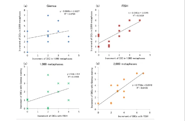
Findings: Dicentric chromosome (Dic) formation increased significantly after a CT scan, and increased Dic formation was found in all patients (Fig 1). The number of Dics formed tended to be higher, but not significantly, in patients with a treatment history compared to those without one as determined using both methods. A correlation was observed between the increment of Dic formation after a CT scan as determined by an analysis of 1,000 metaphase-staged cells using Giemsa staining and 2,000 m e t a p h a s e - s t a g e d c e l l s u s i n g C e n t r o m e r e - F I S H ( F i g . 2 a & 2 b ) . Furthermore, we found a correlation between the increment of Dic formation as determined by an analysis of 2,000 metaphase-staged cells using Giemsa staining and 2,0 00 metaphase spreads using Centromere-FISH
Figure 1. Comparison of the number of DICs formed before and after CT scan (Centromere-FISH).
Figure 2. Relationship between analysis method and observe metaphase number.
【細則様式第1-2号続き】
(Fig. 2c & 2d). However, no correlations were observed between the increment of Dic formation and the effective radiation dose calculated using WAZA-ARI.
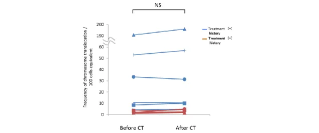
We observe a trend with a steep slope on a graph when CT scan part is wide. But, estimated dose is not consistent with the CT scan parts. Furthermore, the frequency of chromosome translocations was higher than the number of Dics formed before and after a CT scan as determined by a chromosome translocation analysis. However, no significant differences in the frequency of chromosome translocations were detected before and after the CT scan examination (Fig. 3).
Furthermore, though the frequency of translocations in the patients with a treatment history was generally higher than those without a treatment history, no significant differences between the two groups were observed.
Interpretation: Our results suggest that chromosomal cleavage may be induced by a single CT scan. However, we did not observe any significant increases in chromosome translocations.
Therefore, we suggest that the contribution of radiation exposure from CT scans to carcinogenesis is small. However, the CT examination range is considered to be one factor that affects the induction of chromosomal abnormalities. Therefore, we suggest that the CT examination range be reduced as much as possible. These results suggest that DCA is useful for evaluating radiation exposure of less than 100 mSv such as that from a CT scan. In this case, it is also necessary to analyze 2,000 or more metaphases by Giemsa staining or centromere-FISH for DCAs.
Figure 3. Comparison of the number of translocations formed before and after CT scan.
【細則様式第1-2号続き】
学位論文のもととなる研究成果としての筆頭著者原著
論 文 題 目
Increase in dicentric chromosome formation after a single CT scan in adults著 者 名
Yu Abe, Tomisato Miura, Mitsuaki A. Yoshida, Risa Ujiie, Yumiko Kurosu, Nagisa Kato, Atsushi Katafuchi, Naohiro Tsuyama, Takashi Ohba, Tomoko Inamasu, Fumio Shishido, Hideyoshi Noji, Kazuei Ogawa, Hiroshi Yokouchi, Kenya Kanazawa, Takashi Ishida, Satoshi Muto, Jun Ohsugi, Hiroyuki Suzuki, Tetsuo Ishikawa, Kenji Kamiya & Akira Sakai
掲載学術誌名
Scientific Reports巻,号,項
5, 13882掲載年月日
2015年
9月
論 文 題 目
Analysis of chromosome translocation frequency after a single CT scan in adults著 者 名
Yu Abe, Tomisato Miura, Mitsuaki A. Yoshida, Risa Ujiie, Yumiko Kurosu, Nagisa Kato, Atsushi Katafuchi, Naohiro Tsuyama, Fumihiko Kawamura, Takashi Ohba, Tomoko Inamasu, Fumio Shishido, Hideyoshi Noji, Kazuei Ogawa, Hiroshi Yokouchi, Kenya Kanazawa, Takashi Ishida, Satoshi Muto, Jun Ohsugi, Hiroyuki Suzuki, Tetsuo Ishikawa, Kenji Kamiya & Akira Sakai
掲載学術誌名
Journal of Radiation Research巻,号,項
57, 3, 220-226掲載年月日
2016年
2月