ORIGINAL ARTICLE
EFFECT OF COUPLING FACTOR 6 ON CHEMOKINE RECEPTORS IN VASCULAR ENDOTHELIAL CELLS
Akiko Suzuki1),Tomohiro Osanai1,2),Makoto Tanaka2),Tomohide Endo1), Kazuo Murakami1),Hirofumi Tomita1),and Ken Okumura1,2)
Abstract Objectives: Vascular endothelial cells are exposed to an acidic pH, but its influence on chemokine receptors expressed in the vascular endothelial cells is unclear. We investigated the role of coupling factor 6 (CF6), a novel stimulator of proton importer, in the regulation of chemokine receptors in the vascular endothelial cells.
Methods and Results: In microarray analysis, there were the increased expression of CC chemokine receptor 9 (CCR9)
and CX3C chemokine receptor 1 (CX3CR1) and the decreased expression of CXC chemokine receptor 4 (CXCR4) in the human umbilical vascular endothelial cells (HUVEC) that were exposed to CF6. The ratio of CXCR4 to GAPDH mRNA was decreased in HUVEC that were exposed to either CF6 at 10-7M or hypoxia to a similar degree. Apoptotic cells, measured by annexin-V propidium iodide kit, were increased in HUVEC that were exposed to CF6 for 24 hours in normoxia.
Conclusions: CF6 influences the expression of chemokine receptors and induces apoptosis in the vascular endothelial cells.
Hirosaki Med.J. 65:119―127,2014 Key words: coupling factor 6; chemokine receptor; vascular endothelial cells; apoptosis.
原 著
Coupling factor 6 の血管内皮細胞におけるケモカイン受容体発現に 及ぼす影響
鈴 木 晃 子1) 長 内 智 宏1,2) 田 中 真 実2) 遠 藤 知 秀1)
村 上 和 男1) 富 田 泰 史1) 奥 村 謙1,2)
抄録 背景と目的:血管内皮細胞は常に虚血関連酸性状態に暴露されるが,それらのケモカイン受容体に及ぼす影響は 不明である.本研究では細胞内酸性化物質であるcoupling factor 6 (CF6) のケモカイン受容体調節における役割につい て検討した.
方法と結果:ヒト臍帯静脈内皮細胞に10-7M CF6を添加し,24時間後にマイクロアレイ法でケモカイン受容体の遺伝 子発現を検討した.CC chemokine receptor 9とCX3C chemokine receptor 1は増加し,CXC chemokine receptor 4
(CXCR4)は減少した.CXCR4のmRNAおよび蛋白発現の減少を,リアルタイムPCRとWestern Blot法で確認した.ヒ ト臍帯静脈内皮細胞にCF6を添加するとapoptosisが惹起された.
結論:CF6は血管内皮細胞のケモカイン受容体発現に影響し,apoptosisを誘導する可能性が示唆された.
弘前医学 65:119―127,2014 キーワード:カップリングファクター6;ケモカイン受容体;血管内皮細胞;アポトーシス.
1) Department of Cardiology, Respiratory Medicine and Nephrology, Hirosaki University Graduate School of Medicine, Hirosaki, Japan
2) Department of Hypertension and Stroke Medicine, Hirosaki University Graduate School of Medicine, Hirosaki, Japan
Correspondence: T. Osanai
Received for publication, December 16, 2013 Accepted for publication, December 25, 2013
1) 弘前大学大学院医学研究科循環呼吸腎臓内科学講座
2) 弘前大学大学院医学研究科高血圧・脳卒中内科学講座
別刷請求先:長内智宏 平成25年12月16日受付 平成25年12月25日受理
receptor expression and cellular function in the cultured vascular endothelial cells. We report here that many kinds of chemokine receptors are expressed in the vascular endothelial cells, and that CXCR4 expressed most in the vascular endothelial cells is attenuated by CF6 and associated with cell survival.
Materials and Methods
Materials
QIA shredder and RNeasy Protect Mini Kit were from QIAGEN, Valencia, CA, USA. Human CXCR4 and glyceraldehydes 3-phosphate dehydrogenase (GAPDH) primers and TaqMan probe, and TaqMan reverse transcription (RT)
reagent and TaqMan universal polymerase chain reaction (PCR) master mix were from Applied Biosystems, Foster City, CA, USA.
HuMedia-EG2 kit was purchased from Kurabo Co., Ltd., Osaka, Japan. Aneropack and hypoxic chamber were obtained from Mitsubishi Gas Chemistry, Tokyo, Japan. Annexin V-FITC Apoptosis Detection Kit was from Abcam, Cambridge, MA, USA. ApopMark™ Apoptosis Detection Kit was from Exalpha Biologicals, Inc., Maynard, MA, USA. Human CF6 was from Phoenix Pharmaceuticals, Inc., Belmont, CA, USA. RIPA lysis buffer and antibodies for human CXCR4 and GAPDH were from Santa Cruz Biotechnology, Santa Cruz, California, USA.
Polyvinilidene difluoride membrane was from Bio-Rad Laboratories, Hercules, California, USA.
Enhanced chemiluminescence plus detection systems were from Amersham Pharmacia Biotech, Piseataway, New Jersey, USA. Amino Allyl MessageAmpTM aRNA kit was from Life Technologies (Ambion, TX, USA). All other reagents were of the finest grade available from Sigma Chemical Co., St. Louis, Missouri, USA.
Cell culture
Human umbilical vein endothelial cells
(HUVEC) were cultured in HuMedia-EG2
Introduction
Chemokine plays an important role in the onset of a process known as chemotaxis that traffics the cell to a desired location within the organism1). At least nineteen chemokine receptors are expressed in many kinds of cell, and are divided into different families, CXC chemokine receptors, CC chemokine receptors, CX3C chemokine receptors and XC chemokine receptors that correspond to the 4 distinct subfamilies of chemokines they bind2). Recently, it was shown that some kinds of chemokine receptors are expressed in the vascular endothelial cells as well as hematopoietic and epithelial cells3). However, the role of these receptors in cell function remains unclear.
Vascular endothelial cells are exposed to an acidic pH in a variety of pathological and physiological conditions including ischemia and/or hypoxia, and shear stress4), and CXC chemokine receptor 4 (CXCR4) was recently reported as a key molecule in response to acidosis5). Coupling factor 6 (CF6) is a novel activator for a proton importer, and elicits sustained decrease in intracellular pH by means of a different manner of hypoxia6). CF6 activates the plasma membrane ATP synthase
(F1Fo complex); the molecular rotary motor F1- ATPase forcefully hydrolyzes ATP in a reverse mode of mitochondria, and inversely rotates Fo
motor against the original clockwise direction, resulting in proton import6). CF6 suppresses prostacyclin generation via inhibition of cytosolic phospholipase A27) and nitric oxide (NO)
generation via upregulation of asymmetric dimethylarginine, an endogenous competitive inhibitor of NO synthase8). Prostacyclin and NO are recognized as the major mediator of the maintenance of vascular homeostasis, and the decrease in these compounds is implicated in endothelial dysfunction. In the present study, we examined the effect of CF6 on chemokine
(complete media) at 37℃ under 5% CO2. HUVEC from the second to sixth passages were used for the study. After treatment with various molecules, the viability of the cells, which was determined by trypan blue exclusion, was generally >95%.
RNA Isolation
The cells were rinsed quickly in ice-cold phosphate buffered saline (PBS) and RNA was isolated by using RNeasy® Mini Kit (Qiagen)
according to the manufacturer's instructions.
RNA quality was insured by spectrophotometric analysis (OD260/280). The RNA was quantitated by spectrophotometric analysis at 260 nm. Linear amplification of mRNA from total RNA was obtained using the Amino Allyl MessageAmpTM aRNA kit (Ambion, TX, USA) with two consecutive amplification steps according to the manufacturer’s recommendations. Two replicates of each experiment were carried out using different microarray slides where the RNA samples from two different sources were labelled with either Cy3- or Cy5-conjugated deoxyribonucleotides (Amersham Biosciences, Germany). The fluorescent dye on probes derived from the experimental aRNA was Cy5, while the dye on control probes was Cy3.
cDNA microarrays
We used a commercially available cDNA microarray, the AceGene-Mouse Oligo Chip 30K 1 Chip Version and the AceGene-Human Oligo Chip 30K 1 Chip Version (Hitachi Software Engineering Co.,Ltd. Kanagawa, Japan), which contained 30,000 cDNA named human genes, to identify genes altered in primary HUVEC exposed to CF6 at 10-7M for 24 hours. Labeled probes were mixed with a hybridization solution (5×sodium chloride and sodium citrate
(SSC), 0.5% sodium dodecyl sulfate (SDS), 4×
Denhardt’s solution, 20% hybridization solution, 0.1 mg/ml denatured salmon sperm DNA and 10% Formamide). After hybridization for 14 h at 46 ℃, the slides were washed in 5×SSC and
0.1% SDS for 2 min at room temperature, 5×
SSC and 0.1% SDS for 10 min at 30 ℃, 0.5×
SSC for 2 min at room temperature. Slides were scanned for Cy3 and Cy5 fluorescence with a 428 ARRAY scanner (AFFYMETRIX), and the fluorescence was quantified with DNASIS Array software version 2.6 (Hitachi Software Engineering Co., Ltd. Kanagawa, Japan). The current analysis used intensity dependent Global Normalization (Mean: 10,000). Intensity dependent normalization is just one technique used to eliminate dye-related artifacts in two- color experiments such as this. The results for each gene were reported as an average obtained from 3 slides. The data are reported as the normalized ratio of Cy5 (for CF6 +) to Cy3 (for CF6 -). We used intensity of 2,000 or above.
Determination of gene expression
Total RNA was extracted from the cells using the QIAamp RNA Kit. A two-step RT- PCR was carried out according to the protocol supplied with the TaqMan Gold RT-PCR kit.
The standard curves of CXCR4 and GAPDH were linear between 0.1 and 250 ng/μl total RNA. Values were averaged from duplicate data and normalized with the human GAPDH.
Determination of protein expression
Cell samples were homogenized in RIPA lysis buffer (20 mmol/l Tris-HCl pH 7.5, 150 mmol/l NaCl, 1 mmol/l EDTA, 1 mmol/l EGTA, 1%
Triton X-100, 1% glycerol, 1 mmol/l dithiothreitol and 0.5 mmol/l phenylmethylsulfonyl fluoride).
Samples were mixed with Laemelli buffer that contained 5% β-mercaptoethanol and were loaded onto SDS-polyacrylamide gel electrophoresis. Protein was transferred electrophoretically to a polyvinilidene difluoride membrane, and was incubated with the primary antibodies for CXCR4 and GAPDH at 4℃
overnight. The protein bands were detected by the enhanced chemiluminescence plus detection systems. Densitometric analysis was performed
with Scion image software, and the relative ratio to the protein bands was calculated in each sample.
Hypoxic stimulation
HUVEC were placed in the hypoxic chamber with the Anaeropack, a disposable oxygen- absorbing and CO2-generating agent, and incubated at 37℃ for the indicated time. The control cells were incubated at 37℃ in an atmosphere of 21% O2 and 5% CO2 for the same duration like the hypoxic cells. The Anaeropack started to absorb oxygen within 1 minute;
oxygen tension inside the box droped to 1 mmHg within 1 hour (O2<1%, CO2 around 5%), and continued for 24 hours.
Annexin V and propidium iodide (PI) assay for apoptosis
Flow cytometric analysis with a detection apoptosis kit was used for assessment of apoptotic cell. Briefly, the cells were trypsinized, washed with cold phosphate buffered saline twice, and incubated in the binding buffer (10 mmol/l HEPES, 140 mmol/l NaCl, 2.5 mmol/l CaCl2) with annexin-V-FITC and PI for 10 minutes on ice in the dark. The sample was analyzed using a dual- laser FACS VantageSE flow cytometer (Becton Dickinson, Mountain View, CA) within one hour
period. The percentage of apoptotic cells for each sample was estimated.
Statistics
All data are shown as mean ± one SEM. A paired or unpaired t test for comparison of two variables, and one-way ANOVA for multiple comparisons followed by Bonferroni’s test were used for statistical analysis. The level of significance was less than 0.05.
Results
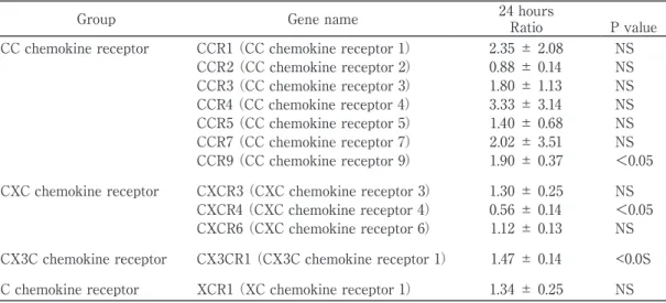
Effect of CF6 on cDNA microarray in HUVEC Table 1 shows the effect of CF6 on the gene expression of four classes of chemokine receptors in HUVEC. The genes with the significantly increased expression after 24- hour exposure to CF6 at 10-7M included CCR9 and CX3CR1. In contrast, the gene with the significantly decreased expression was CXCR4
(all p<0.05). Of these receptors, the most highly expressed one was CXCR4 in HUVEC. Since activation of the chemokine receptor CXCR4 regulates chemotaxis, survival, proliferation, gene transcription and intracellular calcium flux in various kinds of cells1), we examined the expression of CXCR4 by real-time PCR and
Table 1 Coupling factor 6-induced changes in chemokine receptors in human vascular endothelial cells
Group Gene name 24 hours
Ratio P value CC chemokine receptor CCR1 (CC chemokine receptor 1) 2.35 ± 2.08 NS
CCR2 (CC chemokine receptor 2) 0.88 ± 0.14 NS CCR3 (CC chemokine receptor 3) 1.80 ± 1.13 NS CCR4 (CC chemokine receptor 4) 3.33 ± 3.14 NS CCR5 (CC chemokine receptor 5) 1.40 ± 0.68 NS CCR7 (CC chemokine receptor 7) 2.02 ± 3.51 NS CCR9 (CC chemokine receptor 9) 1.90 ± 0.37 <0.05 CXC chemokine receptor CXCR3 (CXC chemokine receptor 3) 1.30 ± 0.25 NS
CXCR4 (CXC chemokine receptor 4) 0.56 ± 0.14 <0.05 CXCR6 (CXC chemokine receptor 6) 1.12 ± 0.13 NS CX3C chemokine receptor CX3CR1 (CX3C chemokine receptor 1) 1.47 ± 0.14 <0.0S C chemokine receptor XCR1 (XC chemokine receptor 1) 1.34 ± 0.25 NS Ratio, CF6+/CF6 - ratios of each gene expression (n=3); NS, not significant
Western blot analysis.
Effects of CF6 on CXCR4 gene and protein expression
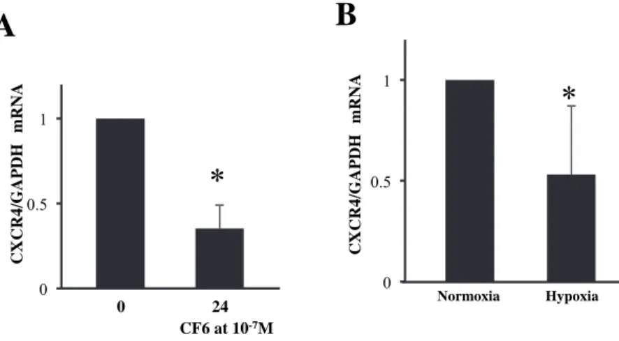
As shown in Figure 1A, the ratio of CXCR4 to GAPDH mRNA by real-time quantitative RT- PCR was decreased at 24 hours by 65±14%
(p<0.05) by CF6 at 10-7M.
Since hypoxia induces intracellular acidosis, we tested the hypothesis that like CF6, hypoxia may attenuate CXCR4 expression through the more or less similar pathway of intracellular acidosis. As shown in Figure 1B, the ratio of
Figure 1 Effects of human coupling factor 6 (CF6) or hypoxia on the gene expression of CXC chemokine receptor type 4 (CXCR4) in human umbilical vein endothelial cells (HUVEC).
A: Effects of CF6 at 10-7M on the ratio of CXCR4 to glyceraldehydes 3-phosphate dehydrogenase
(GAPDH) mRNA at 24 hours (n=6). * p<0.05 vs 0h
B: Effect of hypoxia on the ratio of CXCR4 to GAPDH mRNA at 24 hours (n=6). *p<0.05 vs normoxia without CF6.
Figure 1
A
0 0.5 1
0 24
CF6 at 10-7M
CXCR4/GAPDHmRNA
B
0 0.5 1
CXCR4/GAPDHmRNA
Normoxia Hypoxia
Figure 2 Effects of human coupling factor 6 (CF6) on the protein expression of CXC chemokine receptor type 4 (CXCR4) in human umbilical vein endothelial cells (HUVEC).
Representative bands for CXCR4 protein and the ratio of CXCR4/GAPDH in the presence and absence of CF6 at 10-7M for 24 hours (n=4). *p<0.05 vs CF6 (-).
Figure 2
0 0.5 1.0
Ratio of CXCR4/GAPDH protein
CF6 at 10-7M
(-) (+) CXCR4
GAPDH
CXCR4 to GAPDH mRNA was decreased by hypoxia (p<0.05) to a similar degree with CF6.
Figure 2 illustrates the representative bands for CXCR4 proteins in the presence or absence of CF6 at 10-7M for 24 hours. The immunoreactive band for CXCR4 at 40-47kD was decreased after treatment with CF6.
The ratio of CXCR4 to GAPDH protein was decreased by 41±10% in HUVEC treated with CF6 at 10-7M for 24 hours (p<0.05).
Effect of CF6 on apoptosis in HUVEC
As shown in Figure 3A, the percentage of apoptotic cells was slightly but significantly increased from 4.3±0.7 % to 5.6±1.7 % by CF6 at 10-7M in normoxia for 24 hours (p<0.05).
Discussion
The present study aimed at investigating the effect of CF6 on widespread chemokine receptor expression in the vascular endothelial cells. The result showed that CF6 increased the gene expression of CCR9 and CX3CR1 and decreased that of CXCR4, that either CF6 or hypoxia decreased the gene expression of CXCR4, and that CF6 decreased the protein expression of CXCR4 and induced apoptosis.
Effect of CF6 on chemokine receptor expression CF6 upregulated both CC and CX3C chemokine receptors in HUVEC, namely CCR9
and CX3CR1. The chemokine receptor CCR9 is one of the key molecules in leukocyte homing to gut mucosa. The pathological features of inflammatory bowel disease are associated with leukocyte cell infiltrates, which contribute to disease progression and persistence by production of proinflammatory mediators9). Accumulating evidences further showed that the CCR9/CCL25 axis participated in a variety of disease processes such as breast cancer, hepatitis, and rheumatoid arthritis10). Thus, the present finding of upregulated CCR9 suggests that CF6 may be associated with vascular inflammation such as atherosclerosis.
As to CX3CR1, a large body of evidence suggests that interaction between CX3CL1 and CX3CR1 plays a role in allergic diseases and inflammatory diseases in which CX3CL1 expression was increased, such as psoriasis, chronic obstructive pulmonary disease, pulmonary hypertension, and pulmonary fibrosis11). Like CCR9, CF6-induced upregulation of CX3CR1 may participate in vascular inflammation, but it should be clarified in the future study.
In contrast to upregulated genes, CXCR4 was downregulated by CF6 in HUVEC. Activation of the chemokine receptor CXCR4 was shown to regulate chemotaxis, survival, proliferation, transcription and intracellular calcium flux in various kinds of cells3). Thus, CF6 may be
Figure 3 Flow cytometric analysis of apoptosis with annexin-V-FITC and propidium iodide (PI) label in HUVEC.
Representative charts under normoxia for 24 hours with or without CF6 at 10-7M.
*p<0.05 vs Control (n=6).
(-) CF6
Figure 3
0 0.5 1 1.5
Ratioto control
Control CF6
associated with the regulation of vascular endothelial survival.
Effect of CF6 and hypoxia on CXCR4 expression CF6 stimulates ATPase activity at the surface of HUVEC after binding to the β-subunit of plasma membrane ATP synthase and induces intracellular acidosis by a flux of hydrogen ion through Fo6). The present result showed that CF6 decreased the gene and protein expression of CXCR4 in HUVEC, and the decrease in CXCR4 in response to CF6 may be associated with intracellular pH. This notion is supported by the previous report that direct administration of acid into the extracellular fluid decreased CXCR4 expression in HUVEC5). Acidosis occurs in a variety of pathological conditions including ischemia and/or hypoxia.
Thus, we next examined the effect of hypoxia on CXCR4 expression, and found that like CF6, hypoxia decreased the gene expression of CXCR4.
Effect of CF6 on cell death
Finally, we investigated the effect of CF6 on cell survival using annexin V apoptosis detection kit. CXCR4 activation by its chemokine SDF- 1 could lead to the simultaneous activation of both anti- and proapoptotic signaling pathways;
the balance ultimately influencing cell survival.
The anti-apoptotic second messenger signal is the Giα protein-dependent activation of Akt and ERK, whereas the proapoptotic signaling pathway is the Giα protein-independent activation of p38 MAP kinase12). In the present study, we showed that apoptotic cells were slightly but significantly increased at 24 hours by CF6 in normoxia. Since acidosis directly activates p38 MAP kinase13), CF6 might upregulate the activity of p38 MAP kinase as the proapoptotic signaling. Concerning the anti- apoptotic signaling, we previously showed that CF6 downregulated Akt phosphorylation in the heart14). CF6 slightly induced apoptosis in HUVEC. However, it is unclear whether this
effect is dependent on CF6-induced CXCR4 downregulation. Further examination is needed.
Implications of linkage between CF6 and CXCR4 in cardiovascular disorders
In the clinical settings, we and others showed that circulating CF6 is elevated in patients with hypertension, acute myocardial infarction, end-stage renal disease, stroke, and diabetes15-20), all of which predisposed to the development of arteriosclerosis. Given the present finding of proapoptotic effect and the widespread biological actions such as inhibition of prostacyclin and NO7,8), reduction in CF6 level may be important and useful to prevent atherosclerosis. To date, we have reported that salt restriction, vitamin C, and vitamin B12 plus folic acid lowered the plasma level of CF6 in patients with hypertension and stroke15,
16), and that peroxisome proliferator-activated receptor γ ligand attenuated CF6 release from cultured vascular endothelial cells21). Thus, the present finding might provide new insights into our understanding of the effect of CF6 on endothelial cells.
Study limitations
We investigated the effect of CF6 on various chemokine receptors in vascular endothelial cells.
However, neither the underlying mechanism nor the in vivo effect was examined in the present study. Further investigation is needed.
In conclusion, this report first showed the effect of CF6 on the expression of chemokine receptors in the vascular endothelial cells.
The decrease in the most highly expressed CXCR4 in endothelial cells may be involved in endothelial survival function.
Acknowledgements
This study was partly supported by Grant- in-Aid for Scientific Research from the Ministry of Education, culture, Sports, Science and Technology, Japan (No.19590800 for TO).
Reference
1)Murdoch C, Finn A. Chemokine receptors and their role in inflammation and infectious diseases.
Blood 2000;95:3032-43.
2)Murphy PM. International Union of Pharmacology.
XXX. Update on chemokine receptor nomencla- ture. Pharmacol Rev 2002;54:227-9.
3)Teicher BA, Fricker SP. CXCL12 (SDF-1)/CXCR4 pathway in cancer. Clin Cancer Res 2010;16:2927- 31.
4)Ziegelstein RC, Cheng L, Capogrossi MC. Flow- dependent cytosolic acidification of vascular endothelial cells. Science 1992;258:656-9.
5)Melchionna R, Romani M, Ambrosino V, D'Arcangelo D, Cencioni C, Porcelli D, Toietta G, et al. Role of HIF-1alpha in proton-mediated CXCR4 down-regulation in endothelial cells.
Cardiovasc Res 2010;86:293-301.
6)Osanai T, Magota K, Tanaka M, Shimada M, Murakami R, Sasaki S, Tomita H, et al.
Intracellular signaling for vasoconstrictor coupling factor 6: Novel function of β-subunit of ATP synthase as receptor. Hypertension 2005;46:1140-6.
7)Osanai T, Kamada T, Fujiwara N, Katoh T, Takahashi K, Kimura M, Satoh K, et al. A novel inhibitory effect on prostacyclin synthesis of coupling factor 6 extracted from the heart of spontaneously hypertensive rats. J Biol Chem 1998;273:31778-83.
8)Tanaka M, Osanai T, Murakami R, Sasaki S, Tomita H, Maeda N, Satoh K, et al. Effect of vasoconstrictor coupling factor 6 on gene expression profile in human vascular endothelial c e l l s : e n h a n c e d r e l e a s e o f a s y m m e t r i c dimethylarginine. J Hypertens 2006;24:489-97.
9)Koenecke C, Förster R. CCR9 and inflammatory bowel disease. Expert Opin Ther Targets 2009;
13:297-306.
10)Li J, Xiong T, Xiao R, Xiong A, Chen J, Altaf E, Zheng Y, et al. Anti-CCL25 antibody prolongs skin allograft survival by blocking CCR9 expression and impairing splenic T-cell function. Arch Immunol
Ther Exp 2013;61:237-44.
11)Julia V. CX3CL1 in allergic diseases: not just a chemotactic molecule. Allergy. 2012;67:1106-10.
12)Vlahakis SR, Villasis-Keever A, Gomez T, Vanegas M, Vlahakis N, Paya CV. G protein-coupled chemokine receptors induce both survival and apoptotic signaling pathways. J Immunol 2002;
169:5546-54.
13)Zheng M, Reynolds C, Jo SH, Wersto R, Han Q, Xiao RP. Intracellular acidosis-activated p38 MAPK signaling and its essential role in cardiomyocyte hypoxic injury. FASEB J 2005;
19:109-11.
14)Sagara S, Osanai T, Itoh T, Izumiyama K, Shibutani S, Hanada K, Yokoyama H, et al.
Overexpression of coupling factor 6 attenuates exercise-induced physiological cardiac hypertrophy by inhibiting PI3K/Akt signaling in mice. J Hypertens 2012;30:778-86.
15)Osanai T, Sasaki S, Kamada T, Fujiwara N, Nakano T, Tomita H, Matsunaga T, et al. Circulating coupling factor 6 in human hypertension: role of reactive oxygen species. J Hypertens 2003;21:2323- 8.
16)Osanai T, Fujiwara N, Sasaki S, Metoki N, Saitoh G, Tomita H, Nishimura T, et al. Novel pro- atherogenic molecule coupling factor 6 is elevated in patients with stroke: A possible linkage to homocysteine. Ann Med 2010;42:79-86.
17)Li XL, Xing QC, Gao YY, Dong B, Pang YZ, Jiang HF, Tang CS. Plasma level of mitochondrial coupling factor 6 increases in patients with type 2 diabetes mellitus. Int J Cardiol 2007;117:411-2.
18)Osanai T, Nakamura M, Sasaki S, Tomita H, Saitoh M, Osawa H, Yamabe H, et al. Plasma concentration of coupling factor 6 and cardiovascular events in patients with end-stage renal disease. Kidney Int 2003;64:2291-7.
19)Ding WH, Chu SY, Jiang HF, Cai DY, Pang YZ, Tang CS, Qi YF. Plasma mitochondrial coupling factor 6 in patients with acute myocardial infarction. Hypertens Res 2004;27:717-22.
20)Chai SB, Hui YM, Li XM, Tang CS. Plasma level of mitochondrial coupling factor 6 increases in
patients with coronary heart disease. Circ J 2007;71:693-7.
21)Tomita H, Osanai T, Toki T, Sasaki S, Maeda N, Murakami R, Magota K, et al. Troglitazone and 15-deoxy-deta 12, 14-prostaglandin J2 inhibit shear- induced coupling factor 6 release in endothelial cells. Cardiovasc Res 2005;67:134-41.