r egul at or of f l agel l ar m
ot i l i t y r eveal s t he
open- c l os ed s t r uc t ur al t r ans i t i on
著者
Shoj i m
a Tom
oki , H
ou Feng, Takahas hi Yus uke,
M
at s um
ur a Yos hi t aka, O
kai M
as ahi ko, N
akam
ur a
Aki r a, M
i z uno Kat s ut os hi , I naba Kaz uo, Koj i m
a
M
as aki , M
i yakaw
a Takuya, Tanokur a M
as ar u
j our nal or
publ i c at i on t i t l e
Sc i ent i f i c r epor t s
vol um
e
8
page r ange
2014
year
2018- 01
権利
( C) The Aut hor ( s ) 2018
Thi s ar t i c l e i s l i c ens ed under a Cr eat i ve
Com
m
ons At t r i but i on 4. 0 I nt er nat i onal
Li c ens e, w
hi c h per m
i t s us e, s har i ng,
adapt at i on, di s t r i but i on and r epr oduc t i on i n
any m
edi um
or f or m
at , as l ong as you gi ve
appr opr i at e c r edi t t o t he or i gi nal aut hor ( s )
and t he s our c e, pr ovi de a l i nk t o t he Cr
e-at i ve Com
m
ons l i c ens e, and i ndi c at e i f c hanges
w
er e m
ade. The i m
ages or ot her t hi r d par t y
m
at er i al i n t hi s ar t i c l e ar e i nc l uded i n t he
ar t i c l e’
s Cr eat i ve Com
m
ons l i c ens e, unl es s
i ndi c at ed ot her w
i s e i n a c r edi t l i ne t o t he
m
at er i al . I f m
at er i al i s not i nc l uded i n t he
ar t i c l e’
s Cr eat i ve Com
m
ons l i c ens e and your
i nt ended us e i s not per - m
i t t ed by s t at ut or y
r egul at i on or exc eeds t he per m
i t t ed us e, you
w
i l l need t o obt ai n per m
i s s i on di r ec t l y f r om
t he c opyr i ght hol der . To vi ew
a c opy of t hi s
. . .
U
RL
ht t p: / / hdl . handl e. net / 2241/ 00151035
doi: 10.1038/s41598-018-19898-7
Cr eat i ve Commons : 表示
Crystal structure of a Ca
2
+
-dependent regulator of lagellar
motility reveals the open-closed
structural transition
Tomoki Shojima
1, Feng Hou
1, Yusuke Takahashi
1, Yoshitaka Matsumura
2, Masahiko Okai
1,
Akira Nakamura
1, Katsutoshi Mizuno
3, Kazuo Inaba
3, Masaki Kojima
2, Takuya Miyakawa
1&
Masaru Tanokura
1Sperm chemotaxis toward a chemoattractant is very important for the success of fertilization. Calaxin, a member of the neuronal calcium sensor protein family, directly acts on outer-arm dynein and
regulates speciic lagellar movement during sperm chemotaxis of ascidian, Ciona intestinalis. Here, we present the crystal structures of calaxin both in the open and closed states upon Ca2+ and Mg2+ binding. The crystal structures revealed that three of the four EF-hands of a calaxin molecule bound Ca2+ ions and that EF2 and EF3 played a critical role in the conformational transition between the open and closed states. The rotation of α7 and α8 helices induces a signiicant conformational change of a part of the α10 helix into the loop. The structural diferences between the Ca2+- and Mg2+-bound forms indicates that EF3 in the closed state has a lower ainity for Mg2+, suggesting that calaxin tends to adopt the open state in Mg2+-bound form. SAXS data supports that Ca2+-binding causes the structural transition toward the closed state. The changes in the structural transition of the C-terminal domain may be required to bind outer-arm dynein. These results provide a novel mechanism for recognizing a target protein using a calcium sensor protein.
In many species, the female gamete or its associated structures release chemoattractants to attract spermatozoa. he chemoattractant gradient provides cues that guide sperm chemotaxis toward the egg1. In the sea urchin sperm, ater binding of the chemoattractant peptide to its receptor, rapid synthesis of cGMP is induced to activate the K+-selective cyclic nucleotide-gate (KCNG) channel, leading to membrane potential (E
m) hyperpolarization. he Em change facilitates the Ca2+ extrusion activity of K+-dependent Na+/Ca2+ exchangers (NCKX) and results in an inlux of Ca2+ into the cell2,3. his transient change in the intracellular Ca2+ concentration ([Ca2+]
i) is nec-essary for the directional changes of the sperm lagellum4–7. Spermatozoa from marine invertebrates have been especially used to study the mechanism to control lagellar motility during sperm chemotaxis8–11. he spermato-zoa of the ascidian Ciona intestinalis clearly exhibit chemotaxis toward the egg and a transient [Ca2+]
i increase in the lagellum accompanying a change of the swimming direction along a chemoattractant gradient ield12–14. he sperm keeps straight swimming at lower [Ca2+]
i, and higher [Ca2+]i induces asymmetric lagellar bending for the turning motion15,16.
Ca2+ ions activate axonemal dyneins through the phosphorylation of the Txtcx-2-related light chain (LC)
of outer-arm dynein and dephosphorylation of an intermediate chain (IC) of inner-arm dynein to induce a change in the lagellar beat, allowing the swimming of sperm along a gradient of the chemoattractant17–20. hese responses are mediated by a sharp increase in [Ca2+]
i from 10−6 M to 10−4 M21. Studies on dynein-driven micro-tubule sliding in isolated axonemes have indicated that the calcium signal may be mediated by calmodulin and a calmodulin-dependent kinase22. he dynein light chain from the outer arm of Chlamydomonas lagella was also
Department of Applied Biological Chemistry, Graduate School of Agricultural and Life Sciences, The University of
Tokyo, - - Yayoi, Bunkyo-ku, Tokyo, - , Japan. Laboratory of Bioinformatics, School of Life Sciences,
Tokyo University of Pharmacy and Life Science, Hachioji, Japan. Shimoda Marine Research Center, University of
Tsukuba, Shizuoka, - , Japan. Correspondence and requests for materials should be addressed to M.T. (email:
amtanok@mail.ecc.u-tokyo.ac.jp)
Received: 4 September 2017 Accepted: 10 January 2018 Published: xx xx xxxx
shown to contain a Ca2+-binding regulatory protein, which is directly associated with the γ dynein heavy chain23. However, the mechanism underlying the Ca2+-dependent regulation of dyneins remains obscure.
Recently, an axonemal Ca2+-binding protein from ascidian C. intestinalis, named calaxin, was demonstrated
to bind to a heavy chain (HC) of outer-arm dynein and tubulin in a Ca2+-dependent manner and to be essential
for the propagation of Ca2+-induced asymmetric lagellar bending24,25. Based on the amino acid sequence, calaxin belongs to the neuronal calcium sensor (NCS) protein family. Members of this protein family include recoverin and frequenin, which undergo conformational changes upon Ca2+ binding26–29, providing the surfaces to interact with their target proteins. To elucidate the relationship between the Ca2+-dependent regulatory mechanism and
structural features of calaxin, we herein solved crystal structures of calaxin in the Ca2+-bound and Mg2+-bound
forms. he crystal structure of calaxin shows an open-closed structural transition in both forms, and small-angle X-ray scattering data provides the evidence of structural transition in solution.
Results
Overall structure of calaxin in the Ca
2+-bound form.
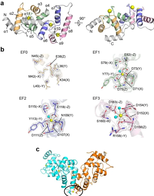
The crystal structure revealed that calaxin was composed of 11 helices (Fig. 1a and Supplementary Table 1) and contained four EF-hand motifs (Fig. 1b). Similarly to other NCS-family proteins, calaxin can be divided into two domains: the N-terminal domain and the C-terminal domain. In the N-terminal domain, EF0 (residues 23–55) interacted with EF1 (residues 60–92), and EF2 (residues 95–129) and EF3 (residues 137–170) formed the C-terminal domain (α6 through α10 helices) by interacting with each other. Both domains adopted their respective hydrophobic pockets, and the extended α11 helix was accommodated in the hydrophobic pocket of the N-terminal domain.he classical EF-hand is characterized by a sequence of 12 residues involved in Ca2+ binding. In calaxin, EF1
and EF2 were composed of the conserved residues for Ca2+ binding that are generally observed as D, D/N, D/N
and E at the positions X, Y, Z and −Z, respectively (Fig. 1b)30. hese residues formed ive coordinate bonds with Ca2+, including bidentate coordination by Glu. he position −Z of EF3 was substituted by Asp, which
coor-dinates Ca2+ ion in a monodentate manner (Fig. 1b). In this type of EF-hand, the shorter side chain of Asp at
the position −Z changes the angle of the canonical loop residue, resulting in a smaller and more compact Ca2+
binding motif31. By contrast, no characteristic residues are observed in EF0 (Supplementary Fig. 1), resulting in an impaired capacity to bind Ca2+ as shown in the crystal structure of calaxin (Fig. 1a and b). Among three
Ca2+-bound EF hands, only EF2 has a Gly112 at the position between Asp111 (Z) and Tyr113 (−Y). he Gly
residue of this position is known to be compatible to the bend of the loop region ater position Z32, indicating that EF2 might allow its conformational changes upon Ca2+ binding (Fig. 1b and Supplementary Fig. 1). Moreover, we
found that another Ca2+ ion is bonded between two calaxin molecules in an asymmetric unit (Fig. 1c). his Ca2+
coordination may contribute to stabilize the crystal packing.
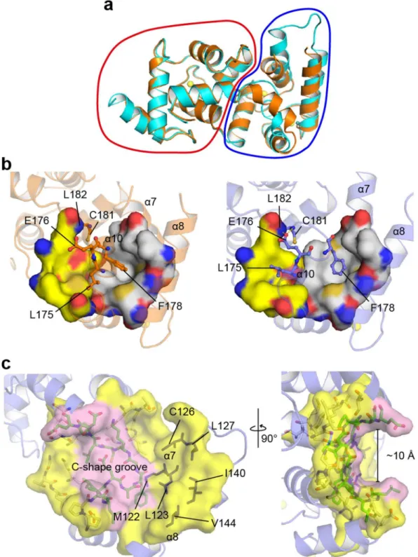
Conformational diferences between two calaxin molecules in an asymmetric unit.
We found that the crystal structure adopts the diferent conformations in the C-terminal domains between two calaxin molecules in an asymmetric unit: One molecule is in the open state and the other is in the closed state (Fig. 2a). Compared with the open state, the closed state exhibited the inward movement of the α7 and α8 helices and reduced the exposure of the hydrophobic surface formed by the α7, α8 and α10 helices (Met122, Leu123, Cys126, Leu127, Ile140 and Val144) (Fig. 2b and c). Unlike the C-terminal domain, the N-terminal domain showed little structural diference between the open and closed states (Fig. 2a). Another conformational change was observed at the α10 helix. he residues (from Phe172 to Cys181), which formed a 3-turn helix in the closed state, relaxed the helical structure, resulting in a 1-turn helix and a longer loop between the α10 and α11 helices in the open state (Fig. 2b). he side chain of Phe178, which is located in the α10 helix, was accommodated in the hydropho-bic surface formed by the α7, α8 and α10 helices in both the closed and open states. Phe178 was dragged by the movement of the α7 and α8 helices, which relaxed a part of the α10 helix in the conformational transition to the open state and induced a C-shaped groove with a width of ~10 Å (Fig. 2c). Furthermore, Glu176 also formed hydrogen bonds with the main-chain imino groups of Cys181 and Leu182 in the open state, resulting in the sta-bilization of the loop between the α10 and α11 helices in the open state. Two molecules in the open and closed states contacted each other using the α7 and α8 helices in the crystals (Supplementary Fig. 2). his contact may be due to crystal packing, resulting in the appearance of the open and closed states in an asymmetric unit. he simultaneous existence of both states in the Ca2+-bound form as crystal structures indicates that the driving forceof Ca2+ binding to calaxin may be insuicient to move the α7 and α8 helices and induce the conformational
change of the α10 helix.
EF2 and EF3 play pivotal roles in the conformational change between the open and closed
states.
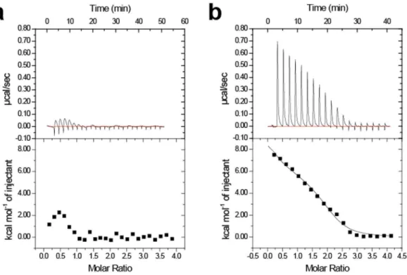
To characterize the conformational diferences in the EF-hands, we measured the interhelical angles between the E-helix and F-helix in each EF-hand (Table 1). EF0 and EF1, which reside in the N-terminal domain, showed slight changes in the interhelical angle between the open and closed states. However, EF2 and EF3 induced a signiicant change in the interhelical angle in the transition from the closed state to open state, corre-sponding to the large conformational change in the C-terminal domain (Fig. 2a). his implies that EF2 and EF3 contribute to produce the driving force to convert between the open and closed states.To validate the importance of EF2 and EF3, we performed isothermal titration calorimetry experiments using two mutants, E118A and D163A (in the −Z position of EF2 and EF3, respectively) (Fig. 3). Some studies have reported that EF-hand proteins show large enthalpy changes by Ca2+ titration33,34. Regarding calaxin, it has pre-viously been reported that Ca2+ binding to WT at 4 °C exhibits an endothermic enthalpy change25. E118A and D163A are predicted to be disabled for Ca2+ binding to each EF-hand. Ca2+ titrations to D163A represent
endo-thermic binding similar to that of WT. However, Ca2+ binding to E118A showed a remarkably lower endothermic
Ca2+. he circular dichroism spectroscopy showed that both E118A and D163A mutants retained their secondary
structures despite respective mutation (Supplementary Fig. 3). he results indicate that E118A mutation afects the sensitivity of calaxin to Ca2+ ions without the disruption of the native conformation.
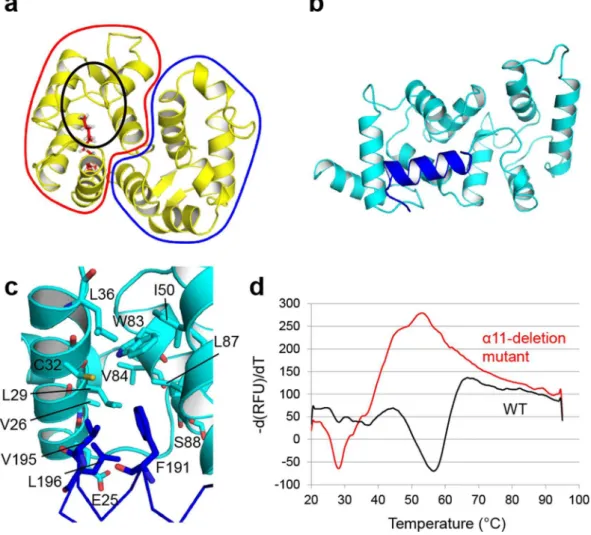
The
α
11 helix contributes to the thermostability of calaxin in the Ca
2+-bound form by
hydro-phobic interaction.
NCS-family proteins have an N-terminal myristoylation motif, such as recoverin, andFigure 1. Structure of the Ca2+-bound form of calaxin. (a) Ribbon diagrams of the overall structure of calaxin
in the Ca2+-bound open state. EF0, EF1, EF2 and EF3 are shown in light orange, pale green, slate and pink,
respectively. Ca2+ ions are indicated by a yellow sphere model. (b) Close-up view of individual EF-hands
binding a Ca2+ ion. Ca2+-binding residues are shown as a stick model. Ca2+ ions and water molecules are
indicated by yellow and cyan spheres, respectively. he coordination bonds with Ca2+ ions are highlighted with
dashed lines. he Fo−Fc omit maps of EF1, EF2 and EF3 are shown as gray meshes contoured at 3σ. (c) Calaxin dimer in the open state (cyan) and the closed state (orange) in an asymmetric unit. Ca2+ ions are represented as
yellow spheres. Side chains coordinating Ca2+ between dimers are represented as sticks (surrounded by a black
the myristoyl group is buried in the hydrophobic pocket in the Ca2+-free form (apo form or Mg2+-bound form)
(Fig. 4a). Calaxin has no myristoylation motif in the N-terminus. Instead, in the crystal structure of calaxin, the hydrophobic core in the N-terminal domain interacted with hydrophobic residues (Phe191, Val195 and
Figure 2. Structural basis for the open-closed transition of calaxin. (a) Structural comparison between the open state (cyan) and closed state (orange) in the Ca2+-bound form. he N-terminal domains and C-terminal
domains are circled by a red line and a blue line, respectively. (b) Structures of Ca2+-bound calaxin in the closed
Leu196) on the α11 helix (Fig. 4b and c). To assess the role of the α11 helix, we prepared an α11-deletion mutant (residues 1–182) and analyzed the thermostability of WT and this mutant in the Ca2+-bound forms using the
luorescence-based thermal stability assay (Fig. 4d). he irst derivative curve showed the melting temperature (Tm) for WT and mutant calaxin. he irst derivative curve of WT showed a large melting peak at 56 °C. However, the α11-deletion mutation caused a remarkable decrease in the melting peak (Tm= 28 °C). Furthermore, the irst derivative curve of the mutant showed a wide positive peak near 50 °C, indicating aggregation. Although the thermal stability was decreased, the protein folding was hardly afected by the α11-deletion compared to WT (Supplementary Figs 3 and 4). he results indicate that the hydrophobic interaction of the α11 helix with the N-terminal domain contributes to the thermostability of calaxin in the Ca2+-bound form.
Comparison of the structures between the Ca
2+-bound and Mg
2+-bound forms.
To clarify the structural features in Ca2+-bound form, we determined the crystal structure of calaxin in the Mg2+-bound formand compared the structures between these forms. Although calaxin in the Mg2+-bound form was puriied in
the presence of 100 mM magnesium chloride, the crystallizing conditions contained 10 mM barium chloride. We used isothermal titration calorimetry to determine which ion was bound in calaxin. Titrations of Ba2+ to
calaxin in the presence of Mg2+ showed no heat, indicating that Mg2+ bound to calaxin was not replaced by
Ba2+ (Supplementary Fig. 5). In addition, the anomalous signals of Ba2+ ions were not observed (Supplementary
Fig. 6). herefore, we obtained the crystal structure of Mg2+-bound calaxin.
he crystal structures are similar between the Ca2+-bound and Mg2+-bound form (RMSD 0.516 Å), and
the open and closed states were observed also in the Mg2+-bound form, supporting that Ca2+ is insuicient for
the driving force to convert between the open and closed states (Fig. 5a and b). Mg2+ ions in EF1 and EF3 are
liganded with octahedral coordination geometry in the open state, due to lack of the monodentate binding at −Z position as compared with Ca2+ ions, which is attributed to the diference of the ionic radii between Mg2+ and
Ca2+. However, no electron density of Mg2+ was observed in EF3 of the closed state, and the side chains of the
loop between E-helix and F-helix, especially Asp152 and Asp154, seem to be lexible because of poor electron density (Fig. 5c). hese data indicate that the ainity of EF3 for Mg2+ is diferent between the open and closed
states: Mg2+ prefers to bind to the open state over the closed state.
EF-hand open state (°) closed state (°) open state − closed state (°)
EF0 63.38 67.01 −3.63
EF1 58.01 61.82 −3.81
EF2 91.41 72.48 18.93
EF3 94.26 80.31 13.95
Table 1. Interhelical angle between the E-helix and F-helix in each EF-hand of Ca2+-bound calaxin.
Figure 3. Isothermal titration calorimetry of Ca2+ binding to the EF2-defective E118A mutant (a) and
Changes in structural transition between the Ca
2+-bound and Mg
2+-bound forms in solution.
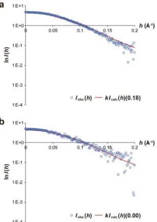
To evaluate the conformation of calaxin in the Ca2+ and Mg2+-bound forms in solution, we carried outsmall-angle X-ray scattering (SAXS) experiments in the presence of Ca2+ or Mg2+ ion. Based on the Guinier
plots (Supplementary Fig. 7), the radius of gyration (Rg) was 19.3 and 20.2 Å for Ca2+ and Mg2+-bound forms,
respectively. hese data show that calaxin exists a monomer in the both states. We further estimated the popula-tion of open and closed conformers under equilibrium in solupopula-tion by itting the intrinsic scattering curves, which were theoretically calculated with crystal structures of the two states of calaxin, to experimental scattering curves of the Ca2+ and Mg2+-bound calaxin (Fig. 6). Fraction of the open state with the lowest R factor was 0.18 in the
presence of Ca2+ ion, which shows that calaxin adopts the closed and open sates with abundance ratio of 18%
and 82% in the Ca2+-bound form, respectively. In contrast, the existing rate of Mg2+-bound calaxin was limited
to only open state (100%). hese results suggest that the Ca2+ binding functions to shit open-closed structural
transition toward the closed state. In addition, the open state may be stabilized by a selective binding of Mg2+ to
EF3 in the absence of Ca2+ ion.
Discussion
In the typical target-recognition mechanism of NCS-family proteins, Ca2+ binding to a protein induces the
extru-sion of the N-terminal myristoyl group, which sequesters the hydrophobic groove and covers up the target bind-ing site in the closed state (Supplementary Fig. 8a). his conformational change results in the exposure of residues interacting with the target proteins26–29. However, there is no myristoylation motif in the N-terminus of calaxin24. Several structures of NCS-family proteins have also been determined and are similar to the structure of calaxin
Figure 4. Contribution of the α11 helix to the thermostability of calaxin. (a) Solution structure of recoverin in the Ca2+-free state (PDB ID: 1IKU). he N-terminal domain and C-terminal domain are surrounded by a red
line and a blue line, respectively. he N-terminal myristoyl group is depicted in red sticks. he black circle shows the hydrophobic pocket interacting with the myristoyl group. (b) Crystal structure of Ca2+-bound calaxin in the
(Fig. 7). he C-terminal regions of the NCS-family proteins provide the surface to receive the target protein in the Ca2+-bound form, such as KChIP135 and AtCBL236 (Supplementary Fig. 8b and c). In contrast to these typical NCS-family proteins, calaxin shows an open-closed transition state in the Ca2+-bound form. In the closed state,
the exposure of the hydrophobic surface of calaxin is reduced by the movement of the α7 and α8 helices instead of the movement of the N-terminal myristoyl group or C-terminal region in the other NCS-family proteins. Typical NCS-family proteins receive the α-helical structure of target proteins with the induced surfaces of the Ca2+-bound forms. herefore, the Ca2+-induced surface in the C-terminal domain of calaxin may participate the
interaction with dynein, whose structure is largely composed of α-helices37.
EF-hand proteins are known to change the conformation and metal-binding pattern between the Ca2+-bound
and Mg2+-bound forms30,38,39. However, calaxin in the Mg2+-bound form shows a similar structure to that of
the Ca2+-bound form, indicating that the conformational transition between the Ca2+-bound and Mg2+-bound
forms is insigniicant and that the efect of the crystal packing may surpass that of the conformational transition. Interestingly, we found that EF3 has a lower ainity for Mg2+ in the closed state due to incompatible
arrange-ments of the acidic residues (Fig. 5c), whereas Ca2+ induces the same arrangements of the EF3 both in the closed
and open states. hese structural diferences in the Ca2+- and Mg2+-bound forms indicate the possibility that
calaxin in the Ca2+-bound form is more compatible with the conformational change than the Mg2+-bound form.
Moreover, SAXS data supports that Ca2+-binding promotes the open-closed structural transition toward the
closed state (Fig. 6). A transient [Ca2+]
i is increased up to 10−4 M in the process of the directional changes of sperm movement21. In addition to the Ca2+ binding to the EF-hands causing structural transition, the interaction
with outer-arm dynein may be essential to complete the conformational change of calaxin.
In the crystal structure of AtCBL2, which is a member of the NCS protein family, the helices in the C-terminal domain form the hydrophobic crevice and accommodate the regulatory domain of AtCIPK14 (Supplementary Fig. 8c)36. Furthermore, Ca2+ ions are bound by AtCBL2 with and without AtCIPK14, supporting that a
confor-mational transition between the ligand-bound and ligand-free forms can occur in other NCS-family proteins, including calaxin, when the proteins adopt their Ca2+-bound states. However, the binding patterns of Ca2+ ions
are diferent between the ligand-bound and ligand-free forms of AtCBL2. All four EF-hands are illed with Ca2+
ions in the ligand-bound form, whereas Ca2+ ions bind only to the irst and fourth EF-hands in ligand-free form.
On the other hand, opposite Ca2+ binding patterns are shown in the structure of SOS3 complexed with SOS2.
Figure 5. Structure of the Mg2+-bound form of calaxin. (a) Overall structure of calaxin in the Mg2+-bound
form. he open state and closed state are depicted in magenta and ruby, respectively. Mg2+ ions are shown in
green spheres. (b) Structural comparison between the Ca2+-bound forms (open state: cyan, closed state: orange)
and Mg2+-bound forms (open state: magenta, closed state: ruby) of calaxin. (c) Enlarged views of EF3 in the
SOS3 in the SOS2-bound form binds Ca2+ ions only at the irst and fourth EF-hands, although all four EF-hands
are occupied with Ca2+ ions in the SOS2-unbound form40. Calaxin adopts both its open state and closed state in the same Ca2+-binding pattern in which three Ca2+ ions are coordinated to EF1, EF2 and EF3, indicating that its
open-closed transition is independent of the Ca2+-binding pattern.
Dynein family proteins use a common principle to generate movement in which they bind to their track, undergo a force-producing conformational change, are released from the track and then return to their original conformation. herefore, corresponding to the action of dynein family members, conformational change will help calaxin recognize dynein efectively. Although the core structures constituted by four EF-hands (α2 through α9) are similar among NCS-family proteins, their C-terminal helices and loops of each protein adopt various con-formations (Supplementary Fig. 9). his indicates that the conformational diferences in the C-terminal helices are involved in the selectivity of NCS-family proteins toward associating partners. Also, our luorescence-based thermal stability assay demonstrated that the C-terminal helix of calaxin contributes the conformational stability by interaction with the hydrophobic core in the N-terminal domain. Although further demonstration requires the identiication of the target binding region of dynein and subsequent structural analyses of calaxin complexed with dynein or its target region, our indings provides a plausible conformational change consistent with outer-arm dynein mechanism of the calaxin-mediated regulation of dynein induced by a transient [Ca2+]
i increase.
Materials and Methods
Expression and Puriication of Ca
2+-bound and Mg
2+-bound Calaxin.
For the preparation of calaxin (UniProt ID: Q8T893), the calaxin gene was cloned at NdeI and BamHI sites of the pET-28a vector (Novagen), and the vector was transformed to the Escherichia coli K12 strain KRX cells (Promega). he cells were grown in Lysogeny Broth (LB) medium containing 30 µg mL−1 kanamycin at 37 °C. Ater the addition of 0.1% L-rhamnose (Wako) at an optical density at 600 nm (OD600) of 0.6, the cell culture was further incubated at 20 °C for 18 h. he cells were harvested by centrifugation at 2,290 ×g for 15 min at 4 °C. he pellet was resuspended in sonication bufer containing 50 mM Tris-HCl (pH 8.0), 300 mM NaCl and 10 mM imidazole. Ater sonication, the suspen-sion was centrifuged at 40,000 ×g for 30 min at 4 °C to remove cell debris and insoluble fractions. he supernatant was loaded onto a His-tag ainity column prepared by illing a 20-mL chromatography column (BioRad) with Ni Sepharose 6 Fast Flow (GE Healthcare). Ater the column was washed with washing bufer containing 50 mM Tris-HCl (pH 8.0), 300 mM NaCl and 50 mM imidazole, calaxin was eluted with elution bufer containing 50 mM Tris-HCl (pH 8.0), 300 mM NaCl and 250 mM imidazole. hrombin was added to the eluted protein to remove the N-terminal His-tag, and the protein solution was then dialyzed overnight against chelate bufer contain-ing 50 mM Tris-HCl (pH 8.0), 1 mM DTT and 1 mM EDTA. he solution was further dialyzed against 50 mMFigure 6. Structural transition between the open and closed state of calaxin observed in SAXS experiments. (a) Experimental (blue circles) and calculated (fraction of closed state = 0.18) (red line) scattering curves of Ca2+-bound calaxin. (b) Experimental (blue circles) and calculated (fraction of closed state = 0.00) (red line)
Tris-HCl (pH 8.0), 1 mM DTT and 1 mM CaCl2 to prepare the calcium binding protein. he protein was puriied using a Resource Q (GE Healthcare) anion exchange column equilibrated with equilibration bufer containing 25 mM Tris-HCl (pH 8.0), 1 mM DTT and 1 mM CaCl2 and was eluted by increasing the NaCl concentration from 0 to 500 mM in the equilibration bufer. For the preparation of Mg2+-bound calaxin, the protein solution
was dialyzed against 20 mM Tris-HCl (pH 8.0), 20 mM MgCl2 and 1 mM DTT. Furthermore, the dialyzed Mg2+ -bound calaxin was eluted with a 0 to 300-mM NaCl gradient from a Resource Q column, followed by bufer exchange with a Superdex 75 10/300 HR (GE Healthcare) column using 10 mM MES-NaOH (pH 6.0), 100 mM MgCl2, 150 mM NaCl and 1 mM DTT.
Crystallization, Data Collection and Structure Determination.
Crystallization of the Ca2+-boundprotein was performed using the sitting drop vapor difusion method. Each sitting drop was prepared by mixing the protein solution (0.75 µL) with the equal volume of reservoir solution (0.75 µL) containing 0.1 M MES (pH 6.4), 0.2 M calcium acetate and 17.5% PEG8000 and then was equilibrated against the reservoir solution at 20 °C. he concentration of protein for crystallization was 10 mg mL−1, and 24% ethylene glycol was used as the cry-oprotectant. he X-ray difraction data were collected with an in-house X-ray difractometer equipped with an FR-E SuperBright X-ray generator and an R-AXIS VII imaging plate. For crystallographic phasing, the crystal was soaked in a solution containing 0.1 M MES (pH 6.2), 10 mM SmCl3 and 16% PEG8000, and the single anom-alous difraction (SAD) data were collected at the samarium peak wavelength of 1.6 Å on the BL-17A beamline at Photon Factory (Tsukuba, Japan).
The collected data sets were indexed and integrated using XDS41, and were scaled using XSCALE41. Experimental phasing was performed with the SAD data and the program package autoSHARP42. he initial Model was automatically built with ARP/wARP43 in the CCP4 suite44. Ater automatic modeling, manual model building was carried out with Coot45, and Refmac546 was used for the reinement of the obtained model. To deter-mine the structure of Ca2+-bound protein, the molecular replacement method was carried out using Molrep47 and the samarium-bound structure as a template. Model building and reinement were performed using Coot and Refmac5, respectively. he quality of the models was veriied by PROCHECK48. he images of the protein struc-ture were created by PyMol (http://www.pymol.org/). he sequence alignment and visualization were performed using ClustalW and ESPript, respectively.
Crystallization of the Mg2+-bound protein was also performed using the sitting drop vapor difusion method
at 20 °C by mixing 0.3 µL of 12 mg mL−1 protein solution with 0.3 µL of reservoir solution (containing 0.2 M ammonium sulfate, 0.1 M MES, pH 6.3, 26% (w/v) PEG 5000 MME and 10 mM barium chloride). he X-ray difraction data were collected at a wavelength of 1.0 Å at Photon Factory beamline AR-NW12A. he data were indexed and integrated with XDS41, and were scaled with AIMLESS49. he crystal structure of Mg2+-bound
Figure 7. Crystal structures of Ca2+-bound NCS-family proteins. Structural comparison among NCS-family
calaxin was determined by Molrep using the structure of the Ca2+-bound calaxin without Ca2+ ions as a template
model. Coot was used for model building, and Phenix.reine50 and Refmac5 were used for reinement. Twin reinement was applied because twinning fraction of structure factor data for Mg2+-bound calaxin was 0.155.
Preparation of Mutant Proteins.
Two mutant proteins (E118A and D163A) were prepared for isothermal titration calorimetry experiments. he expression vectors were created using PrimeSTAR Max DNA Polymerase (Takara), the expression vector for wild-type protein as a template and primers (Supplementary Table 2). he protein expression and puriication were carried out with the same procedure as that of the wild type.Isothermal Titration Calorimetry (ITC) Experiments.
ITC experiments were carried out using an iTC200 calorimeter (GE Healthcare) at 4 °C. he protein solutions were prepared in aqueous bufer containing 25 mM MOPS-KOH (pH 7.8), 1 mM DTT and 200 mM NaCl. Calcium titrations were carried out with 1 mM CaCl2 and a 50-µM protein solution. he experiments were carried out with each injection consisting of 1.5 µL of the titrant against 200 µL of the protein solution. Data analysis was performed using Origin 7 sotware. he titration curve of the D163A mutant was itted using a two sequential binding model. Barium titrations were carried out using 0.8 mM BaCl2 and a 35-µM calaxin solution in the bufer containing 25 mM MOPS-KOH (pH 7.8), 1 mM DTT, 200 mM NaCl and 1 mM MgCl2. Data analysis was conducted in the same way as that for Ca2+ titrations.Fluorescence-based thermal stability assay.
The assay was carried out using the CFX Connect Real-Time System (Bio-Rad) as previously described51. he sample solutions were prepared by mixing 2.22 µL of 100× SYPRO Orange (Life Technologies) and 20 µL of 0.3 mg/mL calaxin solution (20 mM Tris-HCl, pH 8.0, 150 mM NaCl, 1 mM DTT and 1 mM CaCl2). Each sample was heated from 20 to 95 °C in increments of 0.5 °C.Circular dichroism measurements.
Wild type calaxin and its mutants (E118A, D163A and the α11-deletion mutant) were analyzed by circular dichroism to compare their secondary structures. he decal-ciied WT, E118A and D163A samples (11 µM) were prepared in a bufer of 25 mM MOPS-KOH (pH 7.8) and 1 mM DTT, and the α11-deletion mutant (11 µM) was prepared in a bufer containing 20 mM Tris-HCl (pH 8.0), 150 mM NaCl, 1 mM DTT and 1 mM CaCl2. he measurements were performed at room temperature by using a Jasco J-720 spectropolarimeter (WT, E118A and D163A) and a Jasco J-820 spectropolarimeter (the α11-deletion mutant) with a quartz cuvette of 0.1 cm path length. CD spectra were recorded and analyzed between 200–260 nm.Small-angle X-ray scattering experiments and analyses.
he SAXS experiments were carried out on beamline BL-10C at the Photon Factory. All data were collected using X-ray of wavelength of 1.488 Å with a PILATUS3 2 M detector (Dectris) and processed with the FIT2D program (http://www.esrf.eu/computing/sci-entiic/FIT2D/). he sample-to-detector distance was 1.0 m. Equilibrium experiments were performed at 25 °C. he SAXS intensities were accumulated for a total of 30 s by repeating the measurements for a period of 1.0 s each time in order to ensure enough statistical precision. X-ray scattering data were obtained from protein and the corresponding bufers. he scattering data of the bufers were subtracted from those of the protein solutions. X-ray scattering data were analyzed by Guinier approximation, as assuming an exponential dependence of the scattering intensity on h2, where h= 4πsinθ/λ and θ is half of the scattering angle52. Rg and zero angle scattering
intensity I(0) were determined using Guinier approximation52. Molecular weight was determined from I(0) by measuring bovine serum albumin (BSA) as a calibration standard53. he sample concentrations of calaxin were 0.25, 0.5, 1.0 and 1.75 mg/mL for the Ca2+-bound form and 0.25, 0.5, 1.0 and 2.0 mg mL−1 for the Mg2+-bound
form. he samples of the Ca2+- and Mg2+-bound forms were prepared in 20 mM Tris-HCl (pH 8.0), 150 mM
NaCl and 1 mM DTT containing 10 mM CaCl2 and MgCl2, respectively.
he intrinsic scattering intensities of open and closed conformers were theoretically calculated from atomic coordinates of their crystal structures by CRYSOL54 program for default parameter settings. he scattering inten-sity Icalc(h) of equilibrium mixture of open and closed forms with a ratio of (1 −α): α was expressed as:
α α
= − +
Icalc( )h (1 )Iopen( )h Iclosed( )h
where Iopen(h) and Iclosed(h) are intrinsic scattering intensities of open and closed conformers, respectively. We evaluated optimal value of α, fraction of the closed conformer under equilibrium in solution, which yielded the lowest R factor such as:
R I h kI h h
I h h
( ) ( ) ( )
h obs calc
h obs
2
2
= ∑ −
∑
where Iobs(h) is the experimental scattering intensity, and k is the scaling factor between observed and calculated scattering intensities as:
= ∑ ∑
k I h I h h
I h h
( ) ( ) ( )
h obs calc
h calc 2
2 2
References
1. Eisenbach, M. & Giojalas, L. C. Sperm guidance in mammals – an unpaved road to the egg. Nat. Rev. Mol. Cell. Biol.7, 276–285 (2006).
2. Strünker, T. et al. A K+-selective cGMP-gated ion channel controls chemosensation of sperm. Nat. Cell Biol.8, 1149–1154 (2006).
3. Gauss, R., Seifert, R. & Kaupp, U. B. Molecular identiication of a hyperpolarization-activated channel in sea urchin sperm. Nature
393, 583–587 (1998).
4. Morisawa, M. Cell signaling mechanisms for sperm motility. Zool. Sci.11, 647–662 (1994).
5. Darszon, A., Guerrero, A., Galindo, B. E., Nishigaki, T. & Wood, C. D. Sperm-activating peptides in the regulation of ion luxes, singal transduction and motility. Int. J. Dev. Biol.52, 595–606 (2008).
6. Kaupp, U. B., Kashikar, N. D. & Weyand, I. Mechanisms of sperm chemotaxis. Annu. Rev. Physiol.70, 93–117 (2008).
7. Guerrero, A., Wood, C. D., Nishigaki, T., Carneiro, J. & Darszom, A. Tuning sperm chemotaxis. Biochem. Soc. Trans.38, 1270–1274 (2010).
8. Shingyoji, C., Murakami, A. & Takahashi, K. Local reactivation of Triton-extracted lagella by iontophoretic application of ATP. Nature265, 269–270 (1977).
9. Kamimura, S. & Kamiya, R. High-frequency nanometer-scale vibration in ‘quiescent’ lagellar axonemes. Nature340, 476–478 (1989).
10. Ogawa, K. & Mohri, H. A dynein motor superfamily. Cell Struct. Funct.21, 343–349 (1996).
11. Gibbons, B. H., Baccetti, B. & Gibbons, I. R. Live and reactivated motility in the 9 + 0 lagellum of Anguilla sperm. Cell Motil.5, 333–350 (1985).
12. Miller, R. L. Chemotaxis of the spermatozoa of Ciona interstinalis. Nature254, 244–245 (1975).
13. Yoshida, M., Inaba, K. & Morisawa, M. Sperm chemotaxis during the process of fertilization in the ascidians Ciona savignyi and Ciona intestinalis. Dev. Biol.157, 497–506 (1993).
14. Shiba, K., Baba, S. A., Inoue, T. & Yoshida, M. Ca2+ bursts occur around a local minimal concentration of attractant and trigger
sperm chemotactic response. Proc. Natl. Acad. Sci. USA105, 19312–19317 (2008).
15. Cook, S. P., Brokaw, C. J., Muller, C. H. & Babcock, D. F. Sperm chemotaxis: Egg peptides control cytosolic calcium to regulate lagellar responses. Dev. Biol.165, 10–19 (1994).
16. Nishigaki, T. et al. A sea urchin egg jelly peptide induces as cGMP-mediated decrease in sperm intracellular Ca2+ before its increase.
Dev. Biol.272, 376–388 (2004).
17. Inaba, K., Morisawa, S. & Morisawa, M. Proteasomes regulate the motility of salmonid ish sperm through modulation of cAMP-dependent phosphorylation of an outer arm dynein light chain. J. Cell Sci.111, 1105–1115 (1998).
18. Inaba, K., Kagami, O. & Ogawa, K. Tctex2-related outer arm dynein light chain is phosphorylated at activation of sperm motility. Biochem. Biophys. Res. Commun.256, 177–183 (1999).
19. Nomura, M., Inaba, K. & Morisawa, M. Cyclic AMP- and calmodulin-dependent phosphorylation of 21 and 26 kDa proteins in axoneme is a prerequisite for SAAF-induced motile activation in ascidian spermatozoa. Dev. Growth Difer.42, 129–138 (2000). 20. Hozumi, A., Padma, P., Toda, T., Ide, H. & Inaba, K. Molecular characterization of axonemal proteins and signaling molecules
responsible for chemoattractant-induced sperm activation in Ciona intestinalis. Cell Motil. Cytoskeleton65, 249–267 (2008). 21. Bessen, M., Fay, R. B. & Witman, G. B. Calcium control of lagellar waveform. J. Cell Biol.86, 446–455 (1980).
22. Elizabeth, F. S. Regulation of lagellar dynein by calcium and a role for an axonemal calmodulin and calmodulin dependent kinase. Mol. Biol. Cell13, 3303–3313 (2002).
23. King, S. M. & Patel-King, R. S. Identiication of a Ca2+-binding light chain within Chlamydomonas outer arm dynein. J. Cell Sci.108,
3757–3764 (1995).
24. Mizuno, K. et al. A novel neuronal calcium sensor family protein, calaxin, is a potential Ca2+-dependent regulator for the outer arm
dynein of metazoan cilia and lagella. Biol. Cell101, 91–103 (2009).
25. Mizuno, K. et al. Calaxin drives sperm chemotaxis by Ca2+-mediated direct modulation of a dynein motor. Proc. Natl. Acad. Sci.
USA109, 20497–20502 (2012).
26. Ames, J. B., Levay, K., Wingard, J. N., Lusin, J. D. & Slepak, V. Z. Structural basis for calcium-induced inhibition of rhodopsin kinase by recoverin. J. Biol. Chem.281, 37237–37245 (2006).
27. Ames, J. B. et al. Molecular mechanics of calcium-myristoyl switches. Nature389, 198–202 (1997).
28. Lim, S., Strahl, T., Thorner, J. & Ames, J. B. Structure of a Ca2+-myristoyl switch protein that controls activation of a
phosphatidylinositol 4-kinase in ission yeast. J. Biol. Chem.286, 12565–12577 (2011).
29. Tanaka, T., Ames, J. B., Harvey, T. S., Stryer, L. & Ikura, M. Sequestration of the membrane-targeting myristoyl group of recoverin in the calcium-free state. Nature376, 444–447 (1995).
30. Nara, M., Morii, H. & Tanokura, M. Coordination to divalent cations by calcium-binding proteins studied by FTIR spectroscopy. Biochim. Biophys. Acta1828, 2319–2327 (2013).
31. Vijay-Kumar, S. & Cook, W. J. Structure of a sarcoplasmic calcium-binding protein from Nereis diversicolor refined at 2.0 Å resolution. J. Mol. Biol.224, 413–426 (1992).
32. Falke, J. J., Drake, S. K., Hazard, A. L. & Peersen, O. B. Molecular tuning of ion binding to calcium singal proteins. Q. Rev. Biophys.
27, 219–290 (1994).
33. Miyakawa, T. et al. Diferent Ca2+-sensitivities between the EF-hands of T- and L-plastins. Biochem. Biophys. Res. Commun.429,
137–41 (2012).
34. Suzuki, N. et al. Calcium-dependent structural changes in human reticulocalbin-1. J. Biochem.155, 281–93.35 (2014).
35. Pioletti, M., Findeisen, F., Hura, G. L. & Minor, D. L. jr. hree-dimensional structure of the KChIP1-Kv4.3 T1 complex reveals a cross-shaped octamer. Nat. Struct. Mol. Biol.13, 987–995 (2006).
36. Akaboshi, M. et al. he crystal structure of plant-speciic calcium-binding protein AtCBL2 in complex with the regulatory domain of AtCIPK14. J. Mol. Biol.377, 246–257 (2008).
37. Kon, T. et al. he 2.8 Å crystal structure of the dynein motor domain. Nature484, 345–50 (2012).
38. Seamon, K. B. Calcium- and magnesium-dependent conformational states of calmodulin as determined by nuclear magnetic resonance. Biochemistry19, 207–215 (1980).
39. Andersson, M. et al. Structural basis for the negative allostery between Ca2+- and Mg2+-binding in the intracellular Ca2+-receptor
calbindin D9k. Protein Sci.6, 1139–1147 (1997).
40. Sánchez-Barrena, M. J. et al. he structure of the C-terminal domain of the protein kinase AtSOS2 bound to the calcium sensor AtSOS3. Mol. Cell26, 427–35 (2007).
41. Kabsch, W. X. D. S. Acta Crystallogr. D Biol. Crystallogr.66, 125–132 (2010).
42. Vonrhein, C., Blanc, E., Roversi, P. & Bricogne, G. Automated structure solution with autoSHARP. Methods Mol. Biol.364, 215–230 (2007).
43. Cohen, S. X. et al. ARP/wARP and molecular replacement: he next generation. Acta Crystallogr. D Biol. Crystallogr.64, 49–60 (2008).
44. Collaborative Computational Project, Number 4 he CCP4 suite: Programs for protein crystallography. Acta Crystallogr. D Biol. Crystallogr. 50, 760–763 (1994).
46. Murshudov, G. N. et al. REFMAC5 for the reinement of macromolecular crystal structures. Acta Crystallogr. D Biol. Crystallogr.67, 355–367 (2011).
47. Vagin, A. & Teplyakov, A. MOLREP: An automated program for molecular replacement. J. Appl. Cryst.30, 1022–1025 (1997). 48. Laskowski, R. E., Mac Arthur, M. W., Moss, D. S. & hornton, J. M. PROCHECK: A program to check the stereochemical quality of
protein structures. J. Appl. Cryst.26, 283–291 (1993).
49. Evans, P. R. & Murshudov, G. N. How good are my data and what is the resolution? Acta Crystallogr. D Biol. Crystallogr.69, 1204–1214 (2013).
50. Afonine, P. V. et al. Towards automated crystallographic structure reinement with phenix.reine. Acta Crystallogr. D Biol. Crystallogr.
68, 352–367 (2012).
51. Miyakawa, T. et al. Structural basis for the Ca2+-enhanced thermostability and activity of PET-degrading cutinase-like enzyme from
Saccharomonospora viridis AHK190. Appl. Microbiol. Biotechnol.99, 4297–307 (2015). 52. Guinier, A. & Fournet, G. In Small Angle Scattering of X-rays (John Wiley & Sons, 1955).
53. Tashiro, M. et al. Characterization of ibrillation process of α-synuclein at the initial stage. Biochem. Biophys. Res. Commun.369, 910–914 (2008).
54. Svergun, D., Barberato, C. & Koch, M. H. J. CRYSOL – a program to evaluate X-ray solution scattering of biological macromolecules from atomic coordinates. J. Appl. Crystal.28, 768–773 (1995).
Acknowledgements
We would like to thank the beamline staff at Photon Factory. The synchrotron radiation experiments were performed at BL-17A, AR-NW12A and BL-10C in the Photon Factory, Tsukuba, Japan (2010G640, 2013G654 and 2016R-56). his work was supported by a Grant-in-Aid for Scientiic Research (S) from the Japan Society for the Promotion of Science (JSPS) for M.T.
Author Contributions
M.T. and K.I. designed the research; T.S., F.H., Y.T. and Y.M. performed the experimental research; T.S., F.H., Y.M., M.O., A.N., K.M., M.K. and T.M. analyzed the data; T.S., F.H., K.I., M.K, T.M. and M.T. wrote the paper; M.T. edited the paper. T.S., H.F. and Y.T. contributed equally to this work.
Additional Information
Supplementary information accompanies this paper at https://doi.org/10.1038/s41598-018-19898-7.
Competing Interests: he authors declare that they have no competing interests.
Publisher's note: Springer Nature remains neutral with regard to jurisdictional claims in published maps and institutional ailiations.
Open Access This article is licensed under a Creative Commons Attribution 4.0 International License, which permits use, sharing, adaptation, distribution and reproduction in any medium or format, as long as you give appropriate credit to the original author(s) and the source, provide a link to the Cre-ative Commons license, and indicate if changes were made. he images or other third party material in this article are included in the article’s Creative Commons license, unless indicated otherwise in a credit line to the material. If material is not included in the article’s Creative Commons license and your intended use is not per-mitted by statutory regulation or exceeds the perper-mitted use, you will need to obtain permission directly from the copyright holder. To view a copy of this license, visit http://creativecommons.org/licenses/by/4.0/.