Brief Report
https://academic.oup.com/jes 1
ISSN 2472-1972
This is an Open Access article distributed under the terms of the Creative Commons Attribution License (http://creativecommons.org/licenses/by/4.0/), which permits unrestricted reuse, distribution, and reproduction in any medium, provided the original work is properly cited.
© The Author(s) 2020. Published by Oxford University Press on behalf of the Endocrine Society.
Brief Report
Reevaluation of the Effect of Iodine on Thyroid Cell Survival and Function Using PCCL3 and Nthy-ori 3-1 Cells
Tomomi Kurashige,
1Mika Shimamura,
1and Yuji Nagayama
11Department of Molecular Medicine, Atomic Bomb Disease Institute, Nagasaki University, Nagasaki, 852-8523, Japan
ORCiD numbers: 0000-0002-5216-5785 (T. Kurashige); 0000-0002-3830-2173 (M. Shimamura); 0000-0002-5058-9349 (Y. Nagayama).
Abbreviations: bp, base pair; FBS, fetal bovine serum; IB, immunoblotting; IF, immunofluorescence; LC3, microtubule- associated protein 1 light chain 3; mRNA, messenger RNA; NIS, sodium/iodine symporter; PCR, polymerase chain reaction;
ROS, reactive oxygen species; TSH, thyrotropin; TSHR, thyrotropin receptor; U/L, units per liter.
Received: 20 August 2020; Accepted: 23 September 2020; First Published Online: 28 September 2020; Corrected and Typeset:
21 October 2020.
Abstract
The appropriate amount of iodine is critical for normal function of thyroid cells synthesizing thyroid hormones. Although normal thyroid cell lines such as rat PCCL3 and FRTL5 and human Nthy-ori 3-1 have been widely used for in vitro studies on physiological and pathophysiological effects of iodine on thyroid cells, we have recently pointed out the critical differences between FRTL5/PCCL3 cells and Nthy-ori 3-1 cells. Therefore, we here directly compared some of the cellular characteristics—iodine uptake, differentiated status, iodine-induced cytotoxicity, and iodine-regulation of autophagy—between PCCL3 and Nthy-ori 3-1 cells. PCCL3 cells express messenger RNAs for thyrotropin receptor and sodium/iodine symporter and incorporate iodine in a thyrotropin-dependent manner, whereas Nthy-ori 3-1 cells do not either. Nevertheless, both cells were comparably re- sistant to iodine cytotoxicity: Only far excess iodine (5 × 10⁻² M) killed 20% to 40% cells in 24 hours with perchlorate exhibiting no effect, suggesting this cytotoxic effect is due to extracellular iodine. In contrast, a wide range of iodine (5 × 10⁻⁹ to 5 × 10⁻² M) induced autophagy in PCCL3 cells, which was abolished by perchlorate, indicating intracellular iodine-induction of autophagy, but this effect was not observed in Nthy-ori 3-1 cells. In conclusion, it is critical to discriminate the effect of iodine incorporated into cells from that of extracellular iodine on thyroid cells. Iodine-uptake competent thyroid cells such as PCCL3 and FRTL5 cells, not Nthy-ori 3-1 cells, should be used for studies on iodine effect on thyroid cells.
Key Words: thyroid, iodine, autophagy, NIS, TSHR
Downloaded from https://academic.oup.com/jes/article/4/11/bvaa146/5912612 by Nagasaki University Library user on 04 December 2020
The thyroid follicular epithelial cells actively incorporate iodine from the bloodstream to synthesize thyroid hor- mones, but excessive and deficient iodine both cause thy- roid dysfunction. Normal thyroid cell lines are very useful to study in vitro physiological and pathophysiological ef- fects of iodine on thyroid cell survival and function. They include rat normal thyroid cell lines, PCCL3 [1] and FRTL5 [2], and a human cell line, Nthy-ori 3-1 [3]. Nthy-ori 3-1 cells have recently been used to show iodine-inductions of cell death [4-6] and of endoplasmic reticulum stress [7], and inhibition of autophagy [8]. However, we have recently pointed out the critical differences between FRTL5/PCCL3 cells and Nthy-ori 3-1 cells and expressed our concern that the latter may not be suitable for studies on in vitro effect of iodine on thyroid cells [9]. For example, we and others have previously shown that the former take up iodine [10, 11], whereas the latter do not [12, 13]. Therefore, we here directly compared some cellular characteristics such as the ability to take up iodine, differentiated status, the sensi- tivity to iodine-induced cytotoxicity, and iodine-regulation of autophagy between PCCL3 and Nthy-ori 3-1 cells.
1. Materials and Methods A. Cell Line Used
A rat normal thyroid cell line PCCL3 [1] was previously described [14] and maintained in Coon’s modified F-12 medium supplemented with 5% fetal bovine serum (FBS), antibiotics and 3H (2 U/L bovine thyrotropin [TSH], 5 U/L insulin, and 5 mg/L transferrin) or 2H (insulin and trans- ferrin) (all from Sigma-Aldrich). A human normal cell line Nthy-ori 3-1 [3], obtained from Health Protection Agency Culture Collections, was cultured in RPMI (Roswell Park Memorial Institute) medium 1640 with 10% FBS and anti- biotics (and 2 U/L TSH in Fig. 1). The former was spon- taneously immortalized and its growth is totally TSH dependent, whereas the latter is derived from an HTori-3 cell line that was immortalized by transfection of simian virus 40 T-antigen and can grow without TSH [9]. Human anaplastic thyroid cancer cell line 8505C and human hepatocellular cancer cell line HepG2 were obtained from RIKEN Bioresource Center, cultured in RPMI 1640 with 10% FBS and antibiotics, and used as controls.
B. 131Iodine Uptake
PCCL3 cells were maintained in the presence of 3H or in- cubated with 2H for 3 days, and Nthy-ori 3-1 cells were maintained in the regular medium or incubated with 2 U/L TSH for 3 days. A total of 5 × 10⁴ cells in a 96-well plate were incubated with 10 μCi/mL (37 kBq/μCi) 131iodine
(131I; PerkinElmer) for 30 minutes. Following extensive washing with phosphate buffered saline and resuspension by trypsinization, 131I incorporated into the cells was meas- ured using the automatic gamma counter 2470 WIZARD2 (PerkinElmer). Perchlorate (NaClO4) was added to the cul- ture medium at the final concentration of 5 μM [15] 1 hour before 131I addition to block 131I uptake.
C. Quantitative Real-Time Polymerase Chain Reaction for Thyrotropin Receptor and Sodium/Iodine Symporter Expression
Total RNA extraction and complementary DNA synthesis were performed with PCCL3 cells maintained with 3H and Nthy-ori 3-1 cells incubated with 2 U/L TSH for 3 days as previously described [16]. Complementary DNA synthesized from total RNA from normal human thyroid tissue in the previous study [16] was also used. Polymerase chain reaction (PCR) was then carried out on a Thermal Cycler Dice Real- time system (Takara). The primer pairs used were previously described; human thyrotropin receptor (TSHR) (length of a PCR product; 213 base pairs [bp]) [17], human sodium/
iodine symporter (NIS) (420 bp) [18], rat TSHR (302 bp) [19], rat NIS (529 bp) [20], and β-actin (105 bp) [21]. The PCR condition was 40 cycles of denature at 95°C for 15 seconds, annealing at 55°C for 15 seconds, and an extension at 72°C for 30 seconds. The cycle threshold values, which were determined using a second derivative, were used to cal- culate the normalized expression of the indicated messenger RNAs (mRNAs) using Q-Gene software using β-actin for normalization. The PCR product sizes were also confirmed with 1.5% agarose gel electrophoresis.
D. Cell Viability Assay
A total of 1 × 10⁴ cells in a 96-well plate were incubated for 24 to 48 hours and then treated with up to 5 × 10⁻² M NaI for 24 hours. In PCCL3 cells maintained with 3H, per- chlorate was added to the culture medium (final concentra- tion: 5 μM) 1 hour before NaI addition. Cell viability was then measured using a Cell Counting Kit-8 (Dojindo) in accordance with the manufacturer’s protocol.
E. Monitoring of Autophagy
Autophagic activity was monitored in PCCL3 main- tained with 3H and Nthy-ori 3-1 cells maintained in the regular medium by immunofluorescence (IF) and immuno- blotting (IB), as previously described [22]. The primary and secondary antibodies for p62, LC3 and anti‒β-actin are also shown in [22-28]. When autophagy is induced, LC3-I is converted to LC3-II by lipidation and recruited
Downloaded from https://academic.oup.com/jes/article/4/11/bvaa146/5912612 by Nagasaki University Library user on 04 December 2020
to autophagosome from the cytoplasm. Therefore, the amount of LC3-II increases in IB, and punctate staining of LC3 appears in IF. Furthermore, the amounts of p62, a substrate of autophagy, decrease by degradation in the autolysosome, which can be determined by IB and IF (see [22] for more details).
F. Statistical Analyses
Experiments were repeated twice with essentially same re- sults. Statistical differences between different groups were
examined with analysis of variance test and Games-Howell test using SPSS. P less than .05 was used to identify statis- tically significant differences.
2. Results
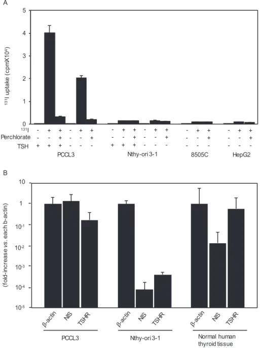
We first confirmed the ability/inability of 131I uptake in PCCL3 and Nthy-ori 3-1 cells cultured in the presence or absence of TSH [10, 12, 13]; an anaplastic thyroid cancer cell line 8505C and a hepatocellular carcinoma cell line HepG2 were used as negative controls. We have A
0 1 2 3 4 5
131I uptake (cpmX104)
131I Perchlorate
TSH
PCCL3 + ++ + + +
-- -
- - - - + + - - +
Nthy-ori 3-1 + ++ + + + -- -
- - - - + +
+ - -
8505C - - - - + + - - +
HepG2 - - - - + + - - +
Relative mRNA levels (fold-increasevs. each b-actin) B
10
1
10-1
10-2 10-3 10-4 10-5
PCCL3 Nthy-ori 3-1 Normal human
thyroid tissue
Figure 1. 131Iodine (131I) uptake in PCCL3, Nthy-ori 3-1, 8505C, and HepG2 cells, and thyrotropin receptor (TSHR) and sodium/iodine symporter (NIS) messenger RNA (mRNA) expression in PCCL3 and Nthy-ori 3-1 cells. A, Cells were cultured in the presence or absence of thyrotropin (TSH) and/or perchlorate and then incubated with 10 μCi/mL 131I for 30 minutes. 131I taken up into the cells was measured as described in “Materials and Methods.”
Data are means ± SD (n = 3). B, Total RNA was extracted from PCCL3 and Nthy-ori 3-1 cells and subjected to RT-PCR for TSHR, NIS, and β-actin mRNAs as described in “Materials and Methods.” Data are means ± ranges (n = 2).
Downloaded from https://academic.oup.com/jes/article/4/11/bvaa146/5912612 by Nagasaki University Library user on 04 December 2020
previously shown that PCCL3 cells incorporated 131I in a dose-dependent manner, reaching the peak at 30 minutes in the presence of TSH [10]. Therefore, 131I uptake was de- termined 30 minutes after the addition of 131I into the cul- ture medium. As shown in Fig. 1A, PCCL3 cells maintained with 3H clearly incorporated 131I, which was completely abolished by 5 μM NaClO4 (perchlorate), a competitive inhibitor of NIS, confirming NIS-mediated iodine uptake.
Removal of TSH for 3 days reduced 131I uptake by ap- proximately 50%, indicating the TSH-dependent action of iodine uptake. On the other hand, Nthy-ori 3-1 cells, irre- spective of the presence or absence of TSH, showed almost negligible 131I uptake; these low levels of iodine uptake are likely nonspecific because the similarly low levels of 131I up- take were also observed in 8505C and HepG2 cells, both of which have been shown not to express NIS [29, 30].
We next examined mRNA expression of thyroid-specific molecules critical for TSH-dependent iodine uptake;
namely TSHR and NIS. Thus, mRNAs for TSHR and NIS were readily detectable in PCCL3 cells and human thy- roid tissue (a positive control), but extremely low levels of their expression was barely observed in Nthy-ori 3-1 cells (Fig. 1B), supporting the 131I uptake results in Fig. 1A.
These results clearly demonstrate the substantial dif- ferences in the differentiated status between PCCL3 and Nthy-ori 3-1 cells. PCCL3 cells are well differentiated, expressing TSHR and NIS mRNAs, and able to take up iodine, whereas Nthy-ori 3-1 cells are conversely less differ- entiated, expressing few mRNAs for these 2 molecules, and unable to take up iodine. Nevertheless, both cell lines have long been used comparably as normal thyroid cell lines.
These results then caused doubts about the previous studies on the effects of iodine on the survival and function of thyroid cells using Nthy-ori 3-1 cells (see “Introduction”) and prompted us to reevaluate and compare some of these results—iodine cytotoxicity and iodine modulation of autophagic activity—using PCCL3 and Nthy-ori 3-1 cells in parallel in our own hands. The concentrations of iodine we used are 5 × 10⁻⁹ M, the physiological concen- tration in human sera; 10–6 M, the concentration that sup- presses iodine uptake in cultured thyroid cells; 10–3 M, excess iodine; and 1 and 5 × 10⁻² M, far excess iodine [10].
Fig. 2 demonstrates the results of the study on iodine cyto- toxicity. Up to 10–2 M iodine had no effect and only the highest dose of iodine (5 × 10⁻² M) induced 20% to 40%
cell death in 24 hours in both cell lines as well as 8505C and HepG2 cells. Perchlorate had no effect on cell death in PCCL3 cells, indicating that this mild cytotoxic effect of iodine is attributed to extracellular iodine present in the culture medium, not intracellular iodine incorporated into the cells. Thus, both thyroid cell lines as well as 8505C and HepG2 cells are comparably resistant against extracellular iodine cytotoxicity, and iodine incorporated into PCCL3 cells is likely harmless, even when the cells were exposed to far excess iodine (10–2 M). To the best of our knowledge, this is the first study on iodine cytotoxicity in PCCL3 cells.
Similar results were reported in Nthy-ori 3-1 cells [4-6], human thyroid cells, and FRTL5 cells [31, 32]. Although the mechanism(s) for cytotoxic effect of far excess iodine (5 × 10⁻² M) in the culture medium is at present unknown, some interaction between iodine and membranous pro- teins/lipids may explain this effect [33].
Cell survival rate (%)
0 50 100 150
NaI (M) Perchlorate
PCCL3
- +
Nthy-ori 3-1 -
8505C -
HepG2 -
Figure 2. Cytotoxicity of iodine in PCCL3, Nthy-ori 3-1, 8505C, and HepG2 cells. The cells, cultured in the presence or absence of perchlorate, were incubated with up to 5 × 10⁻² M iodine for 24 hours and cell viability was determined as described in “Materials and Methods.” Data are means ± SD (n = 3).
Downloaded from https://academic.oup.com/jes/article/4/11/bvaa146/5912612 by Nagasaki University Library user on 04 December 2020
The results of autophagy experiments are shown in Figs. 3 and 4. A wide range of iodine (5 × 10⁻⁹ M to 10–3 M) increased LC3 puncta and deceased p62 fluorescence intensity 0.5 to 2 hours after the addition of iodine in IF in PCCL3 cells (Fig. 3A and 3B), clearly demonstrating induc- tion of autophagy by iodine. The data are also confirmed by IB in cells incubated for 1 hour with 10–3 M iodine (Fig. 3C). Perchlorate abolished the effect of iodine (10–6
M) on autophagy almost completely, indicating that this effect is exerted by intracellular iodine (Fig. 3D). Far excess iodine (5 × 10⁻² M) induced the lower degree of autophagy as compared to the lower doses of iodine, which is likely attributed to the cytotoxic effect of far excess iodine as shown in Fig. 2. In contrast, 5 × 10⁻⁹ M to 10–3 M iodine had no effect, and 5 × 10⁻² M iodine reduced LC3 puncta and increased p62 levels in Nthy-ori 3-1 cells (Fig. 4), again
0 1 2 3
** **
fluorescence intensity Of p62 (105/per cell)
NaI 5 x 10⁻⁹M NaI 10-6M
A
B
** **
NaI 10-3M NaI 5 x 10⁻²M
0 10
Number of LC3 puncta (per cell)
0 0.5 1 2
** **
0 0.5 1 2
** **
0 0.5 1 2
0 0.5 1 2 (h)
NaI 1 mM LC3
p62
0 1 (h)
1 mM Na for 1 h ( - ) ( + )
LC3 14
16
C
p62 β-action
62 42(kDa)
0 1 2
0 5 10 Fluorescence intensity of p62 (105/per cell)Number of LC3 puncta (per cell)
NaI Perchlorate -
-
- -
+ + + +
* *
* *
D
Figure 3. Control of autophagic activity by iodine in PCCL3 cells. A, Cells were incubated with up to 5 × 10⁻² M iodine for up to 2 hours, and LC3 puncta and P62 fluorescence intensities were quantified by immunofluorescence (IF) as described in “Materials and Methods.” B, Representative photographs for LC3 puncta and p62 fluorescence in the cells incubated with 10–3 M iodine for up to 1 hour are shown. C, Western blotting of LC3 and p62 in the cells incubated with 10–3 M iodine for 1 hour are shown. D, Cells, cultured in the presence or absence of perchlorate, were incubated with 10–3 M iodine for 1 hour, followed by quantification of LC3 puncta and p62 fluorescence intensities by IF as described in “Materials and Methods.” *P less than .05. **P less than .01.
Downloaded from https://academic.oup.com/jes/article/4/11/bvaa146/5912612 by Nagasaki University Library user on 04 December 2020
suggesting that far excess iodine suppressed autophagy by its cytotoxicity. Although our data are somewhat different from the previous study, in which 10–4 to 5 × 10⁻² M iodine decreased LC3-II in Western blotting and autophagosome and autolysosome formation with the LC3-RFP-GFP vector in Nthy-ori 3-1 cells [8], our results demonstrate that in- duction of autophagy by iodine ranging from 5 × 10⁻⁹ M to 10–3 M in PCCL3 cells are due to iodine incorporated into the cells, which cannot be seen in Nthy-ori 3-1 cells that are incapable of incorporating iodine.
The pathophysiological significance of iodine-induced autophagy is unknown. We first thought that iodine in- crease in reactive oxygen species (ROS) might cause autophagy induction because it is well known that oxida- tive stress induces autophagy through the PERK-eLF2α- ATF4-CHOP pathway and mitogen-activated protein kinases such as c-Jun N-terminal kinase 1 [34, 35]. Indeed ROS elevation by iodine has previously been reported in FRTL5 and PCCL3 cells; an approximately 2-fold increase in intracellular ROS by 10–3 M iodine in 30 minutes and a huge increase in mitochondrial ROS by 10–2 M iodine in 2 hours [36] and a biphasic increase (at 2 and 24, not 4, hours) by 10–4 M iodine [37]. However, we could not de- tect ROS increase in iodine-treated PCCL3 cells using 2’,7’- dichlorofluorescin diacetate in our hands (data not shown).
We also found one report inconsistent with our data [38], in which low doses of iodine (10–8 to 10–5 M) stimu- lated cell proliferation but a higher dose (10–3 M) induced apoptosis and autophagy in the thyroid cancer cell line BCPAP. BCPAP cells were established from a differentiated
papillary thyroid cancer, but likely gained an undifferen- tiated phenotype during long culture [39, 40]. Indeed, BCPAP cells are reported not to express NIS nor take up iodine [12, 39]. Therefore, the previously reported data may indicate a possibility for high sensitivity of BCPAP cells to extracellular iodine.
In conclusion, our results indicate that the in vitro ef- fects of high doses of iodine on thyroid cells so far reported using Nthy-ori 3-1 cells are likely due to iodine present in the culture medium, not iodine incorporated into the cells.
Although cultured cell lines are useful tools for in vitro studies on cell behavior and function, appropriate one(s) should be chosen for a certain experiment in general, and in particular, the effect of iodine, especially that incorpor- ated into thyroid cells, should be studied with functional, iodine-uptake competent thyroid cells such as PCCL3 and FRTL5 cells, not Nthy-ori 3-1 cells.
Acknowledgments
Financial Support: This work was supported in part by the Japan Society for the Promotion of Science (KAKENHI Grant No.
16K00548 to T.K. and 19K09028 to Y.N.).
Additional Information
Correspondence: Yuji Nagayama, MD, PhD, Department of Mo- lecular Medicine, Atomic Bomb Disease Institute, Nagasaki Uni- versity, 1-12-4 Sakamoto, Nagasaki 852-8523, Japan. E-mail:
Disclosure Summary: The authors declare no conflict of interest.
fluorescence intensity Of p62 (106/per cell)Number of LC3 puncta (per cell)
NaI 5 x 10⁻⁹M NaI 10-6M NaI 10-3M NaI 5 x 10-2M
*
0 10
0 0.5 1 2 0 0.5 1 2 0 0.5 1 2 0 0.5 1 2
**
0 1
Figure 4. Control of autophagic activity by iodine in Nthy-ori 3-1 cells. Cells were incubated with up to 5 × 10⁻² M iodine for up to 2 hours, and LC3 puncta and p62 fluorescence intensities were quantified by immunofluorescence as described in “Materials and Methods.” *P less than .05. **P less than .01.
Downloaded from https://academic.oup.com/jes/article/4/11/bvaa146/5912612 by Nagasaki University Library user on 04 December 2020
Data Availability: All data generated during this study are in- cluded in this published article.
References
1. CVCL_6712. CVCL_6712 https://web.expasy.org/cellosaurus/
CVCL_6712.txt.
2. CVCL_0265. CVCL_0265 https://web.expasy.org/cellosaurus/
CVCL_0265.txt.
3. CVCL_2659. CVCL_2659 https://web.expasy.org/cellosaurus/
CVCL_2659.txt.
4. Liu H, Zeng Q, Cui Y, et al. The effects and underlying mech- anism of excessive iodide on excessive fluoride-induced thyroid cytotoxicity. Environ Toxicol Pharmacol. 2014;38(1):332-340.
5. Liu H, Zeng Q, Cui Y, et al. The role of the IRE1 pathway in excessive iodide- and/or fluoride-induced apoptosis in Nthy-ori 3-1 cells in vitro. Toxicol Lett. 2014;224(3):341-348.
6. Liu J, Mao C, Dong L, et al. Excessive iodine promotes pyroptosis of thyroid follicular epithelial cells in Hashimoto’s thyroiditis through the rOS-NF-κB-NLRP3 pathway. Front Endocrinol (Lausanne). 2019;10:778.
7. Chen X, Huang H, Liang B, Zhou J. Abnormal iodine nutrition- induced ER stress upregulates MCP-1 expression through P38/
MAPK signaling pathway in thyroid cells. Biol Trace Elem Res.
2019;191(1):98-103.
8. Xu C, Wu F, Mao C, et al. Excess iodine promotes apoptosis of thyroid follicular epithelial cells by inducing autophagy sup- pression and is associated with Hashimoto thyroiditis disease. J Autoimmun. 2016;75:50-57.
9. Nagayama Y. General commentary: excessive iodine promotes pyroptosis of thyroid follicular epithelial cells in Hashimoto’s thyroiditis through the ROS-NF-κB-NLRP3 pathway. Front Endocrinol 2020;11:581.
10. Kurashige T, Shimamura M, Nagayama Y. N-Acetyl-L- cysteine protects thyroid cells against DNA damage induced by external and internal irradiation. Radiat Environ Biophys.
2017;56(4):405-412.
11. Grollman EF, Smolar A, Ommaya A, Tombaccini D, Santisteban P. Iodine suppression of iodide uptake in FRTL-5 thyroid cells. Endocrinology. 1986;118(6):2477-2482.
12. Tuncel M, Aydin D, Yaman E, et al. The comparative effects of gene modulators on thyroid-specific genes and radioiodine up- take. Cancer Biother Radiopharm. 2007;22(3):443-449.
13. Lemoine NR, Mayall ES, Jones T, et al. Characterisation of human thyroid epithelial cells immortalised in vitro by simian virus 40 DNA transfection. Br J Cancer. 1989;60(6):897-903.
14. Kurashige T, Shimamura M, Nagayama Y. Differences in quantification of DNA double-strand breaks assessed by 53BP1/γH2AX focus formation assays and the comet assay in mammalian cells treated with irradiation and N-acetyl-L- cysteine. J Radiat Res. 2016;57(3):312-317.
15. Tran N, Valentín-Blasini L, Blount BC, et al. Thyroid- stimulating hormone increases active transport of per- chlorate into thyroid cells. Am J Physiol Endocrinol Metab.
2008;294(4):E802-E806.
16. Kurashige T, Shimamura M, Yasui K, et al. Studies on expres- sion of aldehyde dehydrogenase in normal and cancerous tis- sues of thyroids. Horm Metab Res. 2015;47(3):194-199.
17. Chinnappa P, Taguba L, Arciaga R, et al. Detection of thyrotropin-receptor messenger ribonucleic acid (mRNA) and thyroglobulin mRNA transcripts in peripheral blood of patients with thyroid disease: sensitive and specific markers for thyroid cancer. J Clin Endocrinol Metab. 2004;89(8):3705-3709.
18. Li W, Ain KB. Human sodium-iodide symporter (hNIS) gene ex- pression is inhibited by a trans-active transcriptional repressor, NIS-repressor, containing PARP-1 in thyroid cancer cells.
Endocr Relat Cancer. 2010;17(2):383-398.
19. Endo T, Kobayashi T. Thyroid-stimulating hormone receptor in brown adipose tissue is involved in the regulation of thermogen- esis. Am J Physiol Endocrinol Metab. 2008;295(2):E514-E518.
20. Chai W, Yin X, Ren L, et al. Sodium/iodide symporter gene transfection increases radionuclide uptake in human cisplatin-resistant lung cancer cells. Clin Transl Oncol.
2015;17(10):795-802.
21. Knauf JA, Ma X, Smith EP, et al. Targeted expression of BRAFV600E in thyroid cells of transgenic mice results in papil- lary thyroid cancers that undergo dedifferentiation. Cancer Res.
2005;65(10):4238-4245.
22. Kurashige T, Nakajima Y, Shimamura M, Yamada M, Nagayama Y. Hormonal regulation of autophagy in thyroid PCCL3 cells and the thyroids of male mice. J Endocr Soc.
2020;4(7):bvaa054.
23. RRID:AB_2274121, http://scicrunch.org/resolver/AB_2274121.
24. RRID:AB_2099233, http://scicrunch.org/resolver/AB_2099233.
25. RRID:AB_2687531, http://scicrunch.org/resolver/AB_2687531.
26. RRID:AB_88247, http://scicrunch.org/resolver/AB_88247.
27. RRID:AB_2714189, http://scicrunch.org/resolver/AB_2714189.
28. RRID:AB_330924, http://scicrunch.org/resolver/AB_330924.
29. Bauriaud-Mallet M, Vija-Racaru L, Brillouet S, et al. The cholesterol-derived metabolite dendrogenin A functionally re- programs breast adenocarcinoma and undifferentiated thyroid cancer cells. J Steroid Biochem Mol Biol. 2019;192:105390.
30. Xia W, Li D, Wang G, et al. Small activating RNA upregulates NIS expression: promising potential for hepatocellular carcinoma endoradiotherapy. Cancer Gene Ther. 2016;23(10):333-340.
31. Vitale M, Di Matola T, D’Ascoli F, et al. Iodide excess induces apoptosis in thyroid cells through a p53-independent mechanism involving oxidative stress. Endocrinology. 2000;141(2):598-605.
32. Golstein J, Dumont JE. Cytotoxic effects of iodide on thyroid cells: difference between rat thyroid FRTL-5 cell and primary dog thyrocyte responsiveness. J Endocrinol Invest. 1996;19(2):119-126.
33. Pereira A, Braekman JC, Dumont JE, Boeynaems JM.
Identification of a major iodolipid from the horse thyroid gland as 2-iodohexadecanal. J Biol Chem. 1990;265(28):17018-17025.
34. B’chir W, Maurin AC, Carraro V, et al. The eIF2α/ATF4 pathway is essential for stress-induced autophagy gene expres- sion. Nucleic Acids Res. 2013;41(16):7683-7699.
35. Son Y, Cheong YK, Kim NH, Chung HT, Kang DG, Pae HO.
Mitogen-activated protein kinases and reactive oxygen species:
how can ROS activate MAPK pathways? J Signal Transduct.
2011;2011:792639.
36. Serrano-Nascimento C, da Silva Teixeira S, Nicola JP, Nachbar RT, Masini-Repiso AM, Nunes MT. The acute inhibi- tory effect of iodide excess on sodium/iodide symporter ex- pression and activity involves the PI3K/Akt signaling pathway.
Endocrinology. 2014;155(3):1145-1156.
Downloaded from https://academic.oup.com/jes/article/4/11/bvaa146/5912612 by Nagasaki University Library user on 04 December 2020
37. Yao X, Li M, He J, et al. Effect of early acute high concentrations of iodide exposure on mitochondrial superoxide production in FRTL cells. Free Radic Biol Med. 2012;52(8):1343-1352.
38. Zhang D, Xu X, Li J, et al. High iodine effects on the prolifer- ation, apoptosis, and migration of papillary thyroid carcinoma cells as a result of autophagy induced by BRAF kinase. Biomed Pharmacother. 2019;120:109476.
39. van Staveren WC, Solís DW, Delys L, et al. Human thyroid tumor cell lines derived from different tumor types present a common de- differentiated phenotype. Cancer Res. 2007;67(17):8113-8120.
40. Landa I, Pozdeyev N, Korch C, et al. Comprehensive gen- etic characterization of human thyroid cancer cell lines:
a validated panel for preclinical studies. Clin Cancer Res.
2019;25(10):3141-3151.
Downloaded from https://academic.oup.com/jes/article/4/11/bvaa146/5912612 by Nagasaki University Library user on 04 December 2020