INTRODUCTION
Angiosarcomas are vascular endothelium-derived malignant tumors that arise in blood vessel walls, and account for only 2.3% of all soft tissue sarcomas in adults. Angiosarcomas mainly arise in the skin, breast,
soft tissues, bone, and viscera, with the skin and breast as the most common site of occurrence for primary an- giosarcoma1). Primary hepatic angiosarcoma(PHA)is rare and accounts for less than 5% of all angiosarco- mas1,2). We report here in a rare case of PHA in which the patient presented with a chief complaint of hemop- tysis.
CASE REPORT
A 61-year-old man was evaluated by the Depart- ment of Gastroenterology at Dokkyo Medical Universi- ty Koshigaya Hospital for a chief complaint of right- sided abdominal pain. He had a past history of Received August 13, 2014;accepted October 7, 2014
Reprint requests to:Toshikuni Suda
Department of Gastroenterology, Dokkyo Medical University Koshigaya Hospital 2-1-50 Minami-Koshigaya, Koshigaya-Shi, Saitama 343-8555, Japan
A Case of Hemangiosarcoma of the Liver which led to a Diagnosis with Hemoptysis
Toshikuni Suda1), Yasumi Katayama1), Tatsuhiko Sekiyama1), Hidetsugu Yamagishi2), Akihiro Nakamoto1), Akihiro Kitahama1), Yumi Kusano1), Itsuo Terauchi1), Koji Toyoda1), Masaya Tamano1)
1)Department of Gastroenterology, Dokkyo Medical University Koshigaya Hospital
2)Anatomic and Diagnostic Pathology, Dokkyo Medical University
SUMMARY
Angiosarcoma is a vascular endothelium-derived malignant tumor that arises in blood vessel walls, ac- counting for only 2.3% of soft tissue sarcomas in adults. Primary hepatic angiosarcoma(PHA)is rare, com- prising<5% of all angiosarcomas. We report a case of PHA in a 61-year-old man evaluated by another clin- ic around our hospital for a chief complaint of hemoptysis in May 2012. Chest computed tomography(CT)
showed abnormal shadows in bilateral lung fields, so he was referred to Department of Respiratory Medi- cine at our hospital in late July. However, no definitive diagnosis was not made, even after bronchoscopy. In mid-August, he presented to the outpatient clinic of the respiratory department with a chief complaint of right-sided abdominal pain. Abdominal CT showed a liver lesion, and he was urgently admitted to our de- partment. Initial physical examination was unremarkable except for palpable liver in the right hypochondri- um. Tumor markers for liver and biliary cancers were all within normal limits, and negative results were obtained for hepatitis B and C virus. CT, ultrasonography, and(MRI)identified a large lesion replacing the right hepatic lobe and medial segment of the left hepatic lobe. Hemangioma or hepatic angiosarcoma was suspected, accompanied by intraperitoneal rupture. Transcatheter arterial embolization was attempted, but had to be discontinued, and the patient died from hemorrhagic shock due to tumor rupture after onset of abdominal pain. The PHA which assumes hemoptysis primary symptom is extremely rare, and by reports for the past ten years searched using PubMed, this is the second report in the world.
Case Report
Chest CT showed ground glass opacities with indis- tinct borders bilaterally, predominantly in the lower lung fields(Fig. 1). Plain abdominal CT showed a 15×
12-cm lesion that had replaced the right hepatic lobe and medial segment of the left hepatic lobe(Fig. 2a).
The tumor showed slightly lower density than normal liver parenchyma, with faint high-density areas interi- orly. The tumor increased in comparison with the last CT clearly. The tumor interior showed patchy en- hancement early in the arterial phase(Fig. 2b), and heterogeous faint enhancement in the portal/equilibri- um phase( Fig. 2 c). Besides the tumor described appendicitis at 9 years old and gastric ulcer at 57 years
old. The family history was positive for diabetes in both parents. He had been smoking 25 cigarettes/day for 40 years, did not drink alcohol, and was employed as a high school teacher.
The patient was evaluated by another clinic around our hospital for a chief complaint of hemoptysis in May 2012. Chest computed tomography(CT)showed ab- normal shadows bilaterally in the lung fields, so he was referred to Department of Respiratory Medicine at our hospital in late July. It was not shown in a figure for an indistinct image, but about 10 cm tumors were found in liver at this time. However, no definitive diagnosis was made even after bronchoscopy. In mid-August, he pre- sented to the outpatient clinic of the Department of Respiratory Medicine with a chief complaint of right- sided abdominal pain, and abdominal CT showed a le- sion in the liver. He was therefore urgently admitted to our department.
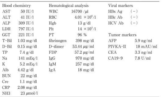
Initial physical examination was unremarkable ex- cept for a palpable liver 3 fingerbreadths in the right hypochondriac region. Table 1 shows blood test results from the initial physical examination at our depart- ment. Levels of LDH, g-GTP, WBC count, CRP, and co- agulation/fibrinolytic factors were all elevated. Tumor markers for liver cancer and biliary cancer, including AFP, PIVKA-II, CEA, and CA19-9 were all within normal limits, and negative results were obtained for HBs antigen and HCV antibody.
GGT 221 IU/l PT 96 % Tumor markers
T-Bil 1.03 mg/dl fibrinogen 398 mg/dl AFP 5.9 ng/ml
D-Bil 0.15 mg/dl D-dimer 53.44mg/ml PIVKA-II 18 mAU/ml
TP 7.4 g/dl FDP 57.2mg/ml CEA 3.3 ng/ml
Na 141 mEq/l IgG 970 mg/dl CA19-9 7.8 U/ml
K 5.2 mEq/l IgM 257 mg/dl
Alb 4.42 g/dl IgA 18 mg/dl
BUN 22 mg/dl Cre 1.1 mg/dl CRP 2.08 mg/dl NH3 23mmol/l
Figure 1 Plain chest computed tomography(CT)
Plain chest CT shows ground glass opacities with indistinct borders predominantly in the lower lung fields bilaterally.
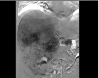
es in the anterior segment of the right hepatic lobe and countless cotton wool-like areas corresponding to the giant tumor in the liver S6(Fig. 3).
Based on these imaging findings, hepatic tumor was diagnosed and suspected as either hepatic hemangioma or hepatic angiosarcoma, with accompanying intraperi- toneal rupture. Transcatheter arterial embolization
(TAE)was attempted on the 11th day after admission, but had to be discontinued due to worsening respirato- ry status. Further TAE was scheduled for a later time, but during systemic management, the patient died from hemorrhagic shock due to tumor rupture after onset of abdominal pain on the 14th day after admis- sion. Autopsy was performed.
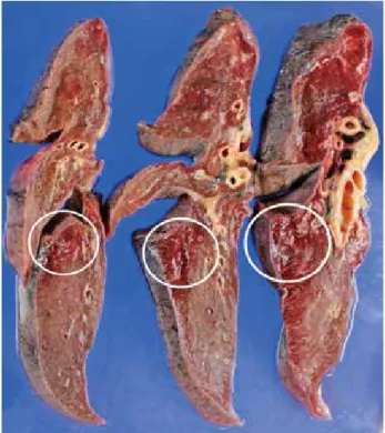
Figure 4 shows the excised liver at autopsy. The liv- er was enlarged, weighing 2780 g, and a soft, dark-red tumor lesion measuring about 19.5×16.5 cm was pres- ent, mainly involving the anterior segment of the right lobe from the surface. In addition, a fissure in the he- patic capsule was observed, measuring about 7.0×
above, multiple tumors with ring-like enhancement were evident in both hepatic lobes, and accumulation of hyperdense fluid surrounded the liver.
Abdominal ultrasonography showed the neoplastic lesion which almost accounted for the whole of liver right lobe.
Abdominal magnetic resonance imaging( MRI)
showed tumor replacing the anterior segment of the right hepatic lobe and medial segment of the left he- patic lobe. T1-weighted imaging showed signal hypoin- tensity of the tumor with a septum-like structure in the interior. T2-weighted imaging showed signal hy- perintensity throughout. Diffusion-weighted imaging revealed a mixture of high and low signal intensity.
Angiography was performed for further evaluation.
CT during transarterial portography showed the tu- mor lesion as a large perfusion defect in liver S6. CT during hepatic arteriography showed enhancement of the entire tumor from the margins. Digital-subtraction angiography(DSA)showed marked dilation of branch-
Figure 2 Abdominal CT
Plain abdominal CT shows a 15×12-cm tumor replacing the right hepatic lobe and medial segment of the left hepatic lobe. a)The tumor shows slightly lower density than the normal liver parenchyma, with faint high- density areas interiorly. b)On a dynamic study, the tumor interior showed patchy enhancement early in the arterial phase(b), and heterogenous faint enhancement in the portal/equilibrium phase(c).
a
b
phic cells and atypical round cells among the vascular endothelial cells(Fig. 7a). Immunohistological staining of these cells yielded positive results for factor VIII and CD34(Fig. 7b). These lesions were diagnosed as lung metastases from hepatic angiosarcoma.
DISCUSSION
PHA is more common in men, and the mean age of patients with PHA is 59 years3). The 50 cases in our survey ranged in age from 29 to 81 years, with a mean age of 58.2 years. These 50 cases included 39 men(78
%)and 11 women(22%). Initial symptoms include abdominal pain in 24(48%), malaise and anorexia in 11(22%), jaundice in 3(6%), fever in 2(4%), hemop- tysis in 1(2%), and anemia in 1(2%), while 8 patients
(16%)were asymptomatic. The cause of abdominal pain is usually intraperitoneal rupture of the PHA, and intraperitoneal hemorrhage has been reported in 15-27% of cases of PHA4,13). Among the 50 patients with PHA in our survey, 13(26%)had intraperitoneal hemorrhage.
PHA may be difficult to diagnose, with some cases, as in our patient, diagnosed on autopsy12,14). In addition, specific tumor markers for PHA have yet to be identi- fied, and levels of AFP, CEA, and CA19-9 have been within normal limits, or only slightly elevated, in the reported cases. From a morphological perspective, PHA occurs as multiple nodules, dominant masses, or a 4.5 cm in the posterior segment of the right hepatic
lobe(lateral S6)and showing adherent blood clots around this site. About 2700 ml of blood, together with blood clots, was seen in the peritoneal cavity at autop- sy, due to intraperitoneal hemorrhage because of lesion rupture.
Figure 5 shows microscopic images of the liver. His- tologically, low magnification showed dilation of various sized blood vessels, with partial sloughing, hemorrhage, and degeneration of blood vessel walls(Fig. 5a). The other nodular lesions showed similar findings. High magnification showed a high nucleus/cytoplasm ratio, with proliferation of large polymorphic cells and atypi- cal spindle cells and round cells predominantly in the vascular lumens(Fig. 5b). Hepatic angiosarcoma was suspected based on these findings. Immunohistological staining yielded negative results for pan-cytokeratin markers(AE1/AE3), and epithelial tumor was ruled out(Fig. 5c). Findings were positive for both factor VIII and CD34 as vascular endothelial cell markers
(Fig. 5d). These immunohistological findings confirmed the hepatic tumor as angiosarcoma.
Figure 6 shows the excised right lung. Dark-red nodular lesions measuring about 12×11 mm and about 8×5 mm were present in the right lung lower lobe.
The histological appearance was similar to the liver le- sions, including hemorrhage, proliferation of various sized blood vessels, and proliferation of large polymor-
Figure 4 Liver excised at autopsy
The liver is enlarged, weighing 2780 g, and a dark red tumor lesion measuring about 19.5×16.5 cm is present mainly in liver segment 6.
Figure 3 Digital-subtraction angiography(DSA)
DSA shows countless cotton wool-like areas corresponding to the giant tumor in liver segment 5.
diffusely infiltrating lesion, and exhibits a spectrum of appearances on CT or MRI15). Abdominal contrast-en- hanced ultrasonography may also be useful11), but is not commonly performed. Liver biopsy, which is re- quired for histological diagnosis, is not currently rec- ommended because of an increased risk of hemor- rhage5,6,12,14). Diagnosis is currently based on imaging findings showing a rapidly enlarging hemangioma-like tumor, in which case PHA should be strongly suspect- ed. Moreover, the risk of spontaneous rupture of he- patic hemangioma is small, estimated as less than 1%.
Hemorrhage from a hepatic vascular tumor may thus suggest a diagnosis of angiosarcoma7).
Hemoptysis as the initial symptom in patients with PHA is rare, having only been reported once previous- ly7), and our patient is the second such case to be re- ported. Our patient was a high school teacher, and al- though he had a history of smoking, previous chest X- rays during medical check-ups had shown no abnormalities. The patient only developed his main
a b c d
Figure 5 Histological findings of the liver
a)Histologically, low magnification shows dilation of various sized blood vessels, with partial sloughing, hemorrhage, and degeneration of blood vessel walls. b)High magnification shows a high nucleus/cytoplasm ratio, with proliferation of large polymorphic cells and atypical spindle cells and round cells predominantly in the vascular lumens. c)Immunohistological staining showed negative results for pan-cytokeratin markers
(AE1/AE3). d)Findings were positive for both factor VIII and CD34 as vascular endothelial cell markers.
Figure 6 Right lung excised at autopsy
Dark red nodular lesions measuring about 12×11 mm and about 8×5 mm are present in the right lung lower lobe.
complaint of hemoptysis in May 2012, at which time abnormal chest shadows were noted. Subsequently, the patient experienced abdominal pain due to intraperito- neal hemorrhage of the PHA within about 3 months, and died 14 days after the onset of abdominal pain. The clinical course was thus rapid.
Our patient had not undergone abdominal imaging studies previously, but the tumor was large(15 cm)at the time of diagnosis, and is thought to have shown rapid growth based on the clinical history. Among the 50 cases that we surveyed, survival time ranged from 3 days to 40 months among the 35 cases for which out- comes were documented,(mean survival time, 11.1 months). However, most patients with PHA die within 6 months after diagnosis16). The prognosis differs sig- nificantly between patients without and with organ metastases(9 months vs. 3 months, p=0.172), probably due to increased treatment options in patients without metastases8).
Because PHA is a rare disease with very rapid pro- gression, systematic treatments have yet to be estab- lished. The first option for treatment after definitive diagnosis is usually surgical resection. Duan et al. re- ported a median survival time of 41 months( 2 3 - 84 months)in 5 patients who underwent liver resec- tion1 0). Improved prognosis with postoperative chemotherapy or the addition of transarterial chemo- embolization(TACE)of the liver has also been re- ported6,9). Although chemotherapy has been tried for unresectable cases, few reports have been made. Kim et al. described a median survival time of 86 days
(range, 8-439 days)in 4 patients with unresectable PHA treated using chemotherapy7). Transcatheter ar- terial embolization of the liver for intraperitoneal hem- orrhage of PHA, and TACE to control hemorrhage, was reported as effective, but did not improve progno- sis 4,8). Unfortunately, TACE was not performed in our case, but even if it had been, prognosis would most Figure 7 Histological findings of the right lung
a)Histological appearance of the lung sample was similar to that of the liver lesions, including hemorrhage, proliferation of various sized blood vessels, and proliferation of large polymorphic cells and atypical round cells among the vascular endothelial cells. b)Immunohistological staining of these cells was also positive for factor VIII and CD34. These findings were consistent with lung metastases from hepatic angiosarcoma.
b
likely have been poor. Liver transplantation has also been abandoned because of a high recurrence rate and poor post-transplantation survival17).
REFERENCES
1) Lahat G, Dhuka AR, Hallevi H, et al:Angiosarcoma:
clinical and molecular insights. Ann Surg 251:1098- 1106, 2010.
2) Fayette J, Martin E, Piperno-Neumann S, et al:An- giosarcomas, a heterogeneous group of sarcomas with specific behavior depending on primary site:a retro- spective study of 161 cases. Ann Oncol 18:2030- 2036, 2007.
3) Zheng YW, Zhang XW, Zhang JL, et al:Primary He- patic Angiosarcoma and Potential Treatment Options, J Gastroenterol Hepatol, 2013.
4) Leowardi C, Hormann Y, Hinz U, et al:Ruptured an- giosarcoma of the liver treated by emergency cathe- ter-directed embolization. World J Gastroenterol 12:
804-808, 2006.
5) Stambo GW, Guiney MJ:Hepatic angiosarcoma pre- senting as an acute intraabdominal hemorrhage treat- ed with transarterial chemoembolization, Sarcoma, 2007, 90169, 2007.
6) Cioffi-Pretti JL, Kalof AN, Ebert G, et al:Hepatic an- giosarcoma five years following spontaneous intraperi- toneal bleed of a hepatic mass. Rare Tumors 1:e33, 2009.
7) Kim HR, Rha SY, Cheon SH, et al:Clinical features and treatment outcomes of advanced stage primary hepatic angiosarcoma. Ann Oncol 20:780-787, 2009.
8) Park YS, Kim JH, Kim KW, et al:Primary hepatic angiosarcoma:imaging findings and palliative treat-
ment with transcatheter arterial chemoembolization or embolization. Clin Radiol 64:779-785, 2009.
9) Zhou YM, Li B, Yin ZM, et al:Results of hepatic re- section for primary hepatic angiosarcoma in adults.
Med Sci Monit 16:CR61-66, 2010.
10) Duan XF, Li Q, Primary hepatic angiosarcoma:a ret- rospective analysis of 6 cases. J Dig Dis 13:381-385, 2012.
11) Wang L, Lv K, Chang XY, et al:Contrast-enhanced ultrasound study of primary hepatic angiosarcoma:a pitfall of non-enhancement. Eur J Radiol 81:2054- 2059, 2012.
12) Wiland HOt, Pai RK, Purysko AS:Hepatic angiosar- coma mimicking sinusoidal obstruction syndrome/ve- noocclusive disease:a pathologic-radiologic correla- tion. Ann Diagn Pathol 16:275-279, 2012.
13) Molina E, Hernandez A:Clinical manifestations of primary hepatic angiosarcoma. Dig Dis Sci 48:677- 682, 2003.
14) Lee SW, Song CY, Gi YH, et al:Hepatic angiosarco- ma manifested as recurrent hemoperitoneum. World J Gastroenterol 14:2935-2938, 2008.
15) Koyama T, Fletcher JG, Johnson CD, et al:Primary hepatic angiosarcoma:findings at CT and MR imag- ing. Radiology 222:667-673, 2002.
16) Bioulac-Sage P, Laumonier H, Laurent C, et al:Be- nign and malignant vascular tumors of the liver in adults. Semin Liver Dis 28:302-314, 2008.
17) Orlando G, Adam R, Mirza D, et al:Hepatic heman- giosarcoma:an absolute contraindication to liver transplantation--the European Liver Transplant Reg- istry experience. Transplantation 95:872-877, 2013.