Acta Med. Nagasaki 38:232 - 236
Effects of Nicardipine and Prostaglandin E1 on Coronary Hemodynamics in Dogs with Coronary Artery Stenosis.
Masahiko Miyako
Department of Anesthesiology, Nagasaki University School of Medicine, 1-7-1, Sakamoto, Nagasaki 852, Japan
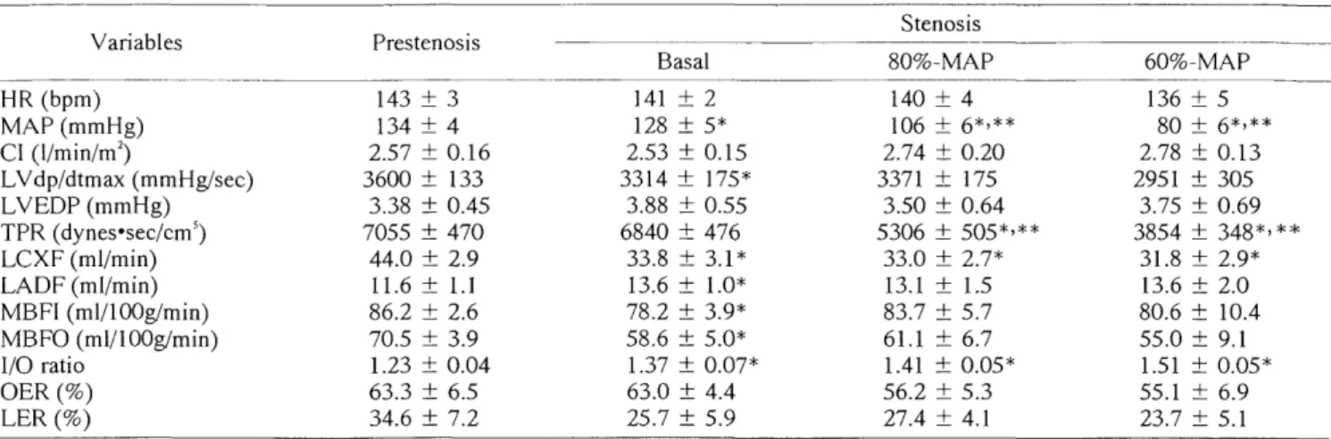
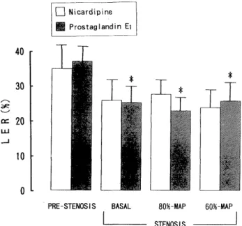
Summary: This study was undertaken in dogs to investigate the effects of nicardipine (NCR)- and prostaglandin E1 (PGE1)- induced hypotension on left ventricular myocardial function, and on coronary hemodynamics in the presence of a partially obstructed left coronary artery. A stenosis of left circumflex coronary artery (LCX) was induced to reduce LCX blood flow by 20%. Mean arterial pressure (MAP) was lowered by infusion of NCR or PGE1 to 80% and 60% of the control. Heart rate, cardiac index, left ventricular dp/dt max, myocardial oxygen extraction ratio, inner/outer blood flow ratio and myocardial lactate extraction ratio showed no significant change at either 80%- or 60%-MAP during infusion of either drug. PGE1 -induced hypotension reduced left ventricular end-diastolic pressure at both 80%- and 60%-MAP. The results indicate that the moderate hypotension induced by NCR or PGE1 does not exert either myocardial depression or adverse effects on the myocardial oxygen supply-demand relationship in the presence of a critical coronary stenosis.
Introduction
Deliberate hypotension is an anesthetic technique to de- crease blood loss during surgery and to provide a dry field for the surgeon."'-" Intravenous nicardipine (NCR)4.5' 6) and prostaglandin E, (PGE,)''8.9' have been introduced in recent years to control blood pressure during clinical anesthesia, and have proved to be useful for deliberate hypotension.
NCR lowers blood pressure resulting from vasodilation due to inhibition of calcium influx into the vascular smooth muscle. NCR mainly dilates resistance vesseles and also has an effect to increase coronary blood flow.'°' PGE, exerts direct relaxing action on the vascular smooth muscle and causes hypotension. PGE, also has been shown to cause coronary vasodilation and reduction of both preload and afterload of the left ventricle.". 12. 13)
The blood flow to ischemic myocardium depends on coronary perfusion pressure, and it is risky to lower arterial pressure in the patients with ischemic heart disease."' Although NCR and PGE, are considered to be beneficial for the ischemic heart, it is not clear how these compounds affect the heart with critical coronary-artery stenosis14' when administered to lower arterial blood pressure rapidly during general anesthesia.
The present study was designed to investigate the effects of NCR- and PGE,-induced hypotension on left ventricular myocardial function, and on coronary hemodynamics in the presence of partially obstructed left coronary circulation.
Materials and Methods.
Surgical preparation
Sixteen mongrel dogs of either sex, weighing 11.4 to 15.6 (mean 13.1) kg were used in this study. Anesthesia was induced with 30mg/kg of sodium pentobarbital i. v., and maintained with continuous infusion of sodium pentobar- bital, 1 mg/kg/min, pancuronium chloride, i. v. appropri- ately, and inhalation of 50% N2O in O2. The animals were intubated with a cuffed endotracheal tube and ventilated mechanically with air at a tidal volume of 15ml/kg. End- tidal CO, concentration was measured continuously with an infrared CO2 analyzer (Datex, Helsinki) and maintained at levels of 35-40mmHg by adjusting the respiratory rate. An infrared heating lamp and a circulating water blanket were used to maintain the esophageal temperature between 37-38.5C. Arterial blood gas analysis and measurement of serum electrolytes were carried out frequently to maintain pH of 7.35 to 7.45, serum Na of 135 to 155mEq/1 and serum K of 3.7 to 5.3mEq/1. Active intervention was not necessary to maintain these ranges except for adjusting pH with 7% NaHCO3 solution in some animals.
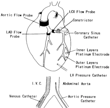
In all dogs, catheters were inserted into the femoral vein, into the abdominal aorta via the left femoral artery (Fig. 1).
Normal saline was administered at a rate of 4-6ml/kg/hr
into the femoral vein into which NCR and PGE, were also
administered. Intraarterial pressure was monitored via the
abdominal aorta. The chest was opened through the sixth
left intercostal space, and the heart was suspended in a
pericardial cradle. Precalibrated electromagnetic flow
probes (Nihon Kohden, Japan) of appropriate sizes to
ensure a snug fit were placed around the ascending aorta,
the left circumflex coronary artery (LCX) approximately
1-2cm distal to its origin, and the left anterior descending
coronary artery (LAD) distal to its first large diagonal
Fig. 1. Schematic diagram of the experimental preparation
branch. The flow probes were connected to flowmeters (Nihon Kohden, Japan). A catheter was inserted into the left ventricle via the cardiac apex to measure the left ventricular pressure. A damping device (Accudynamics Sorenson Research, Salt Lake City) was used to get an appropriate damping of the pressure wave. Myocardial blood flow was measured with the hydrogen gas clearance method using platinum tissue electrodes (200um in diam- eter) at both the outer layers (2-3mm in depth) and the inner layers (8-9mm in depth).
Experimental protocol
At the end of the surgical preparation, at least 30min were allowed for stabilization. After control readings (presten- osis value) had been obtained, a stenosis of LCX was induced using a stainless steel screw type mechanical occluder placed immediately distal to the flow probes. The stenosis was defined as the constriction necessary to reduce LCX blood flow by 20%. About 30min was allowed after inducing stenosis for the reactive hyperemia to disappear and for hemodynamic stabilization, and the second readings (basal value) were obtained.
Then the dogs were randomly divided into two groups to recieve NCR or PGE,. Both NCR and PGE, were dissolved in normal saline, and NCR was used as a 0.02% solution, PGE, as a 0.0025% solution. NCR and PGE, was infused at a dose which caused decrease in MAP to 80% of the basal value. After maintenance of stable MAP for 30min, the third readings (80% MAP value) were obtained. The dose of NCR or PGE, was then increased to cause decrease in MAP to 60% of the basal value. After maintenance of
stable MAP for 30min, the fourth readings (60% MAP value) were obtained. The hemodynamic data were calcu- lated by the formula shown in Table 1.
Table 1. Hemodynamic Formulas