Acta Med. Nagasaki 39: 82-86
Ventilatory and Pulmonary Vascular Responses to Acute Hypoxia in Patients with Chronic Obstructive Lung Disease
Tatsuji NAITO, Yoshiyuki MIYAHARA and Satoshi IKEDA
The Second Department of Internal Medicine, Nagasaki University School of Medicine, Nagasaki, Japan.
The present study was undertaken to examine the pulmo- nary vascular and ventilatory responses to acute hypoxia in chronic obstructive lung disease. Pulmonary hemodyna- mics, minute ventilation (VE) and oxygen uptake (VO2) were serially measured during inhalation of 13 %O2 for 15 min.
There was a wide variablility in the pulmonary, vascular response to acute hypoxia. ,The initial increase in VE and the magnitude of change in VO2 were significantly lower in subjects developing a 25 % or great increase in mean pulmo- nary arterial pressure during hypoxic breathing. These results suggest that the ventilatory response to acute hypoxia plays a significant role in the pulmonary vascular response to acute hypoxia, and blunted initial ventilatory response to acute hypoxia may be a physiological adaptation to en- hanced responses of pulmonary vessels.
Introduction
A rapid f all in alveolar oxygen tension causes constric- tion of pulmonary vessels and elevation of pulmonary arterial pressure. Since first reported in 1946 by Von Euler and Liljestrandl°, this phenomenon, i. e., hypoxic pulmo- nary vasoconstriction (HPV), has been studied by several investigators. However, the mechanism underlying HPV is still unclear. The response of pulmonary vessels to acute alveolar hypoxia exhibits marked interspecies and intraspecies differences','). These differences have been attributed to the quantity of smooth muscles in the media of the pulmonary arteries4), to differences in the release of mediators induced by the direct effect of acute hypoxia') or to a restricted collateral ventilation"'). Although various studies investigated the response of pulmonary vessels to
acute hypoxia in healthy individuals'-"), the magnitude of the resultant rise in pulmonary arterial pressure varied among different reports. Abraham et al."), who studied patients with chronic bronchitis, reported a negative correlation between arterial oxygen saturation and pulmo- nary arterial pressure during hypoxic inhalation.
Weitzenblum et al."), who studied patients with the same disease, reported that the response of pulmonary vessels to acute hypoxia varied among individual patients, and taht some patients showed little rise in pulmonary arterial
pressure despite a marked reduction in arterial oxygen saturation.
High-altitude pulmonary edema (HAPE) develops when healthy individuals without a past history of cardiopul- monary disease rice rapidly to high altitudes. Factors involved in the onset of HAPE include hypoxia, hypobaria, cold envrironment and exercise (factors prevailing in high altitudes), in addition to other predisposing factors. Of these factors, hypoxia has been the focus of attention, and its effect on the living body has been studied extensively. It has been recognized that the intensity of HPV plays a major role in the development of HAPE. Previous studies examining the hypoxic ventilatory response (HVR) in HAPE-susceptible subjects revealed a significantly reduced HVR in HAPE-susceptible subjects compared with control subjects"-"). This finding suggests that low HVR could be a predisposing factor for HAPE.
The present study was undertaken to serially measure pulmonary hemodynamics and ventilation in patients with chronic obstructive lung disease (COLD) during inhalation
of a hypoxic gas mixture. The protocol allowed assessment of the relationship between the response of pulmonary vessels and ventilatory function.
Subjects and Methods Subjects
The subjects consisted of 21 patients (15 males and 6 females) with COLD such as chronic pulmonary emphy- sema or diffuse panbronchiolitis (excluding bronchial asthma) diagnosed based on physical findings, chest X-ray films, pulmonary function test, selective alveobron- chography, transbronchial lung biopsy or open lung biopsy. The condition of each patient was clinically stable at the time of this study. Table 1 summarizes the age distribution, pulmonary function tests and arterial blood gases. All subjects gave informed consent.
Methods
In the evening before the day of the planned examina- tion, drugs that affect the circulation or central nervous
TABLE 1. Characteristics of subjests
COLD
n 21
Age, yr 63.3±2.5
FEV,/FVC, % 57.5±3.2
VC, % predicted 93.0±5.6 DLco, % predicted 68.8±5.0
PaCO2, Torr 34.5±0.7
Pa02, Torr 77.2±2.4
Sa02, % 94.9±0.4
pH 7.403±0.005
EFV,/FVC, percent of forced VC expired in Is;
VC, vital capacity ; DLco, CO diffusing capacity ; PaCO2 arterial CO, tension ; Pa02 , arterial 02 tension ; Sa02, arterial 02 saturation ;
system were suspended. No food or premedication were given in the morning on the days of examination. Right heart catheterization was performed with the subject in the supine position, using a Swan-Ganz catheter (Model TF002H-7F, Baxter Healthcare Corporation) introduced via the femoral vein. Arterial pressure measurements and arterial blood sampling were made through an introducer in the femoral artery. Intravascular pressure was meas- ured relative to atmospheric pressure with a zero reference point at the mid-axillary line. We measured the mean pulmonary capillary wedge pressure (PCWP), mean pulmonary arterial pressure (MPAP) and mean arterial pressure (MAP). In addition, cardiac output (CO) was determined by the thermodilution technique, using REF-1 ejection fraction/cardiac output computer (Edwards Critical Care Division). Total pulmonary resistance (TPR) was calculated, using the following equation :
TPR = MPAP/COX80 (dyne- sec- cm-9.
Arterial blood gases were measured with a Ciba-Corning pH/blood gas analyzer fitted with a co-oximeter. Minute ventilation (VE) and oxygen uptake (V02) was measured using a respirometer (RM-300 System DHC, Minato Medical Corporation). Using a Douglas bag fitted with a unidirectional valve, it was possible to change the compo- sition of inspired air from that of room air to a hypoxic gas mixture of 13 %02 in N2. Pulmonary function tests were examined one week prior to the measurement of ventilation and hemodynamics, using an Autospirometer System 9.
Protocol
When the Swan-Ganz catheter was checked to be in the proper position, the subject was connected to the res- pirometer via a tightly fitting face mask. A period of ten minutes was allowed for adaptation to the recording system and stabilization of measurement of hemodynamic and ventilation variables during room air breathing.
Measurement of MPAP, MAP,CO commenced thereafter,
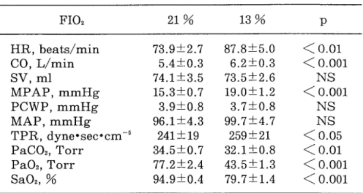
TABLE 2. Hemodynamics and blood gases
FIO2 21% 13% p
HR, beats/min 73.9±2.7 87.8±5.0 < 0.01
CO, L/min 5.4±0.3 6.2±0.3 < 0.001
SV, ml 74.1±3.5 73.5±2.6 NS
MPAP, mmHg 15.3±0.7 19.0±1.2 < 0.001
PCWP, mmHg 3.9±0.8 3.7±0.8 NS
MAP, mmHg 96.1±4.3 99.7±4.7 NS
TPR, dyne•sec°cm-5 241±19 259±21 < 0.05 PaCO2, Torr 34.5±0.7 32.1±0.8 < 0.01 Pa02, Torr 77.2±2.4 43.5±1.3 < 0.001
Sa02, % 94.9±0.4 79.7±1.4 < 0.001
FI02, fractional inspiratory oxygen ; HR, heart rate ; CO, cardiac output ; SV, stroke volume ; MPAP, mean pulonary arterial pressure ; PCWP, mean pulmonary capillary wedge pressure ; MAP, mean arterial pressure ; TPR, total pulmo- nary resistance ;
in addition to VE and V02 Blood samples were simul- taneously withdrawn from the femoral and pulmonary arteries. After measurements in room air were completed, F102 was lowered to 13 % and MPAP, MAP, VE and VO2 were monitored. After 15 minutes, when ventilation and vascular pressures were again steady, measurements of MPAP, MAP, VE, V02 and CO were repeated and blood samples were withdrawn again. The patients were then disconnected from the respirometer.
Statistics
Values are expressed as mean ± SEM. A paired t-test was used to compare measurements within groups while unpaired t-test was used to compare measurements be- tween groups. Comparisons were considered significant at p < 0.05.
Results
During room air inhalation, HR, CO, MPAP, PCWP and MAP were 73.9±2.7 beats/min, 5.4 ± 0.3 L/min, 15.3 ± 0.7 mmHg, 3.9±0.8 mmHg and 96.1±4.3 mmHg, respectively.
Table 2 summarizes the changes in hemodynamic parame- ters and blood gases during hypoxia. Reduced arterial oxygen tension and arterial oxygen saturation levels significantly augmented CO and increased HR. However, stroke volume and MAP remained unchanged during hypoxia, while MPAP and TPR were significantly ele- vated.
Subjects developing a 25 % or greater increase in MPAP during hypoxic breathing (i. e., % 0 MPAP ? 25 %) were classified as responders, while those developing < 25 % increase in this parameter were classified as non- responders. The magnitude of change in TPR (0 TPR) was significantly greater in responders compared with
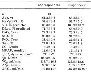
TABLE 3. Comparison of main functional and hemody- namic data between nonresponders and re-
sponders
nonresponders responders
Fig. 1. Comparison of the increase in CO (A CO) and the magnitude of change in TPR (A TPR) between nonrespond- ers and responders
nonresponders, while the change in CO (A CO) did not differ significantly between the two groups (Fig. 1). There were no significant differences in pulmonary function tests, arterial blood gases, hemodynamic parameters, VE or V02 between responders and nonresponders during room air breathing (Table 3). On the commencement of breath- ing of hypoxic gas mixture, VE increased initially but gradually declined to a steady state. When the difference between the baseline VE and peak VE was expressed as
A VE and the magnitude of change in V02 was expressed as A V02, A VE and A V02 were significantly lower in re- sponders than in nonresponders (Table 3).
n 13 8
Age, yr 61.2±3.8 66.8±1.6
FEV,/FVC, % 57.4±4.4 57.7±5.0
VC, % predicted 86.3±5.8 103.9±10.7
DLco, % predicted 71.7±5.5 65.0±9.9
Pa02, Torr 77.3±2.9 76.9±4.5
Sa02, % 95.0±0.5 94.7±0.8
Pv02, Torr 38.4±0.9 37.5±1.1
Sv02, % 71.8±1.5 70.0±1.8
CO, L/min 5.4±0.4 5.4±0.3
MPAP, mmHg 14.6±0.8 15.1±1.7
TPR, dyne•sec•cm-5 241±27 241±25
VE, L/min 10.83±0.6 9.99±0.8
V0;, ml/min 216.7±16.8 225.8±18.6
A VE, L/min 3.66±0.50 2.05±0.22'
A V02, ml/min 19.0±14.8 -37.3±16.22*
VF, minute ventilation ; V02, 02 uptake ; A VF, early increase in ventilation ; A V02, early change in 02 uptake ; *p < 0.05 in
responders compared with nonresponders
Discussion
The findings of the present study suggest that the pulmonary vascular response to hypoxia varied among subjects and that in could not be predicted using tests performed during resting ventilation. In addition, our results suggested that ventilatory response to acute hypoxia plays an important role in the pulmonary vascular response to acute hypoxia.
It is generally accepted that the rise in pulmonary arterial pressure during acute hypoxia in healthy subjects is due to the combined effect of increased pulmonary vascular resistance and increased CO9`°'16)In COLD paients with pulmonary hypertension, Saadjian et al.") showed that hypoxic breathing further increased MPAP but did not affect CO. A significant elevation in MPAP, TPR and CO was observed during hypoxic breathing in ours sub- jects. A TPR was significantly high in responders com- pared with nonresponders, while A CO was not different between the two groups (Fig. 1). This finding suggests that the magnitude of rise in pulmonary arterial pressure during hypoxia depends on pulmonary vascular reactivity.
There was a wide variability in pulmonary vascular response to acute hypoxia in the present study. Sixty-two percent of subjects appeared to be nonresponders to hypoxia. Similarly, a wide variability in pulmonary vascular response was also observed, ranging from a complete lack of response to a marked elevation of pulmo- nary arterial pressure. In 11 normal subjects investigated
by Beard et al.'), 28 % failed to respond to hypoxia (FI02 = 12 %). In addition, 36 % of 17 normal subjects studied by Fritts et al." )were also nonresponders to hypoxia (FIO2 = 12-14 %). Furthermore, 50 % of 10 normal subjects inves- tigated by Fishman et al.'°)also failed to responds to hypoxia (FIO2 =12-14 %). In chronic bronchitis, nearly 50
% of the patients studied by Weitenblum et al.18)appeared to be nonresponders to hypoxia (F102 =13 %). However, such variability in hemodynamic response to acute hypoxia was not observed by Doyle et al.') when testing eight normal subjects where all subjects responded to hypoxia (FIO2= 10%).
Our results indicted that there were no significant differences in pulmonary function tests, arterial blood gases, hemodynamics, VE and VO2 during resting breath- ing, between responders and nonresponders (Table 3).
Thus, differences in the response of pulmonary vessels to hypoxic breathing could not be predicted by test performed before hypoxic inhalation. The response of pulmonary vasculature to acute hypoxia also varied among individual subjects, suggesting that this response is closely related to predisposing factors.
The measurement of HVR often employs maintenance of a stable isocapnia, as first described by Weil et al.'). We
measured VE during 13 %02 inhalation and analyzed A VE
as an index of chemostimulation. PaCO2 was measured under poikilocapnic conditions in our experiment, without adding C02 to the breathing circuit. Since the change in PaCO2 was minimal, C02 seemed to be less affected by hypoxic breathing. In this regard, Easton et al.") reported
that hypoxic ventilatory depression, when assessed under isocapnic conditions, did not differ from that measured under poikilocapnic conditions.
Matsuzawa et al.") reported that HAPE-susceptible subjects showed significantly lower HVR than control subjects. Furthermore, Hackett et al. ")demonstrated that HAPE patients had significantly lower HVR during their illness compared with control subjects. In the present study, A VE were significantly lower in responders than in nonresponders. These results support the view that a blunted ventilatory response to acute hypoxic stimulation makes constriction of pulmonary vessels more likely to occur. It would seem, therefore, that the ventilatory response to acute hypoxia plays an important role in the pulmonary vascular response to acute hypoxia.
Hypoxia basically elevates ventilation by stimulating the respiratory center (the medulla oblongata) mediated by peripheral chemoreceptors. However its direct action on the central nervous system (CNS) or its indirect CNS action mediated by an increased cerebral blood flow leads to suppression of ventilation'. Ventilatory responses to hypoxia are therefore a total of these effects (respiratory stimulation via peripheral chemoreceptors +respiratory suppression due to its effects on the CNS). In the present study, I VE were significantly lower in responders than in nonresponders. This means that HVD was more marked in responders than in responders. Neonatal or juvenile warm- blood animals are more likely to develop HVD and tend to show a reduction in metabolic rate or V02 when exposed to hypoxia27''. In the present study, 0 V02 was lower in responders than in nonresponders. Suppression of ventila- tion in response to hypoxic stimuli appears to be quite unnatural when viewed from the standpoint of oxygen supply and seems to be inappropriate from the viewpoint of homeostasis in the living body. However, analyses of the manner by which cells adapt themselves to hypoxia indicate that it fits the purpose of survival to suppress metabolism so that the energy needed for the membranous ion gradient (most important to keep the cell viable) can be minimized. The blunted initial ventilatory response to acute hypoxia, observed in the present study, may be a physiological adaptation to enhanced responses of pulmo- nary vessels.
Acknowledgements
We gratefully acknowledge Professor Kohei Hara for reading this manuscript and for his invaluable comments.
We thank Dr. Toshiyuki Imamura for his critical com- ments and suggestions.
References
1) Von Euler VS and Liljestrand G.: Observations on the pulmonary artery pressure in the cat. Acta Physiol Scand 23: 301-320 (1946).
2) Grover RF, Wagner WW, McMurty IF and Reeves J'T : Pulmonary circulation : The Cardiovascular System : Handbook of Physiology,
Am Physiol Soc vol 3 pp 103-136 Bethesda (1983).
3) Peake MD, I-larabin AL, Brennan NJ and Sylvester JT : Steady state vascular responses to graded hypoxia in isolated lungs of five species.
J Appl Physiol 51 : 1214-1219 (1981).
4) Tucker A, McMurtry IF, Reeves JT, Alexander AF, Will DI-I and Grover RF : Lung vascular smooth muscle as a determinant of
pulmonary hypertension at high altitude. Am J Physiol 228: 762-767
(1975).
5) Ahmed T, Oliver W Jr and Wanner A : Variability of hypoxic pulmonary vasoconstriction in sheep. Am Rev Respir 1)is 127: 59-62
(1983).
6) Kuriyama T and Wagner WW Jr : Collateral ventilation may protect against high altitude pulmonary hypertension. J Appl Physiol 51 :
1252-1256 (1981).
7) Kuriyama T, Latham LP, Horwitz LD, Reeves JT and Wagner WW Jr : Role of collateral ventilation in ventilation-perfusion balance. J
Appl Physiol 56: 1500-1506 (1984).
8) Beard JT II, Newman JH, Loyd JE and Byrd BF IQ : Doppler estimation of changes in pulmonary artery pressure during hypoxic
breathing. J Am Soc Echo 4 : 121-130 (1991).
9) Doyle JT, Wilson JS and Warren TV: The pulmonary vascular responses to short-term hypoxia in human subjects. Circulation 5:
263-270 (1952).
10) Fishman AP, Fritts HW Jr and Cournand A : Effects of acute hypoxia and exercise on the pulmonary circulation. Circulation 12: 204-215
(1960).
11) Fritts HW Jr, Odell JE, Harris P, Braunwald EW and Fishman AP:
Effests of acute hypoxia on the volume of blood in the thorax.
Circulation 12: 216-219 (1960).
12) Guazzi MD, Alimento M, Berti M, Fiorentini C, Galli C and Tamborini G : Enhanced hypoxic pulmonary vasoconstriction in hypertension.
Circulation 79: 337-343 (1989).
13) Melot C, Naeije R, Hallemans R, Lejeuns P and Mole P : I-Ivpoxic pulmonary vasoconstriction and pulmonary gas exchange in normal
man. Respir Physiol 68: 11-27 (1987).
14) Motley HL, Curnand A, Werko L, Himmelstein A and Dresdale 1.) : The influenece of short period of induced acute anoxia upon pulmonary
artery pressure in man. Am J Physiol 150: 315-320 (1947).
15) Naeije R, Melot C, Mols P and Hallemdns R : Effects of vasodilators
on hypoxic pulmonary vasoconstriction in normal man. Chest 82:
404-410 (1982).
16) Westcott RN, Fowler NO, Scott RC, Hauenstein VD and Mcquire J : Anoxia and human pulmonary vascular resistance. J Clin Invest 30:
957-970 (1951).
17) Abraham AS, Hedworth-whitty RB and Bishop JM : Effects of acute hypoxia and hypervolemia singly and together, upon the pulmonary
circulation in patients with chronic bronchitis. Clin Sci 33 : 371-380
(1967).
18) Weitzenblum E, Schrijen F, Mohan-Kumar T, Colas VF and Lockhart A : Variability of the pulmonary vascular response to acute hypoxia in
chronic bronchitis. Chest 94 : 772-778 (1988).
19) Hackett PH, Roach RC, Schoene RB, Harrison GL and Mille WJ Jr:
Abnormal control of ventilation in high-altitude pulmonary edema. J
Appl Physiol 64: 1268-1272 (1988).
20) Masuyama S, Kimura H, Sugita T, Kuriyama T, Tatsumi K, Kunimoto F, Okita S, Tojima H, Yuguchi Y, WW'atanabe S and Honda
Y : Control of ventilation in extremealtitude climbers. J Appl Physiol
61: 500-506 (1986).
21) Matsuzawa Y, Fujimoto X, Kobayashi T, Namushi VR, Harada .X, Kohno H, Fukushima M and Kusama S : Blunted hypoxic ventilatory
drive in subjects susceptible to high-altitude pulmonary edema. J Appl
Physiol 66: 1152-1157 (1989).
22) Moore LG, Harrison GL, McCullough RE, McCullough RG, Micco AJ, Tucker A, Weil J V and Reeves JT : Low acute hypoxic ventilatory
response and hypoxic depression in acute altitude sickness. J Appl
Physiol 60: 1407-1412 (1986).
23) Saadjian A, Philip-Joet F, Levy S and Arnaud A : Vascular and cardiac reactivity in pulmonary hypertension due to chronic obstructive
lung disease : assessment with various oxygen concentrations. Eur
Respir J 5 : 525-530 (1992).
24) Weil JV, Byrne EQ, Sodal IE, Friesen WO, Underhill B, Filley GF and Grover RF : Hypoxic ventilatory drive in normal man. J Clin Invest
49: 1061-1072 (1970).
25) Easton PA amd Anthonisen NR: Carbon dioxide effects on the
ventilatory response to sustained hypoxia. J Appl Physiol 64:
1451-1456 (1988).
26) Neubauer JA, Melton JE and Edelman NH: Modulation of respiration during brain.hypoxia. J Appl Physiol 68: 441-451 (1990).
27) Haddad GG and Rosen CL: Developmental Neurobiology of breath- ing. Ventilatory response to hypoxia : Integrated, cellular, and
molecular aspects.591-614 New York. Dekker. (1991).
28) Mortola JP: Hypoxic hypometabolism in mammals. Am Physiol Soc 8:79-82 (1993).