Studies on the survival dynamics of tick-borne viruses in the
hard tick Haemaphysalis longicornis and the effects of tick
antimicrobial peptides on viral replication
The United Graduate School of Veterinary Science
Yamaguchi University
Melbourne Rio Talactac
TABLE OF CONTENTS
GENERAL INTRODUCTIONCHAPTER 1: Demonstration of the survival dynamics of Langat virus in
Haemaphysalis longicornis using anal pore microinjection
1.1 Introduction
1.2 Materials and Methods 1.2.1 Ticks and Animals 1.2.2 Cells and virus 1.2.3 Tick infection
1.2.4 Detection of Langat virus RNA
1.2.5 Langat virus titration among LGTV-infected adult ticks
1.2.6 Detection of Langat virus antigens in tick organs using indirect
immunofluorescent antibody test (IFAT)
1.2.7 Langat virus transmission from ticks to mice 1.2.8 Transovarial transmission of Langat virus in ticks 1.2.9 Statistical Analysis
1.3 Results
1.3.1 Langat virus infection in ticks 1.3.2 Langat virus transmission to mice
1.3.3 Transovarial transmission of Langat virus in ticks 1.4 Discussion
Tables and Figures
CHAPTER 2: Vector competence of Haemaphysalis longicornis ticks for a Japanese isolate of the Thogoto virus
2.1 Introduction
2.2 Materials and Methods 2.2.1 Ticks and animals 2.2.2 Cell culture and virus 2.2.3 Infection of ticks with THOV
2.2.4 Detection of THOV RNA in mouse and tick tissues
2.2.5 Isolation and titration of THOV from tick tissues and whole adult ticks 2.2.6 Detection of THOV antigens in tick organs using IFAT
2.2.8 Determination of transovarial transmission of THOV in ticks 2.2.9 Statistical analysis
2.3 Results
2.3.1 THOV replication in ticks
2.3.2 Tick transmission of THOV to mice
2.3.3 Detection of THOV RNA in eggs and larvae 2.4 Discussion
Tables and Figures
CHAPTER 3: Evaluation of the role of antimicrobial peptide, longicin, from
Haemaphysalis longicornis against Langat virus
3.1 Introduction
3.2 Materials and Methods 3.2.1 Cell culture and virus 3.2.2 Ticks and animals 3.2.3 Peptide synthesis 3.2.4 Cell proliferation assay
3.2.5 Focus formation unit reduction assay 3.2.6 Virucidal assay
3.2.7 Prophylactic antiviral assay 3.2.8 Post-adsorption antiviral assay 3.2.9 Virus yield reduction assay
3.2.10 RNA interference (RNAi) and subsequent virus challenge 3.2.11 Statistical analysis
3.3 Results
3.3.1 Cytotoxicity activity of the longicin P4 peptide 3.3.2 Antiviral effect of longicin P4 peptide against LGTV
3.3.3 Antiviral effect of longicin P4 peptide against human adenovirus 3.3.4 Effect of longicin gene silencing in LGTV replication in adult H. longicornis
3.4 Discussion Tables and Figures
CHAPTER 4: Evaluation of the role of antimicrobial peptide, HEdefensin, from
Haemaphysalis longicornis against Langat virus
4.2 Materials and Methods
4.2.1 Ticks and animals
4.2.2 Sequence of the defensin-like peptide and bioinformatic analysis 4.2.3 Expression profile analysis
4.2.4 Peptide synthesis 4.2.5 Cells and virus
4.2.6 Cell proliferation assay
4.2.7 Focus formation unit reduction assay 4.2.8 Direct virucidal assay
4.2.9 Prophylactic and Post-adsorption antiviral assays 4.2.10 Virus yield reduction assay
4.2.11 RNAi and virus challenge 4.2.12 Statistical analysis
4.3 Result
4.3.1 Identification of defensin-like gene
4.3.2 Whole tick and tissue-specific expression profiles of HEdefensin in H. longicornis
4.3.3 Cell growth inhibition effect of the synthetic HEdefensin peptide 4.3.4 Antiviral activity of the HEdefensin peptide against LGTV
4.3.5 Antiviral activity of the HEdefensin peptide against human adenovirus 4.3.6 Effect of HEdefensin gene silencing on LGTV replication in adult H. longicornis
4.4 Discussion Tables and Figures
SUMMARY AND CONCLUSION ACKNOWLEDGEMENTS REFERENCES
LIST OF TABLES AND FIGURES
CHAPTER 1Table 1.1 List of real-time PCR primers used to detect Langat virus RNA
Table 1.2 Detection of Langat virus RNA from ticks injected with LGTV and EMEM via
anal pore microinjection using reverse transcription PCR
Table 1.3 Comparative Langat virus titers from selected organs of unfed adult
H. longicornis at 28 days post-injection (dpi)
Table 1.4 Langat virus transmission from H. longicornis to mice
Table 1.5 Detection of Langat virus RNA in eggs and larvae using reverse transcription
PCR
Fig. 1.1 Replication of Langat virus in H. longicornis after infection via anal pore
microinjection
Fig. 1.2 Localization of Langat virus in selected organs from unfed adult ticks after
infection via anal pore microinjection and immunofluorescence assay detection of LGTV antibodies in serum samples from mice.
CHAPTER 2
Table 2.1 List of real-time PCR primers used for the detection of THOV in ticks and
mice
Table 2.2 Titration of THOV isolated from tick organs of unfed and partially fed infected
ticks via focus formation assay
Table 2.3 Detection and isolation of THOV from whole adult ticks that molted from
nymphs fed on either EMEM- or THOV-injected mice
Table 2.4 Detection and isolation of THOV from whole adult ticks that molted from
nymphs co-fed with either EMEM- or THOV-injected adult ticks
Table 2.5 THOV transmission from ticks injected with THOV through anal pore
microinjection to mice
Fig. 2.1 Replication of the THOV in H. longicornis after infection using anal pore
microinjection
Fig. 2.2 Localization of the THOV in the midgut and salivary glands of unfed adult ticks
after infection via anal pore microinjection
Fig. 2.3 Localization of the THOV in the midgut, salivary glands, and synganglia of
partially fed, infected adult ticks via anal pore microinjection
Fig. 2.4 Detection of THOV antibodies in serum samples from mice using
CHAPTER 3
Table 3.1 List of PCR primers used for the synthesis of double-stranded RNA Table 3.2 List of PCR primers used for the detection of longicin gene
Table 3.3 List of real-time PCR primers used for the determination of longicin gene Fig. 3.1 Cytotoxicity of longicin P1 and P4 against BHK-21 cells
Fig. 3.2 Virucidal effect of longicin P1 and P4 against Langat virus
Fig. 3.3 Prophylactic and post-adsorption antiviral effects of longicin P1 and P4
against Langat virus
Fig. 3.4 Dose-dependent and time-dependent virucidal effects of longicin P4 against
Langat virus
Fig. 3.5 Virucidal activity of longicin P1 and P4 against adenovirus Fig. 3.6 Effect of longicin silencing in tick mortality and virus titer Fig. 3.7 Virucidal effect of full-length (FL) longicin against LGTV CHAPTER 4
Table 4.1 List of PCR primers used for detection of the HEdefensin gene Table 4.2 List of PCR primers used for the synthesis of double-stranded RNA
Fig. 4.1 Characterization of HEdefensin cDNA. Nucleotide and predicted amino acid
sequences of HEdefensin cDNA
Fig. 4.2 Alignment of the amino acid sequences of HEdefensin and defensins from other
ticks
Fig. 4.3 Transcription profiles of HEdefensin analyzed by real-time PCR Fig. 4.4 Cell growth inhibition effect of the HEdefensin peptide on BHK-21 cells Fig. 4.5 Virucidal activity of HEdefensin against Langat virus
Fig. 4.6 Dose- and time-dependent virucidal effects of HEdefensin against LGTV Fig. 4.7 Temperature-dependent virucidal effect of HEdefensin against LGTV Fig. 4.8 Prophylactic and post-adsorption antiviral activity of HEdefensin against
LGTV
Fig. 4.9 Virucidal activity of the HEdefensin peptide against an adenovirus Fig. 4.10 Gene-specific silencing in ticks from each group at 28 d post-dsRNA
ABBREVIATIONS
AMPs: antimicrobial peptidesBHK: baby hamster kidney BSL2, 3: biosafety level 2 or 3 cDNA: complementary DNA
CCHFV: Crimean-Congo hemorrhagic fever virus DAI: days after infestation
DAC: days after challenge DPI: days post-injection DMEM:
EMEM edium
FFA: focus formation assay FFU: foci-forming-unit FL: full length
dsRNA: double-stranded RNA EST: expressed sequenced tag(s) FBS: fetal bovine serum
HPLC: High Performance Liquid Chromatograph IFA: immunofluorescence assay
IFAT: indirect immunofluorescent antibody test IgG: immunoglobulin G
IMD: immune deficiency
JAK/STAT: Janus kinase-signal transducers and activators of transcription Luc: firefly luciferase gene
LGTV: Langat virus
MOI: multiplicity of infection MEM: minimum essential medium NC: negative control
PAMPs: pathogen-associated molecular patterns PBS: phosphate-buffered solution
PC: positive control
qPCR: quantitative polymerase chain reaction percentage of foci reduction
RNAi: RNA interference
SFTSV: severe fever with thrombocytopenia syndrome virus TCID50: 50% tissue culture infectious dose
TAE: Tris-Acetate EDTA buffer TBEV: tick-borne encephalitis virus TBFVs: tick-borne flaviviruses TBVs: tick-borne viruses THOV: Thogoto virus
GENERAL INTRODUCTION
Ticks are important vectors of disease-causing microorganisms, rickettsiae, spirochaetes, protozoa and viruses affecting humans, livestock, wild and companion animals [1]. Among the important pathogens that ticks transmit, viruses form a major constituency of the transmitted pathogens and remain to be a big threat to both human and animal populations as they can produce diseases with high morbidity and mortality [2, 3].
Tick-borne viruses (TBVs) comprise a wide range of viruses classified into six virus families and these include Asfarviridae, Bunyaviridae, Flaviviridae,
Orthomyxoviridae, Rhabdoviridae, and Reoviridae [4]. Among these viral families, Bunyaviridae and Flaviviridae are considered to have the most important TBVs of
public health importance, including tick-borne encephalitis virus (TBEV) and Crimean-Congo hemorrhagic fever virus (CCHFV) which are known to cause severe clinical symptoms in humans [4 6].
A tabular summary of select virus families transmitted by ticks of medical and veterinary importance is shown in the next page.
Family Representative virus Vectorsa Distribution Diseaseb
Asfarviridae African Swine Fever Or. moubata, Africa Fever
Or. erraticus (pigs)
Bunyaviridae CCHFV Hy. marginatum Africa, Asia, Hemor.
De. marginatus Southern fever Europe (humans)
Flaviviridae TBEV Ix. ricinus, Europe, Encepha.
Ix. persulcatus Northern Asia (humans) Orthomyxo- Bourbon Unknown United Hemor.
viridae States fever
(humans)
Rhabdoviridae Sawgrass De. variabilis United Unknown States
Reoviridae Colorado De. andersoni United Fever
Tick Fever States (humans)
aDe.: Dermacentor; Ix.: Ixodes; Hy.: Hyalomma; Or.: Ornithodoros, bEncepha.: Encephalitis; Hemor.;
Hemorrhagic. (Table adapted from Brackney and Armstrong (2016) and dela Fuente et al. (2017).
Of the 900 currently known tick species, only less than 10% are implicated as virus vectors and these included the Ornithodoros and Argas Genera for the argasid ticks and Ixodes, Haemaphysalis, Hyalomma, Amblyomma, Dermacentor, and
Rhipicephalus Genera in ixodid ticks [6, 7].
Although the role of ticks in the transmission of viruses has been known for over a century [3] and ticks being second to mosquitoes in transmitting human diseases [8], the diversity of tick-borne viruses has been less thoroughly studied than that of mosquito-borne viruses [9]. Even until now, detection of new pathogenic viruses are Table 1. Select tick-borne viruses listed by virus family of medical or veterinary importance
still being reported and known viruses continuously spread to new geographical locations [3].
The flaviviruses account for the majority of arthropod-borne viruses worldwide, which includes the TBEV serocomplex [10, 11]. TBEV serocomplex is consists of closely related flaviviruses that cause important diseases in animals and humans
Kyasanur Forest virus, Langat virus (LGTV), Louping-ill virus, Omsk hemorrhagic fever virus, Powassan virus, and TBEV [12]. The transmission of this group of viruses is commonly associated with tick species which include hard ticks
Ixodes ricinus, I. persulcatus, Dermacentor spp., and Hyalomma spp., [10].
Haemaphysalis longicornis is also a hard tick mainly distributed in East Asia
and Australia and a known vector of theileriosis, non-zoonotic babesiosis [13, 14] and was also recently established as the potential reservoir and vector of a bunyavirus, the severe fever with thrombocytopenia syndrome virus (SFTSV) [15].
Detection and isolation of flaviviruses have been previously reported in H.
longicornis. Powassan encephalitis virus was isolated in H. longicornis in Canada and
USA [16], while TBEV has been molecularly detected in the tick in South Korea [17]. Though these reports suggest that H. longicornis can successfully harbor and transmit flaviviruses, studies showing survival dynamics of any tick-borne flavivirus in H.
longicornis are lacking, most especially experiments demonstrating the capacity of the
tick to maintain and successfully transmit flaviviruses to animals.
Since tick vectors of TBEV and other pathogenic members of TBEV serocomplex circulate in East Asia [10] where H. longicornis is also mainly distributed, it is interesting to know whether H. longicornis is a competent vector of these viruses in East Asia. Vector competence is defined as the ability of an arthropod to become infected with the virus and transmit the virus following blood feeding [18]
Since most TBFVs require at least a biosafety level 3 (BSL3) containment facility, the use of the naturally attenuated LGTV provides a convenient BSL2 model of TBEV and other highly pathogenic TBFVs [19].
LGTV was isolated from pools of I. granulatus from Malaysia [20]. LGTV shares more than 74% nucleotide identity with TBEV which prompted the use of the virus as a vaccine candidate against TBE for several years [21], but the vaccination was later on abandoned due to development of unexpectedly high incidence of neurologic disease in vaccinated patients (around 1:10,000) [21 - 23]. In natural environment, LGTV does not cause disease in rodents, but in young laboratory mice inoculated intracerebrally with the virus, encephalitis develops [24].
On the other hand, Thogoto virus (THOV), a tick-borne virus not previously reported in East Asia, was recently isolated from a H. longicornis in Kyoto, Japan [9]. THOV is a type of species of the genus Thogotovirus in the family Orthomyxoviridae [25]. The virus was first isolated in 1960 from Rhipicephalus (Boophilus) decoloratus and Rhipicephalus spp. ticks collected on cattle in Thogoto Forest, Nairobi, Kenya [26], and is reported to affect vertebrate hosts such as cattle, camels, and, sporadically, humans. Other ticks that are known to be vectors of this virus include R. annulatus, R.
sanguineus, R. appendiculatus, R. bursa, R. evertsi, Amblyomma variegatum, H. truncatum, and H. a. anatolicum [27]. The virus can cause afebrile leucopenia in cattle
and abortion in sheep [28]. THOV can also affect humans, with one fatality already recorded from two reported human cases [29].
Another aspect of TBVs that is still at an early stage of understanding is the tick-virus interaction. As carriers of several pathogenic microorganisms, protozoa, rickettsiae, spirochaetes, and viruses [1, 30], ticks need to employ broad spectrum innate immunity mechanisms that will allow them to maintain the pathogens and commensal microbes without impairing their viability and further development [31, 32]. With the exceptional longevity of ticks, they can harbor TBVs over prolonged periods of time, making them not only vectors but also excellent reservoir hosts for the viruses
[6]. Although tick immune responses against bacteria have been widely studied, the knowledge about antiviral immunity pathways is generally limited in ticks [33, 34]. RNA interference (RNAi) is considered to be the most important antiviral response in mosquitoes and also the most likely important antiviral response in all arthropods [35]. It is a conserved biological response to double-stranded RNA (dsRNA) which results in the production of small RNAs that mediate regulation of expression of genes by degradation or by translation inhibition of homologous messenger RNA [36]. Aside from RNAi, the other signaling pathways in arthropods which respond to virus infection include Immune deficiency (IMD), Janus kinase-signal transducers and activators of transcription (JAK/STAT), and Toll [35, 37 39]. In ticks however, the only antiviral innate immune response described to date is RNAi [40, 41]. In the case of melanisation, it remains unclear whether phenoloxidase (PO) cascade is present in ticks [35], although, the Toll signaling pathway can be activated in ticks after a flavivirus infection [42].
In this study, I explored the role of humoral defenses in ticks particularly the antimicrobial proteins and peptides which play a major role in protecting ticks against microorganisms [43, 44]. Antimicrobial peptides (AMPs) are ancient immune molecules that are important in invertebrate and vertebrate host defenses [45, 46]. These peptides display broad-spectrum biological activity against bacteria, yeast, fungi, protozoan
parasites, and enveloped viruses [47 - 49] and have been demonstrated to possess immunomodulatory properties [50]. Numerous small molecules such as defensins, lysozymes or by tick-specific antimicrobial compounds such as microplusin provide the direct antimicrobial defense in ticks [31].
This dissertation describes studies on survival dynamics of TBVs in H.
longicornis and the effects of tick antimicrobial peptides in viral replication with the
following specific objectives:
1. To demonstrate the survival dynamics of Langat virus in H. longicornis;
2. To evaluate the vector competence of H. longicornis for Thogoto virus isolated in Japan;
3. To evaluate the antiviral activity of H. longicornis antimicrobial peptide, longicin;
4. To identify and characterize a new antimicrobial peptide, HEdefensin, with focus on antiviral activity.
CHAPTER 1
Demonstration of the survival dynamics of Langat virus in
Haemaphysalis longicornis using anal pore microinjection
This work has been published as: Talactac, M.R., Yoshii, K., Hernandez, E.P., Kusakisako, K., Galay, R.L., Fujisaki, K., Mochizuki, M., and Tanaka, T. (2017). Synchronous Langat virus infection of Haemaphysalis longicornis using anal pore microinjection. Viruses. 9, 189.
1.1 Introduction
Majority of arthropod-borne viruses worldwide belongs to Flaviviruses which also include the TBEV serocomplex that can cause important diseases in animals and humans [10, 11]. Tick species of the hard ticks Ixodes ricinus, I. persulcatus,
Dermacentor spp., and Hyalomma spp., commonly transmit this group of viruses [10]. Haemaphysalis longicornis is also a hard tick mainly distributed in East Asia
and Australia and a known vector of theileriosis and non-zoonotic babesiosis [13, 14]. The hard ticks was also the established reservoir and vector of the severe fever with thrombocytopenia syndrome virus (SFTSV) [15]. The detection and isolation of flaviviruses have been reported previously in H. longicornis [17, 51]; however, studies showing the survival dynamics of any tick-borne flavivirus in H. longicornis are still lacking. While ticks can be naturally infected with tick-borne viruses by feeding them on viraemic animals, this method requires a sufficient level of viraemia for transmission to a naïve tick [52]. Moreover, synchronization of tick infection with a defined viral inoculum is a notable limitation in this method [2]. Another method of tick infection is through percoxal microinjection, however, it bypasses the midgut barrier which makes it non-representative of natural route of infection and may not ensure consistent infection rates among fed ticks [53]. Immersion method, on the other hand, can also successfully
infect ticks. This infection method is simpler and relatively inexpensive; however, generating cohorts of infected ticks with equal pathogen burden is its major limitation [54].
In this study, I have demonstrated a consistent infection and maintenance of Langat virus (LGTV), a naturally attenuated member of the TBEV serocomplex of flaviviruses, in adult H. longicornis using anal pore microinjection originally used to infect ticks with Borrelia burgdorferi [54]. Although no transovarial transmission was observed in this study, the ticks infected by this method successfully established horizontal transmission of LGTV to mice making this method an additional tool in studying tick virus host interactions.
1.2 Materials and Methods
1.2.1 Ticks and animals
Parthenogenetic H. longicornis (Okayama strain) ticks were maintained for several generations by feeding on the ears of Japanese white rabbits (KBT Oriental Co., Saga, Japan) at the Experimental Animal Center, Joint Faculty of Veterinary Medicine, Kagoshima University, Kagoshima, Japan [55]. Alternatively, ticks were capsule/tube
fed using six-week-old, female, ICR mice (Kyudo, Fukuoka, Japan) [56]. The animals were kept in a temperature- and humidity-controlled room, with a constant supply of water and commercial feeds. The use and care of animals in this study was in accordance with approved guidelines (approval numbers VM 15055 and VM 15058) from the Animal Care and Use Committee of Kagoshima University.
1.2.2 Cells and virus
Baby hamster kidney (BHK-21) cells (ATCC CCL-10) (ATCC, Manassas, VA,
with 5% fetal bovine serum (FBS) (Equitech-Bio, Kerrville, TX, USA) and 1% antibiotic/antimycotic (Nacalai Tesque, Kyoto, Japan). Cell cultures were maintained at 37 °C under 5% CO2 until use. To amplify the LGTV TP21 strain, BHK-21 cells were utilized in this study. The LGTV stock titer was determined through focus formation assay (FFA), as described previously [57
1.2.3 Tick infection
Adult ticks were infected with LGTV by anal pore microinjection [54], basically as described. Briefly, several 10 µL calibrated capillary tubes (Drummond
Scientific Co., Broomall, PA, USA) were fabricated into microinjection needles by heating and pulling in a capillary pipette puller (model PN-30) (Narishige, Tokyo, Japan), which were eventually stored on adhesive tape in a petri dish. The ticks were then immobilized using a
double-ventra
aperture area was focused. Then, after connecting the microinjection needle in the IM 300 microinjector (Narishige, Tokyo, Japan) equipped with automated foot control, the tip of the tube was snapped where the diameter is slightly smaller than that of the anal aperture of the tick by gently touching the tip of the needle. Then microinjection needle was loaded with 0.3 µL of virus stock containing approximately 15,000 focus forming units (ffu) of LGTV. With the immobilized ticks under the dissecting microscope and focused on the anal aperture, a very mild pressure was gently applied to any area near the anal aperture using fine forceps, allowing the separation of the anal plates and opening the anal pore. The tip of the needle was then carefully inserted slightly into the anal aperture through the forced opening of the anal plates, while keeping the needle insertion to a minimum to prevent any damage to the hindgut. Then using the microinjector, the virus inoculum was injected to each tick, wherein, each tick received a single injection. For the control group, EMEM was injected. After the injection, the
ticks were held for 24 h in a 25 °C incubator to check for any mortality arising from possible injury due to the injection.
1.2.4. Detection of Langat virus RNA
To determine viral infection, ticks and mouse samples were collected at indicated time points and subsequently homogenized to isolate the total RNA for cDNA synthesis. Real-time PCR using THUNDERBIRD SYBR qPCR Mix (Toyobo, Osaka, Japan) with a 7300 real-time PCR system (Applied Biosystems, CA, USA) was used to detect the viral RNA. LGTV membrane associated glycoprotein precursor (Pre-M) gene-specific primers were used to de -actin-specific primers were used for normalization (Table 1.1). Likewise, to quantify the change of viral RNA in ticks post-infection, real-time PCR was also used. LGTV negative-sense RNA-specific primers described elsewhere [2] were used to detect LGTV RNA, while
H. longicornis ribosomal protein L23 gene-specific primers were used for normalization
(Table 1-1). Alternatively, viral infection in different stages of ticks (egg, larva, and adult) was also determined using reverse transcription PCR (RT-PCR). Homogenization of tick samples, cDNA synthesis, and PCR product visualization were already described elsewhere [58]. Detection of viral RNA was carried out using universal flavivirus
-AATGTACGCTGATGACACAGCTGG
CTGGGACAC--TCCAGACCTTCAGC ATGTCTTCTGTTGTCATCCA- ], while H.
longicornis actin gene-
-ATCCTGCGTCTCGACTTGG--GCCGTGGTGGTGAAAGAGTAG- ] were used as internal control.
1.2.5. Langat virus titration among LGTV-infected adult ticks
Ticks inoculated with LGTV were collected and individually homogenized at 0, 1, 3, 7, 14, 21, 28, 60, and 120 days post-injection (dpi). The collected individual homogenate was eventually titrated as previously described [57].
1.2.6. Detection of Langat virus antigens in tick organs using indirect
immunofluorescent antibody test
The indirect immunofluorescent antibody test (IFAT) was performed to demonstrate the localization of LGTV in salivary glands, midguts, and hemocytes of H.
longicornis (28 dpi via anal pore microinjection), following the method described
1.2.7. Langat virus transmission from ticks to mice
To determine whether LGTV could be transmitted to mice by tick bite, LGTV-infected adult ticks (28 dpi) were allowed to feed on 20 mice (one tick per mouse) until fully engorged by feeding capsule method, as previously described [56]. Blood samples collected from each mouse at 28 days after infestation (dai) were used for LGTV detection using specific real-time PCR primer pairs (Table 1). Then, using the previously described immunofluorescence assay (IFA) [15], I also detected LGTV antibodies in serum samples from mice fed upon by infected adult ticks. Likewise, mice were observed for up to 28 dai for any clinical signs, including hunchback posture, ruffled fur, and hind-limb paralysis [63, 64]. Brain samples were collected from mice that exhibited paralysis (considered terminal) and from survivors (live mice 28 dai) to detect viral RNA, as described above. Lastly, five mice infested with EMEM-injected ticks and another five mice injected with 10,000 ffu of LGTV intraperitoneally served as negative and positive controls, respectively.
1.2.8. Transovarial transmission of Langat virus in ticks
All the fully engorged ticks collected from the tick infestation experiment using feeding capsule method were collected and allowed to lay eggs. Fifty percent of the
individual egg clutch produced from both LGTV- and EMEM-injected groups was homogenized separately, while the remaining 50% of each egg clutch was allowed to hatch into larvae. Both egg and larval homogenates were used to isolate total RNA for cDNA synthesis. LGTV RNA was detected in each sample using RT-PCR.
1.2.9. Statistical Analysis
All samples were tested at least in triplicate and statistically analyzed using
t-test in GraphPad Prism version 3.0 software (GraphPad Software, San Diego,
CA, USA), wherein p-values of less than 0.05 and 0.01 were regarded as significant and highly significant, respectively.
1.3 Results
1.3.1 Langat virus infection in ticks
I initially observed that LGTV injected ticks via anal pore microinjection remained positive to LGTV RNA even after 28 dpi (Table 1.2). Additionally, infectious virions can be detected in the midgut (20/20) and carcass (without the midgut and salivary gland; 3/20), but not in the salivary gland (0/20) (Table 1.3).
These observations may suggest that the virus can be maintained in the ticks, specifically in the midgut, even after 28 dpi without any difference in mortality compared to the control. However, since the mere presence of infectious virions may only suggest retention and not necessarily replication, I later on confirmed the LGTV replication through time point determination of LGTV RNA and titer among the anal pore microinjected ticks. The LGTV negative-sense RNA strand, an obligatory marker for virus replication [2], increased over the time course of the infection (Figure 1.1A), and increasing viral titers were also observed beginning 3 dpi, with the maximum titer recorded at 28 dpi, while the virus remained detectable for at least 120 dpi (Figure 1.1B). On the other hand, viral antigens at 28 dpi were consistently detected in the cytoplasm of midgut cells of LGTV-injected ticks (Figure 1.2A). I also detected LGTV antigens in the hemocytes, but not in the salivary glands.
1.3.2 Langat virus transmission to mice
I also tested whether anal pore microinjected, unfed, adult ticks can successfully transmit the virus to a susceptible host. Infected adult ticks (28 dpi) were allowed to feed on mice, and then the presence of both viral RNA and LGTV antibodies was checked 28 dai. Mice were also observed for clinical signs for the entire duration of
the study, wherein paralyzed mice were sacrificed to collect the brains for LGTV RNA detection through real-time PCR. Mice inoculated with LGTV or EMEM through intraperitoneal inoculation served as positive and negative controls, respectively. One mouse from the positive control group and two mice infested with the LGTV-infected ticks that showed hind-limb paralysis were positive for viral RNA in the brain (Table 1.4). In the blood and brain samples collected from the surviving mice at 28 dai, no LGTV RNA was detected; however, 16 of 18 mice (88.8%) infested with LGTV-infected ticks and 4/4 (100%) of the positive control mice showed LGTV-specific antibodies. As expected, no LGTV-specific antibodies were detected from the EMEM-inoculated ticks (Table 1.4). Representative IFA detection images of LGTV antibodies from all experimental groups are shown in Figure 1.2B.
1.3.3 Transovarial transmission of Langat virus in ticks
To observe the vertical transmission of LGTV in ticks, eggs, and larvae from the infected adults were examined for viral RNA using RT-PCR. In summary, 85% (17/20) and 100% (5/5) of the engorged ticks from the LGTV- and EMEM-injected groups successfully laid eggs, respectively. In addition, a 100% hatching rate was observed in both groups. However, as shown in Table 1.5, no LGTV RNA was detected
in both egg (0/17) and larval (0/17) samples from LGTV-injected ticks. As expected, no viral RNA was detected in the control group (0/5).
1.4 Discussion
One of the most important determinants of vector competence is the susceptibility of midgut cells to virus infection [6], and anal pore microinjection method may prove to be an important technique in evaluating vector competency, since the virus will first come in contact with the tick gut. As shown in Figure 1.1, LGTV successfully replicated in the H. longicornis as shown by increasing viral RNA and titer. I also managed to clearly demonstrate that the virus successfully infected the midgut cells and hemocytes (Figure 1.2) and infectious virions can be consistently isolated from the midgut (Table 1.3). The isolation of LGTV in some tick carcasses may suggest infection of some tick organs or may be due to incomplete washing of infected hemocytes.
On the other hand, LGTV localization and isolation in the salivary glands (Figure 1.2 and Table 1.3) was not observed in the current study. I can only speculate that the virus replicated in a sub-detectable level or may have not yet entered the organ, since I have not yet checked for the presence the virus in the salivary glands during or after blood feeding. The latter observation was previously reported in Thogoto virus
(THOV), wherein the virus was not detected in the salivary glands of Rhipicephalus
appendiculatus (transstadially infected) until the ticks had fed on a host for about seven
days [65]. It was previously reported for TBEV that feeding enhances salivary gland infection; thus, partially fed ticks have significantly higher infection prevalences than unfed ticks from the same collection site [66]. It is also during a subsequent meal, that the virus enters the saliva through the salivary gland epithelium for eventual transmission [67]. Thus, despite the absence of detection of LGTV in the salivary glands of the LGTV-injected ticks, the horizontal transmission of the virus was still observed as shown in Table 1.4.
On the other hand, despite the failure to detect viraemia in all the experimental groups of mice, almost 90% of the naïve mice infested with infected ticks seroconverted, suggesting that subclinical infection may have occurred. Such detection of antibodies in mice already suggests virus transmission, since host infection has usually been detected by virus isolation and/or seroconversion [68]. Likewise, the two paralyzed mice infested with infected ticks showed the presence of LGTV RNA in their brains, indicating further the successful transmission of virus from ticks to mice; however, I failed to determine whether meningitis or encephalitis was present in their respective brains. What is also notable in the present study was the absence of transovarial transmission as
determined from the eggs and hatched larvae collected from the engorged infected adult ticks (Table 1.5). Although not all infected ticks successfully transfer the virus to their eggs [53], the absence of detection from eggs and larvae could be explained by previous reports that a large proportion of larvae may become infected by non-viraemic transmission when they co-feed with infected nymph or larvae [69]. Thus, evaluation of the co-feeding transmission between adult infected ticks via anal pore microinjection and naïve nymphs, can further establish if the current method of infection can mimic the natural spread virus of among tick populations.
In summary of this Chapter, H. longicornis can be efficiently infected with LGTV through anal pore microinjection. Moreover, infected H. longicornis can effectively transmit LGTV to a susceptible host, as shown by the presence of viral RNA in the brain of infected mice and the presence of LGTV antibodies in almost 90% of infested mice. However, demonstration of LGTV transmission through co-feeding between an infected adult and immature naïve ticks are needed to demonstrate the possible mechanism on how LGTV can circulate in the tick population, especially that no transovarial transmission was observed in the study using anal pore microinjection. Taken together, the results presented highly suggest that the anal pore microinjection method can be a useful technique in studying tick, virus, and host interaction
Primers Sequence (5 3 )
LGTV Pre-M Forward GGATGGATTGTTGCCCAGGA LGTV Pre-M Reverse CCCAGCTCGAGAACCAATGT LGTV Neg. Forward GTCTCCGGTTGCAGGACTGT LGTV Neg. Reverse CTCGGTCAGTAGGATGGTGTTG
H. longicornis L23 Forward CACACTCGTGTTCATCGTCC
H. longicornis L23 Reverse ATGAGTGTGTTCACGTTGGC Mouse -actin Forward TTCTTTGCAGCTCCTTCGTT Mouse -actin Reverse ATGGAGGGGAATACAGCCC Table 1.1 List of real-time PCR primers used to detect Langat Virus RNA
Inoculum Absolute value LGTV detection Percentage
EMEM 0/20 0
LGTV 20/20 100
Table 1.2 Detection of Langat virus RNA from ticks injected with microinjection using reverse transcription PCR
Tissue No. positive/total (%) Mean titer ± SD (log10 ffu/tick) Midgut 20/20 (100) 3.37±0.16 Salivary gland 0/20 (0) - Carcass 3/20 (15) 2.53 ±0.85 Table 1.3 Comparative Langat virus titers from selected organs of unfed adult Haemaphysalis longicornis at 28 days post-injection (dpi)
aMice were considered terminal and later on sacrificed at the first signs of disease. bReal-time-PCR was used for detection of LGTV in mouse tissues as represented by presence (+), absence (-) or not applicable (N.A.).
Treatment
Moribund micea Survivors Mortality (%) (death/total) Viral RNA in brainb Seroconversion (%) (positive/total) Viral RNA in brainb Viral RNA in bloodb Medium-injected ticks 0 (0/5) N.A. 0 (0/5) - - LGTV-injected ticks 10 (2/20) + (2/2) 88.8 (16/18) - - LGTV Inoculated mice 20 (1/5) + (1/1) 100(4/4) - - Table 1.4 Langat virus transmission from Haemaphysalis longicornis to mice
Sample Group Absolute value LGTV detection Percentage Eggs EMEM LGTV 0/17 0/5 0 0 Larvae EMEM LGTV 0/17 0/5 0 0
Table 1.5 Detection of Langat virus RNA in eggs and larvae using reverse transcription PCR
Fig. 1.1 Replication of Langat virus in Haemaphysalis longicornis after infection via anal pore microinjection. (A) Real-time PCR was used to quantify the changes in the negative-sense strand of LGTV RNA collected from groups of five ticks at each time point. The H. longicornis L23 gene was used to normalize the data at each time point. (B) Virus titration after LGTV infection via anal pore microinjection. Error bars in virus titers indicate the SD in mean values of three ticks at each time point. *P < 0.05, ** P < 0.01, as compared to day 0.
Fig. 1.2 Localization of Langat virus in selected organs from unfed adult ticks after infection via anal pore microinjection and immunofluorescence assay detection of LGTV antibodies in serum samples from mice. (A) Viral antigens were detected using a specific LGTV polyclonal antibody, while normal mouse serum served as a control. Nuclei counterstaining (blue) was done using DAPI, and arrowheads denote ). (B) Sera collected from mouse infested with EMEM-injected tick (1) (1:200, No.1); mouse inoculated with 10,000 ffu of LGTV (2) (1:12,800, No.1); mouse infested with LGTV-injected tick (3) (1:6,400, No.18) reacting with LGTV-infected baby hamster kidney cells. Arrowheads denote LGTV ffu detected by LGTV antibodies (red) (b
CHAPTER 2
Vector competence of Haemaphysalis longicornis ticks for a Japanese
isolate of the Thogoto virus
This work will be published as: Talactac, M.R., Yoshii, K., Hernandez, E.P., Kusakisako, K., Galay, R.L., Fujisaki, K., Mochizuki, M., and Tanaka, T. (2018). Vector competence of Haemaphysalis longicornis ticks for a Japanese isolate of the Thogoto virus.
2.1 Introduction
Ticks are important vectors of viruses of public health importance, including TBEV, CCHFV, and African swine fever virus, which are known to cause severe clinical symptoms in humans and domestic animals [4, 5, 6]. In Japan, the only known endemic tick-borne viruses include TBEV and SFTSV [70, 71]. However, THOV, a tick-borne virus not previously reported in East Asia, was recently isolated from a H. longicornis in Kyoto, Japan [9]. THOV is a type of species of the genus Thogotovirus in the family Orthomyxoviridae [25]. THOV has a genome consisting of 6 negative-sense, single-stranded RNA segments, and it is structurally and genetically similar to influenza viruses [72, 73]. The virus was first isolated in 1960 from Rhipicephalus decoloratus and Rhipicephalus spp. ticks collected on cattle in Thogoto Forest, Nairobi, Kenya [26], and is reported to affect vertebrate hosts such as cattle, camels, and, sporadically, humans. Other ticks that are known to be vectors of this virus include R. annulatus, R.
sanguineus, R. appendiculatus, R. bursa, R. evertsi, Amblyomma variegatum, Hyalomma truncatum, and H. a. anatolicum [27]. The virus can cause afebrile
leucopenia in cattle and abortion in sheep [28], and can also affect humans, with one fatality already recorded from two reported human cases [29].
interested in knowing the survival dynamics of THOV in H. longicornis. Moreover, it is also important to determine whether H. longicornis is the principal vector of THOV in Japan, since the Japanese THOV isolate was originally obtained from H. longicornis, the major tick species at the collection site [9]. Ultimately, I aimed to demonstrate the vector competency of H. longicornis for harboring and transmitting THOV to further enhance our understanding of the public health importance of this newly emerging virus in Japan.
2.2 Materials and Methods
2.2.1 Ticks and animals
Parthenogenetic H. longicornis (Okayama strain) ticks were maintained for several generations by feeding on the ears of Japanese white rabbits (KBT Oriental Co.) at the Experimental Animal Center, Joint Faculty of Veterinary Medicine, Kagoshima University, Kagoshima, Japan [55]. Conversely, ticks were infested on 6-week-old female BALB/c mice (Kyudo) using the feeding capsule method as previously described [56]. Animal experiments were conducted in accordance with approved guidelines (approval numbers VM 15055 and VM 16016) of the Animal Care and Use Committee of Kagoshima University.
2.2.2 Cell culture and virus
BHK-21 cells were grown in EMEM containing 5% FBS and 1% antibiotic/antimycotic and maintained at 37 °C under 5% CO2 until use. The THOV Kamigamo strain was amplified in BHK-21 cells, and the virus stock titer was determined by FFA as previously described [57] with some modifications.
2.2.3 Infection of ticks with THOV
Adult ticks were infected experimentally with THOV by anal pore microinjection as previously described [54, 74]. Infection by microinjection was accomplished by injecting 0.3 µl of virus stock containing approximately 100 ffu of microinjector (Narishige Group). In contrast, EMEM was injected in the control group. After the injection, ticks were kept for 24 h in a 25 °C incubator to observe for any mortality arising from possible injury due to the injection.
Alternatively, naïve nymphs were also evaluated to determine whether they can acquire THOV through feeding on THOV-injected mice or via co-feeding with an adult infected via anal pore microinjection. For feeding in THOV-injected mice, 3 mice were injected subcutaneously with 10,000 ffu of THOV, and then immediately infested with
30 naïve nymphs each. Three EMEM-injected mice, which served as the negative controls, were infested with the same number of nymphal ticks per mice. For the co-feeding experiment, 5 THOV-infected adult ticks (28 dpi) were infested individually onto 5 mice together with 20 naïve nymphs/mouse. Four EMEM-injected adult ticks served as negative controls with the same number of nymphs/mouse. After feeding, all engorged nymphs were collected and allowed to molt. Twenty-one days post-molting, adult ticks were examined for either the presence of THOV RNA or infectious virions via real-time PCR and FFA, respectively.
2.2.4 Detection of THOV RNA in mouse and tick tissues
Total mRNA extraction and analysis of THOV RNA in mice and ticks through real-time PCR using THUNDER ) with a 7300 real-time PCR system (Applied Biosystems) were performed as previously described [60]. Briefly, gene-specific primers were designed to target THOV segment 6 (matrix -actin (internal control) genes. To generate standard curves, eightfold serial dilutions of the cDNA from THOV, adult ticks, or mouse tissue were used. Each sample was run in triplicate, and the data were analyzed using 7300 System SDS software (Applied Biosystems). Lastly, normalized
gene expressions were computed by dividing the amount of THOV gene expression by the amount of L -actin expressions for each sample.
2.2.5 Isolation and titration of THOV from tick tissues and whole adult ticks
Ticks inoculated with THOV were collected and individually homogenized at 0, 1, 3, 7, 14, 21, 28, 60, and 120 dpi). The collected individual homogenate was eventually titrated as previously described [57].
On the other hand, collected tick organs such as salivary glands, midguts, and carcasses (without midguts and salivary glands) were washed 3 times with PBS, homogenized individually, and then diluted with 300 µl of E-MEM with antibiotics, centrifuged, and subsequently filtered as previously described [74]. The collected supernatants were directly used for titration
2.2.6 Detection of THOV antigens in tick organs using IFAT
IFAT was performed to demonstrate the localization of THOV in salivary glands and midguts of unfed, THOV-injected ticks, while, for partially fed ticks, synganglia were also used, following the method described previously [61, 62].
2.2.7 Tick transmission of THOV to mice
To determine whether THOV introduced into ticks via anal pore microinjection can be transmitted to mice by tick bite, I allowed THOV-infected adults to feed on mice via the feeding capsule/tube method [56]. Twenty-two infected adult ticks (28 dpi) were allowed to feed individually on 22 naive mice until engorgement. EMEM-injected ticks were also allowed to feed on 8 mice for a negative control (NC), while another 8 mice were injected with 10,000 ffu of THOV to serve as a positive control (PC). Seven days after infestation (dai), spleen and liver tissues were obtained from 10 mice infested with THOV-injected ticks and examined by real-time PCR to detect THOV RNA as described above. Three NC and PC mice were also sacrificed for spleen and liver collection. Then, the remaining mice from each group were observed for up to 28 dai. At the end of the observation period (28 dai), blood samples were collected for the detection of THOV-specific antibodies from the different experimental groups using IFA [15]. Briefly, serum samples were assayed by using THOV-infected BHK cells as antigens. The cells were cultivated in 48-well plates, fixed with 4% paraformaldehyde, and used as antigens for IFAs to detect THOV antibodies in the collected serum samples. Each serum was initially assayed at a 1:200 dilution (5% skimmed milk in PBS), and then a two-fold diluted, thereafter.
Alternatively, I also allowed the molted adult ticks from nymphs co-infested with THOV-infected adults via anal pore microinjection to feed on mice. Twenty (20) molted adult ticks (28 days post molting) were allowed to feed individually on 20 naive mice until engorgement via the feeding capsule/tube method. EMEM-injected ticks were also allowed to feed on 5 mice for a negative control (NC), while another 5 mice were injected with 10,000 ffu of THOV to serve as a positive control (PC). At 28 dai, blood samples were collected for the detection of THOV-specific antibodies from the different experimental groups using IFA as described above.
2.2.8 Determination of transovarial transmission of THOV in ticks
All fully engorged ticks collected from the tick infestation experiments (experimental anal pore microinjection and experimental virus acquisition through co-feeding) using the feeding capsule method were allowed to lay eggs. Fifty percent of the individual clutch of eggs produced from both LGTV- and EMEM-injected groups was homogenized separately, while the remaining 50% of each clutch of eggs was allowed to hatch into larvae and, later, homogenized 14 days after hatching. Both egg and larval homogenates were used to isolate total RNA for cDNA synthesis. LGTV RNA was detected in each sample using real-time PCR as described above.
2.2.9 Statistical analysis
All samples were tested at least in triplicate unless otherwise stated and statistically analyzed u t-test in GraphPad Prism software, wherein P values
of less than 0.05 and 0.01 were regarded as significant and highly significant, respectively.
2.3 Results
2.3.1 THOV replication in ticks
Figure 2.1 shows that THOV can successfully replicate in H. longicornis. Increasing viral RNA levels can be observed beginning at 3 dpi, and eventually peaking at 21 dpi (Figure 2.1A). On the other hand, Figure 2.1B, also shows an increasing virus titer pattern, peaking at 28 dpi. Even 4 months post-THOV inoculation, infectious virions can still be detected. I also conducted organ titration to determine which organ is the target site for virus replication in unfed and feeding states. In unfed adult ticks, THOV was consistently isolated from the midguts of infected ticks (20/20) (Table 2.2). On the other hand, I can also isolate infectious virions from 10% of the collected salivary glands (2/20), while no virus was isolated from the carcass (0/20). However,
the percentage of infected salivary glands and carcasses increased notably during blood feeding, at 93.3% (14/15) and 100% (15/15), respectively. The midgut remains 100% (15/15) infected during blood feeding.
For IFAT in the unfed ticks, THOV was consistently detected in the midgut (Figure 2.2B) and salivary glands (Figure 2.2D). During blood feeding, the virus can still be detected both in the midgut (Figure 2.3B) and salivary glands (Figure 2.3D), although the synganglia were also notably infected (Figure 2.3 F).
Meanwhile, acquisition experiments showed that H. longicornis can experimentally acquire THOV in an experimentally infected mouse, wherein 3.3% (1/30) of molted adult ticks from engorged nymphs fed on THOV-injected mice turned positive with THOV RNA, while 2.7% (1/36) turned positive for infectious virions (Table 2.3). In contrast, co-feeding between an infected adult and naïve nymphs showed much higher detection rates in the molted adults for viral RNA at 22.5% (9/40) and infectious virions at 7.9% (3/38) (Table 2.4).
2.3.2 Tick transmission of THOV to mice
To establish that H. longicornis infected with THOV through anal pore microinjection can transmit the virus to a susceptible host, I tested the vector capacity of
H. longicornis in transmitting THOV to mice. At the end of the virus transmission
experiment (28 days), all mice were apparently healthy, and no mortality was recorded in any experimental group. As shown in Table 2.5, THOV-injected ticks managed to infect the infested mice, as manifested by the presence of THOV RNA in both the spleen and liver, as determined by real-time PCR. Moreover, all mice (12/12) infested with THOV-injected adult ticks also showed the presence of THOV-specific antibodies after 28 dai. As expected, all mice (5/5) inoculated with THOV seroconverted, while those of the NC group did not. Among the positive serum samples from mice infested with THOV-infected ticks and the positive control group, the maximum detectable IFA titer was determined to be 1:25600. Representative IFA detection images of THOV antibodies from sera collected from all experimental groups are shown in Figure 2.4.
Since the ticks injected with THOV using anal pore microinjection were not infected via the normal tick infection route, I then alternatively utilized adult ticks that had molted from nymphs co-fed with THOV-injected ticks to also demonstrate the transmission of the virus to mice. As observed previously in our initial virus
transmission experiment, no sign of disease and mortality was recorded in any experimental animal group during the entire duration of the study. However, only one infested mouse (1/18) showed THOV-specific antibodies (titer at 1:12800) at 28 dai from the THOV co-feeding group. As expected, all mice inoculated with THOV seroconverted (5/5) (maximum at titer 1:25600), while those in the EMEM co-feeding group did not (0/5).
2.3.3 Detection of THOV RNA in eggs and larvae
Lastly, eggs and larvae collected from THOV- and EMEM-injected engorged ticks were also checked for THOV RNA to establish transovarial transmission; however, no viral RNA detection was observed in either group (0/22) and (0/5), respectively. The same results were also observed in the infected ticks via experimental acquisition, wherein no THOV RNA was detected (0/18) in eggs and larvae. As expected, molted adult ticks from nymphs co-infested with EMEM-injected ticks did not produce infected eggs and larvae.
2.4 Discussion
In this study, anal pore microinjection proved to be an effective method for infecting ticks with THOV with no apparent mortality. As shown in Figure 2.1, THOV can successfully infect and replicate in H. longicornis as observed in increasing levels of THOV RNA and virus titers.
On the other hand, IFAT results show that THOV mainly localizes in the midgut and salivary glands and, additionally, in the synganglia during feeding. However, organ titration revealed that digestive cells could be the primary replication site of the virus, since THOV was only consistently isolated from the midgut, especially in the unfed state. Such an observation is crucial, since one of the most important determinants of vector competence is the susceptibility of midgut cells to virus infection [6].
However, blood feeding greatly influenced the distribution of the virus in tick organs, most especially in the salivary gland. In the unfed state, only 10% of salivary glands were positive for infectious virions, as compared to 93.3% in partially fed ticks. These observations show that the virus readily transfers from the midgut to the salivary gland during feeding to facilitate the transmission of the virus to the host. For most tick-borne pathogens present in the tick gut, dissemination into the hemolymph and migration to the salivary glands can happen immediately after acquisition or after the
stimulus of a new blood meal [76]. Such a phenomenon was previously reported for TBEV, wherein feeding enhances salivary gland infection; thus, partially fed ticks have significantly higher infection prevalences than unfed ticks from the same collection site [66]. Infected synganglia, which could have also been infected from the virus present in the hemolymph during blood feeding, are consistent with the tropism of the virus in the organ. Synganglion infection by tick-borne viruses has already been reported previously, especially for THOV in R. appendiculatus [65] and for Langat virus and Powassan virus in I. scapularis [76].
Virus transmission experiments also showed that H. longicornis is a competent vector of THOV, as virus-injected ticks managed to infect the infested mice, as manifested by THOV RNA expression in both the spleen and liver and THOV-specific antibodies, but with no sign of disease and mortality for the whole duration of the study. Likewise, the absence of clinical signs and deaths in the present results still remains consistent with the previous results indicating that THOV-Kamigamo strain has a low-pathogenic characteristic [9], as previously shown by the absence of mortality in the challenged groups.
The current study also managed to demonstrate that naïve nymphs can be infected by THOV through natural routes of infections. The infection of nymphs
through feeding on THOV-injected mice proved to be possible in the present study; however, co-feeding the naïve nymphs with an infected adult showed a higher infection rate, at 22.5%. This occurrence was previously reported for THOV that was transmitted more efficiently by R. appendiculatus via non-viraemic guinea pigs (co-feeding between infected and uninfected ticks) than via highly viraemic hamsters [77]. The transmission between co-feeding infected and uninfected ticks without systemic infection was also demonstrated in other tick-borne viruses, such as the Crimean-Congo hemorrhagic fever virus, Kyasanur Forest Disease virus, Louping ill virus, and TBEV [78]. Transstadial maintenance of THOV in H. longicornis was also established in the present study, since the engorged nymphs produced infected adults, suggesting that the virus can survive the harsh molting process. Moreover, the non-detection of THOV in eggs and larvae in the present study suggests that no vertical transmission occurred; however, the infection of nymphal ticks through co-feeding with infected adults was clearly established. It was previously reported that in the presence of weak vertical transmission, the infection of nymphal ticks plays an important role in the virus transmission cycle that is critical in pathogen maintenance [79]. On the other hand, as compared to the experimentally infected ticks, the group of transstadially infected adults showed a considerably lower transmission rate as compared to that of experimentally
infected adults.
In summary, this chapter demonstrated that H. longicornis, a widely distributed tick in Japan, is a competent vector or even possibly the main tick carrier of the Japanese THOV isolate. However, additional studies are needed to identify which animals potentially serve as the reservoir host of the virus and to elucidate animal susceptibility and the geographic distribution of THOV infections in Japan. Our findings would also be of great help since emerging tick-borne viruses warrant better understanding for potential disease and vector control.
Primer name
-THOV Forward CGGATGGCAACAAGAAGCTG THOV Reverse AATCAGCACAACATCCCGGT Mouse -Actin Forward TTCTTTGCAGCTCCTTCGTT Mouse -Actin Reverse ATGGAGGGGAATACAGCCC
H. longicornis L23 Forward CACACTCGTGTTCATCGTCC
H. longicornis L23 Reverse ATGAGTGTGTTCACGTTGGC
Table 2.1 List of real-time PCR primers used for the detection of THOV in ticks and mice
Organs Positive/Total (%) Mean titer ± SD (log10 ffu/tick) Unfed Partially fed Unfed Partially fed Midgut 20/20 (100) 15/15 (100) 2.85 ± 0.30 1.66 ± 0.57 Salivary gland 2/20 (10) 14/15 (93.3) 2.83 ± 0.29 1.38 ± 0.34 Carcass 0/20 (0) 15/15 (100) 1.42 ± 0.45 Table 2.2 Titration of THOV isolated from tick organs of unfed and partially fed infected ticks via focus formation assay
Group Viral RNA detection (%) Virus isolation (%) EMEM-injected mice 0/30 (0) 0/32 (0) THOV-injected mice 1/30 (3.3) 1/36 (2.7)
Table 2.3 Detection and isolation of THOV from whole adult ticks that molted from nymphs fed on either EMEM- or THOV-injected mice
Co-feeding group of nymphs Viral RNA detection (%) Virus isolation (%) EMEM-injected adult ticks 0/20 (0) 0/45 (0) THOV-injected adult ticks 9/40 (22.5) 3/38 (7.9) Table 2.4 Detection and isolation of THOV from whole adult ticks that molted from nymphs co-fed with either EMEM- or THOV-injected adult ticks
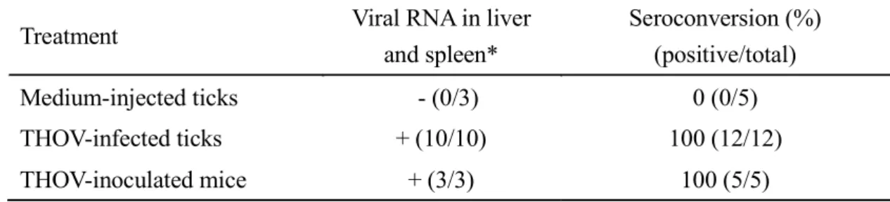
Treatment Viral RNA in liver and spleen* Seroconversion (%) (positive/total) Medium-injected ticks - (0/3) 0 (0/5) THOV-infected ticks + (10/10) 100 (12/12) THOV-inoculated mice + (3/3) 100 (5/5)
Table 2.5 THOV transmission from ticks injected with THOV through anal pore microinjection to mice
Fig. 2.1 Replication of the Thogoto virus (THOV) in Haemaphysalis longicornis after infection using anal pore microinjection. To quantify the changes in THOV RNA collected from groups of five ticks at each time point, real-time PCR was used (A). Virus titration after THOV infection, wherein error bars indicate the SD in mean values of five ticks at each time point (B). * P < 0.05, ** P < 0.01, as compared to day 0.
Fig. 2.2 Localization of the Thogoto virus (THOV) in the midgut (A,B) and salivary glands (C,D) of unfed adult ticks after infection via anal pore microinjection. Viral antigens were detected using a specific THOV polyclonal antibody (B,D), while normal mouse serum served as a control (A,C). Nuclear counterstaining (blue) was done using DAPI, and arrowheads denote THOV antigen
Fig. 2.3 Localization of the Thogoto virus (THOV) in the midgut (A,B), salivary glands (C,D), and synganglia (E,F) of partially fed, infected adult ticks via anal pore
microinjection. Viral antigens were detected using a specific THOV polyclonal antibody (B,D,F), while normal mouse serum served as a control (A,C,E). Nuclei counterstaining (blue) was done using DAPI, and arrowheads denote THOV antigens (red) (bar = 20
Fig. 2.4 Detection of Thogoto virus (THOV) antibodies in serum samples from mice using immunofluorescence assay. Sera collected from a mouse infested with
EMEM-injected ticks (A) (1:200, No.1); mouse inoculated with 10,000 ffu of THOV (B) (1:3200, No.1); mouse infested with THOV-injected ticks (C) (1:3200, No.6) reacting with THOV-infected baby hamster kidney BHK-21 cells. Arrowheads denote
CHAPTER 3
Evaluation of the role of antimicrobial peptide, longicin, from
Haemaphysalis longicornis against Langat virus
This work was published as: Talactac, M.R., Yoshii, K., Maeda, H., Hernandez, E.P., Kusakisako, K., Tsuji, N., Galay, R.L., Fujisaki, K., Tanaka, T., and Mochizuki, M. (2016). Virucidal activity of Haemaphysalis longicornis longicin P4 peptide against tick-borne encephalitis virus surrogate Langat virus. Parasit Vectors. 9:59.
3.1 Introduction
Ticks are hematophagous arachnids capable of transmitting several disease-causing pathogens in domestic and wild animals, including humans [8, 31]. As carriers of several pathogenic microorganisms, protozoa, rickettsiae, spirochaetes, and viruses [1, 30], ticks need to employ broad spectrum innate immunity mechanisms that will allow them to maintain the pathogens and commensal microbes without impairing their viability and further development [32, 31]. As previously demonstrated, antimicrobial proteins and peptides play a major role in protecting ticks against microorganisms [43, 44].
Antimicrobial peptides are ancient immune molecules that are important in invertebrate and vertebrate host defenses [45, 46]. These peptides display broad-spectrum biological activity against bacteria, yeast, fungi, protozoan parasites and enveloped viruses [47 - 49] and have been demonstrated to possess immunomodulatory properties [50]. Numerous small molecules such as defensins, lysozymes or by tick-specific antimicrobial compounds such as microplusin provide the direct antimicrobial defense in ticks [31].
Longicin, a defensin-like peptide identified from the midgut epithelium in the hard tick Haemaphysalis longicornis, is a promising cationic antimicrobial peptide.
Many studies have shown that longicin and one of its synthetic partial analog (longicin P4) have antimicrobial, fungicidal, and parasiticidal properties [32, 80, 81]. Thus, making them attractive molecules to be used as therapeutic agents, not only against tick-borne pathogens, but also to important human and animal disease-causing agents. On the other hand, interest in the therapeutic applications of antimicrobial peptides or their synthetic analogues is increasing due to the rise in resistance to commonly used antibiotics [30, 82].
Tick-borne flaviviruses (TBFVs) cause considerable disease and death worldwide, wherein infections are characterized by mild to severe neurological symptoms, such as meningitis and encephalitis [19, 83]. For Europe, Russia and up to the eastern coast of Japan, TBEV is considered as one of the most medically important arboviruses with 10,000 to 15,000 cases recorded each year [83, 84]. Since most TBFVs require at least a biosafety level 3 (BSL3) containment facility, the use of the naturally attenuated LGTV provides a convenient BSL2 model of TBEV and other highly pathogenic TBFVs [19]. In this study, I investigated the virucidal activity of longicin P4 against LGTV, a member of TBEV serocomplex of the Flaviviridae family.