Material report: A case report on human skeletal remains suggesting the penis removal in the Edo period
Kazuhiro Sakaue
Department of Anthropology, National Museum of Nature and Science 4–1–1 Amakubo, Tsukuba-city, Ibaraki Prefecture 300–0005, Japan
E-mail: k-sakaue@kahaku.go.jp
Abstract This paper reports on a specific sharp force trauma observed in skeletons of the Edo period. A cut mark can be recognized at the inferior margin of the pubic symphysis and on the pos- terior surface of the left innominate bone of “No. 583” excavated from the Shyoken-ji site. The morphological features of this cut mark suggest that this individual suffered the removal of his penis. Some historical reports show the existence of penis removal in the Edo period. This is the first report strongly suggesting the penis removal in Japanese skeletal remains.
Key words: Edo, cut mark, sharp force trauma, penis removal
Introduction
This paper reports on a specific sharp force trauma observed in skeletons of the Edo period.
There are some reports about some sharp force traumas, which reveal historical and cultural tra- ditions in ancient Japanese (Morimoto, 1987;
Morimoto and Hirata, 1992; Sakaue, 2010).
However, the position and direction of the cut mark reported here were very unusual, and dif- ferent from any of those described in the previ- ous studies.
Material
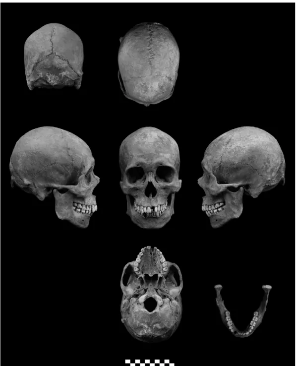
The human skeletal remains with unique sharp force trauma is stored at the National Museum of Nature and Science as “No. 583” of the Shyoken- ji site, which was excavated at 24, Nangen-cho, Shinjuku-ku, Tokyo in 2003 (Taisei Engineering Co., Ltd, 2005). This individual was buried in a circular wooden coffin (Hayaoke), which means this individual is thought to be a townsman or samurai of the lower class in the city of Edo (Tanigawa, 2004; Sakaue, 2012). The preserva- tion of “No. 583” is relatively good, as shown in Figures 1–2. The sex of this individual was esti-
mated as male, diagnosed by the narrow greater sciatic notch of the innominate bone and promi- nent supraorbital ridge and mastoid process of the skull. The age-at-death was estimated as 30–50 years old because of the macroscopic dis- appearance of the epiphyseal lines of the clavi- cle, no appearance of degenerative change in the vertebral body, and the pubic symphysis showing the morphological characteristics of Phase 4 in the Suchey–Brooks system (Brooks and Suchey, 1990).
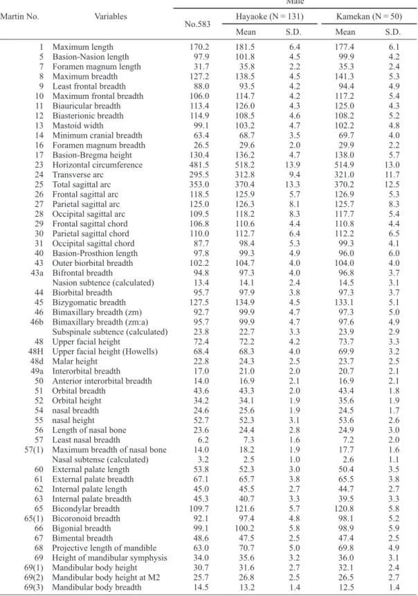
The skull measurements of this individual are shown in Table 1. The morphological features of this skull were a relatively narrow and long face, a prominent high-bridged nose and a weak alveo- lar prognathism. In order to elucidate the charac- teristics of the skull of this individual, principal component analysis on a correlation matrix was performed with 186 male skulls of the Edo period, including 5 of the “daimyo (territorial lords),” 131 of townsmen of the Middle to Late Edo period, and 50 of “samurai” of the middle–
late Edo (Sakaue, 2012). This statistical analysis
was carried out with SYSTAT 13 (Systat Soft-
ware Inc., 2009). The scatter plots of the second
and third principal component scores are pre-
sented in Figure 3. In this plot, No. 583 (black
star) was an outlier for the townsmen and samu- rai. However, interestingly, this individual located close to the daimyo. This finding indi- cated that No. 583 might have had distinctive facial characteristics among the townsmen of the
city of Edo.
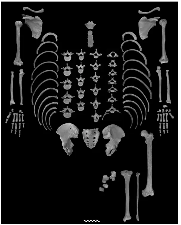
The other notable features of this individual were a cervical rib rising from the seventh cervi- cal vertebra (Figure 2) and that the number of thoracic vertebrae was 11. In Japanese, the fre-
Fig. 1. Photographs of the skull of No. 583.
quency of individuals with cervical ribs is 2.0%
(4 out of 200) (Matsui, 1942), and those with 11 thoracic vertebrae is 0.8% (2 out of 246) (Take- uchi, 1980). This abnormality may be related to no facet for 1st and 2nd costal cartiladges in the
manubrium of sternum in this individual.
Description
A cut mark on this individual can be recog-
Fig. 2. Photographs of the postcranial bones of No. 583.
Table 1. cranial measurements and Indexes of No. 583
Martin No. Variables
Male
No.583 Hayaoke (N=131) Kamekan (N=50)
Mean S.D. Mean S.D.
1 Maximum length 170.2 181.5 6.4 177.4 6.1
5 Basion-Nasion length 97.9 101.8 4.5 99.9 4.2
7 Foramen magnum length 31.7 35.8 2.2 35.3 2.4
8 Maximum breadth 127.2 138.5 4.5 141.3 5.3
9 Least frontal breadth 88.0 93.5 4.2 94.4 4.9
10 Maximum frontal breadth 106.0 114.7 4.2 117.2 5.4
11 Biauricular breadth 113.4 126.0 4.3 125.0 4.3
12 Biasterionic breadth 114.9 108.5 4.6 108.2 5.2
13 Mastoid width 99.1 103.2 4.7 102.2 4.8
14 Minimum cranial breadth 63.4 68.7 3.5 69.7 4.0
16 Foramen magnum breadth 26.5 29.6 2.0 29.9 2.2
17 Basion-Bregma height 130.4 136.2 4.7 138.0 5.7
23 Horizontal circumference 481.5 518.2 13.9 514.9 13.0
24 Transverse arc 295.5 312.8 9.4 321.0 11.7
25 Total sagittal arc 353.0 370.4 13.3 370.2 12.5
26 Frontal sagittal arc 118.5 125.9 5.7 126.9 5.3
27 Parietal sagittal arc 125.0 126.3 8.1 125.7 8.3
28 Occipital sagittal arc 109.5 118.2 8.3 117.7 5.4
29 Frontal sagittal chord 106.8 110.6 4.4 110.8 4.4
30 Parietal sagittal chord 110.0 112.7 6.4 112.2 6.5
31 Occipital sagittal chord 87.7 98.4 5.3 99.3 4.1
40 Basion-Prosthion length 97.8 99.3 4.9 96.0 6.0
43 Outer biorbital breadth 102.2 104.7 4.0 104.0 4.0
43a Bifrontal breadth 94.8 97.3 4.0 96.8 3.7
Nasion subtence (calculated) 13.4 14.1 2.4 14.5 3.1
44 Biorbital breadth 95.7 97.9 3.8 97.3 3.7
45 Bizygomatic breadth 127.5 134.9 4.5 133.1 5.1
46 Bimaxillary breadth (zm) 92.7 99.9 4.7 97.3 5.0
46b Bimaxillary breadth (zm:a) 95.7 99.9 4.7 97.6 4.9
Subspinale subtence (calculated) 23.8 22.7 3.3 23.9 2.9
48 Upper facial height 72.4 72.2 4.2 73.7 3.3
48H Upper facial height (Howells) 68.4 68.3 4.0 69.9 3.2
48d Malar height 22.8 24.3 2.5 23.7 2.5
49a Interorbital breadth 17.0 21.0 2.0 20.7 2.1
50 Anterior interorbital breadth 14.0 16.9 2.1 16.9 2.1
51 Orbital breadth 43.6 43.3 2.0 43.4 1.8
52 Orbital height 34.2 34.1 1.9 35.6 1.9
54 nasal breadth 24.6 25.6 1.9 24.5 1.7
55 nasal height 52.7 52.3 3.1 53.6 2.6
56 Length of nasal bone 23.6 24.4 2.8 24.9 3.0
57 Least nasal breadth 6.2 7.3 1.6 7.2 2.0
57(1) Maximum breadth of nasal bone 14.0 18.2 1.9 17.7 1.6
Nasal subtense (calculated) 3.2 2.5 1.0 2.6 1.1
60 External palate length 53.8 52.3 3.0 50.4 3.5
61 External palate breadth 67.1 65.7 3.8 65.5 3.8
62 Internal palate length 45.0 45.5 2.7 44.7 2.7
63 Internal palate breadth 45.3 40.7 3.3 39.5 3.3
65 Bicondylar breadth 109.7 121.6 5.7 120.8 5.8
65(1) Bicoronoid breadth 92.1 97.4 4.8 98.1 5.2
66 Bigonial breadth 99.1 100.2 5.8 98.9 5.9
67 Bimental breadth 48.6 47.5 2.5 47.4 2.5
68 Projective length of mandible 63.0 70.7 5.0 69.8 4.9
69 Height of mandibular symphysis 34.0 35.6 3.2 36.0 3.1
69(1) Mandibular body height 30.7 31.6 2.7 32.1 2.4
69(2) Mandibular body height at M2 25.7 26.8 2.5 26.5 2.7
69(3) Mandibular body breadth 14.5 13.2 1.4 12.5 1.4
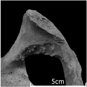
nized at the inferior margin of the pubic symphy- sis in the left innominate bone (Figure 4). This defect has the same color as the surface of the surrounding bone, and has characteristics of sharp force trauma such as “linearity,”
“a well-defined clean edge,” and “a flat smooth cut sur- face” on macroscopic observation (Boylston,
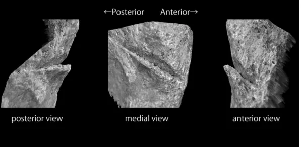
2000). This cut mark is limited to the posterior surface of the left pubic bone (Figure 5). The maximum length of this cut mark was 8.2 mm and the maximum depth was 1.2 mm when mea- sured in a 3D scanner (Keyence VR-3000) (Fig- ure 6).
The smooth surface of this cut mark was
Martin No. Variables
Male
No.583 Hayaoke (N=131) Kamekan (N=50)
Mean S.D. Mean S.D.
69b Mandibular body breadth at M2 16.5 17.1 1.6 16.6 1.3
70 Height of mandibular ramus 66.5 64.9 4.4 64.8 4.3
71a Minimum width of ramus 34.3 34.7 3.3 32.4 2.6
71(1) Condylo-cornoid breadth 35.8 36.2 3.2 33.4 3.9
Mandibular condyle breadth 19.2 20.7 2.0 20.6 1.9
72 Total profile angle 84.3 83.3 3.1 84.4 3.6
74 Alveolar profile angle 74.6 64.8 6.3 67.5 6.7
75 Profile angle of nasal bone 45.3 63.4 6.0 62.2 5.5
79 Mandibular angle 127.2 124.5 6.9 126.5 6.9
8/1 Cranial index 0.75 0.76 0.03 0.80 0.04
17/1 Index 0.77 0.75 0.03 0.78 0.03
17/8 Index 1.03 0.98 0.04 0.98 0.04
(1+8+17)/3 Modulus 142.6 152.03 3.54 152.24 4.09
9/10 Index 0.83 0.82 0.03 0.81 0.03
9/8 Index 0.69 0.68 0.03 0.67 0.04
8/12 Index 1.11 1.28 0.05 1.31 0.06
40/5 Index 1.00 0.98 0.04 0.96 0.05
16/7 Index 0.84 0.83 0.06 0.85 0.06
27/26 Index 1.06 1.00 0.07 0.99 0.07
28/26 Index 0.92 0.94 0.08 0.93 0.06
29/26 Index 0.90 0.88 0.02 0.87 0.02
30/27 Index 0.88 0.89 0.02 0.89 0.03
31/28 Index 0.80 0.83 0.03 0.84 0.02
43/8 Index 0.80 0.76 0.03 0.74 0.03
46/45 Index 0.73 0.74 0.03 0.73 0.03
48/45 Index 0.57 0.54 0.03 0.55 0.03
48/46 Index 0.78 0.72 0.04 0.76 0.04
9/45 Index 0.69 0.69 0.03 0.71 0.03
45/8 Index 1.00 0.97 0.04 0.94 0.04
50/44 Index 0.15 0.17 0.02 0.17 0.02
52/51 Index 0.78 0.79 0.04 0.82 0.05
54/55 Index 0.47 0.49 0.04 0.46 0.03
54/55(1) Index 0.71 0.87 0.10 0.80 0.08
57/57(1) Index 0.45 0.40 0.09 0.41 0.11
61/60 Index 1.25 1.26 0.08 1.30 0.10
63/62 Index 1.01 0.90 0.08 0.87 0.16
68/65 Index 0.57 0.58 0.04 0.58 0.05
69(3)/69(1) Index 0.47 0.42 0.05 0.39 0.05
69b/69(2) Index 0.64 0.64 0.07 0.63 0.08
71/70 Index 0.52 0.54 0.05 0.50 0.05
Frontal index of flatness 0.14 0.14 0.02 0.15 0.03
Zygomatic index of flatness 0.23 0.23 0.03 0.25 0.03
Simotic index 0.51 0.34 0.12 0.37 0.13
“Hayaoke” and “Kamekan” data were cited from Sakaue (2012)
placed in coronal plane and faced in an almost posterior direction, which indicates that the blade of a sharp instrument was used on this individual to scrape the bone surface downward from the
superior to the inferior. Because it was on the posterior surface of the pubic bone, it was possi- ble that this cut mark was made by the insertion of a sharp instrument from the pelvic cavity side
-3 -2 -1 0 1 2 3 4
FACTOR (2)
-3 -2 -1 0 1 2 3 4
FACTOR (3)
★ kamehayaokedaimyo
Shyokenji 583
★
Fig. 3. Plot of the second and third principal component male scores.
The ellipses represent the 68.27% confidence interval for each group.
Fig. 4. Photograph of the cut mark at the inferior margin of the pubic symphysis in the left innominate bone.
Fig. 5. Posterior view of the left innominate bone.
after removal of the pelvic viscera as part of a surgical operation, dissection, or mutilation of the corpse. But this is unlikely, considering the lack of other cut marks on this individual and the rarity of surgical operations and dissection in the Edo period. Additionally, this cut mark could not be made by someoneʼs occasional attack from his front in conflict because this wound cannot be
seen in the frontal view of the reconstructed pel- vis (Figure 7). Moreover, this is the only cut mark observed in this individual. In case of homicide or mayhem by using sharp-edged weapons, cut marks are often observed on the skull, ribs, and upper extremities. This cut mark is located on the internal surface of the left pubic bone, which is just below the pubic symphysis
Fig. 6. 3D images of the cut mark reconstructed in Keyence VR-3000.
Fig. 7. Anterior view of the reconstructed pelvis.
The arrow in the picture shows the position of
the cut mark. Fig. 8. Right anterior view of the reconstructed
pelvis.
The arrow in the picture shows the cut mark.
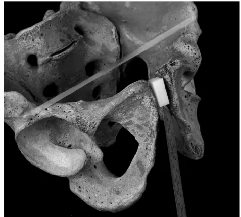
(Figure 8). This area is considered to be too nar- row for a sharp-edged weapon with the width of over 10 mm to have been inserted without any damage to the right pubic bone. Therefore, a thin sharp instrument was inserted from the right anterior to the left posterior of this individual as seen in Figure 9, and moved a little from superior to inferior almost along the frontal surface of the body.
At the point of this cut mark, the arcuate pubic ligament attaches. The ischiocavernosus muscle, the left crus of the penis, the sphincter muscle of the urethra, and the transversus perinei profundus muscle are close to this area. And, most impor- tantly, the root of the penis is around this area. A series of cutting movements estimated from the abovementioned cut mark seem to match the penis removal. The specific options that account for the evidence seem to be one of the following:
1) He cut off his penis himself with a thin sharp instrument like a razor or knife in his right hand;
2) Someone cut his penis off while holding it in his or her right hand and sharp instrument in the left hand. In both cases, the force applied to the sharp-edged weapon was strong enough to injure the bone. No trace of healing in this wound indi- cated that the removal of his penis caused his death or was performed soon after his death.
Interestingly, there are some historical records on the penis removal in the Edo period. Ryoo
DOKAKU
(了翁道覚,1630–1707) was amonk of the Obaku sect of Buddhism and a con- tributor to religious social activities such as edu- cation, culture, social welfare, and public utili- ties. Legend has it that he cut off his own penis in order to escape all carnal desires (Kimura, 2007). The penis removal was performed as a surgical operation (penectomy) for patients with
Fig. 9. Illustration for the insertion of thin sharp instrument in the reconstructed pelvis.
A steel scale with a breadth of 10 mm was placed its long side on the extension of the long axis of the cut mark and its flat surface on the smooth face of the cut mark.
1835) and his disciples (Kure, 1971; Matsuki, 1996). Moreover two incidents regarding the removal of penis where two wives cut off those of their husbands were reported in “Wagakoromo
(我衣),” written by Kato Ebian (加藤曳尾 庵,
1763–?) (Tanigawa, 1971) and “Fujioka-ya nikki
(藤岡屋日記)”written by Sudo Yoshizo
(須藤由蔵,
1793–?) (Suzuki and Koike, 1989). Thus, it is possible that the cut mark observed in No. 583 was caused by the penis removal in the Edo period. This is the first report strongly suggesting the penis removal in Japa- nese skeletal remains.
Acknowledgements
I would like to thank Dr. R. T. Kono, the National Museum of Nature and Science, for her help for VR-3000. I wish to thank Dr. N. Adachi, Professor of the Department of Legal Medicine, University of Yamanashi Interdisciplinary Grad- uate School of Medicine and Engineering. This research is supported by JSPS KAKENHI, Grant Number 15K07242.
References
Boylston A. (2000) Evidence for Weapon-related Trauma in British Archaeological Samples. In: Cox M. and Mays S. (eds.), Human Osteology in Archaeology and Forensic Science. Cambridge University Press, Cam- bridge, pp. 357–380.
Brooks S. and Suchey J.M. (1990) Skeletal age determi- nation based on the os pubis: a comparison of the Acsadi–Nemeskeri and Suchey–Brooks methods.
Human Evolution, 5: 227–238.
Kimura T. (2007) Syoki Obaku ha no sou tachi (Biogra- phies of the monks in the early Obaku sect). Syunbun sya (in Japanese).
Kure S. (1971) Hanaoka Seisyu Snsei oyobi so Geka
Matsui T. (1942) An anthropological study of Japanese skeletons, regarding vertebrae. Kaibogaku Zassi, 19:
427–460 (in Japanese).
Morimoto I. (1987) Note on the Technique of Decapita- tion in Medieval Japan. Journal of the Anthropological Society of Nippon, 95: 477–486.
Morimoto I. and Hirata K. (1992) A Decapitated Human Skull from Medieval Kamakura. Journal of the Anthro- pological Society of Nippon, 100: 349–358.
Sakaue K. (2010) A Case Report of Human Skeletal Remains Performed “Tameshi-giri (Test Cutting with a Japanese sword)”. Bulletin of National Museum Nature and Science Series D, 36: 27–36.
Sakaue K. (2012) Craniofacial variations among the com- mon people of the Edo period. Bulletin of National Museum Nature and Science, Tokyo, Ser. D, 38, 39–49.
Matsuki A. (1996) Hanaoka Seisyu no masui no fukyuni- tuite—Fukui han Hashimoto Sanai ni yoru syujyutusy- ourei no kento (Spread of Hanaokaʼs method of general anesthesia with Mafutsu-san—A discussion of surgical operations by Sanai Hashimoto). Nippon ishigaku zashi 42: 289–302 (in Japanese with English summary) Suzuki T. and Koike A. (1989) Kinsei Syomin Seikatu
Siryo Fujioka-ya nikki6 (Documents of the lives of recent common people, the diary of Fujioka-ya volume 6). Sanichi syobou (in Japanese).
Systat Software, Inc. (2009) SYSTAT 13: Chicago, IL, USA.
Tanigawa A. (2004) edo no haka no maisousisetu to huku- souhin (Burial facilities and accessories in the graves of the Edo period). Edo iseki kennkyuukai (ed.), Haka to Maisou to Edo- jidai. Yoshikawakoubunnkann, Co.
Ltd., Tokyo, pp. 224–250 (in Japanese).
Tanigawa K. (1971) Nihon Syomin Seikatu Siryo Syusei 15 Kan, Toshifuzoku (Documents of the lives of recent common people in Japan, vol. 15, Customs in Cities), Sanichi syobou (in Japanese).
Taisei Engineering Co., Ltd (2005) Kensyutusareta Ikou to Ibutu (Detected relics and remains) In: Taisei Engi- neering Co., Ltd. (ed.), Sugenji Syoukenji Ato, Meiji- Jingu, Tokyo, pp. 22–143 (in Japanese).
Takeuchi S. (1980) A study of the number of Japanese vertebrae. Jikeikai Medical Journal, 95: 584–597 (in Japanese with English summary).