Comparison of Anti-Inflammatory Analgesics for Mechanical Stress-induced Inflammation in a Human Synovial Sarcoma Cell Line
Haruna S
HIRAKO*1,2), Yuko U
DAKA1), Akiko S
ASAKI1), Shota N
AKAMURA1,2), Mayumi T
SUJI1)and Yuji K
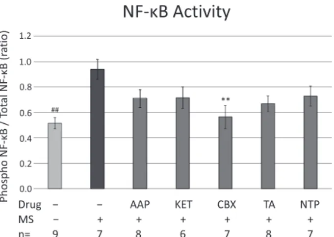
IUCHI1)Abstract : Osteoarthritis is a complicated clinical condition affected by age, mechanical stress, cartilage hypertrophy, cytokines, and genetic predisposition. In this study, we compared the effects of various anti-inflammatory analgesics on mechanical stress-induced inflammation in a synovial sarcoma cell line (SW982 cells) . SW982 cells exposed to mechanical stress by shaking with hydroxyapatite- simulating bone chips were treated with acetaminophen, ketoprofen, triamcinolone acetonide, celecoxib, or neurotrophin for 48 hr. The expression of integrin α 5 β 1 receptor, observed in fibroblasts and synovium, was evaluated. Levels of the transcription factor, nuclear factor- κ B, the inflammatory cytokine, tumor necrosis factor-α , the proteolytic enzyme, matrix metalloproteinase-3, and prostaglandin E
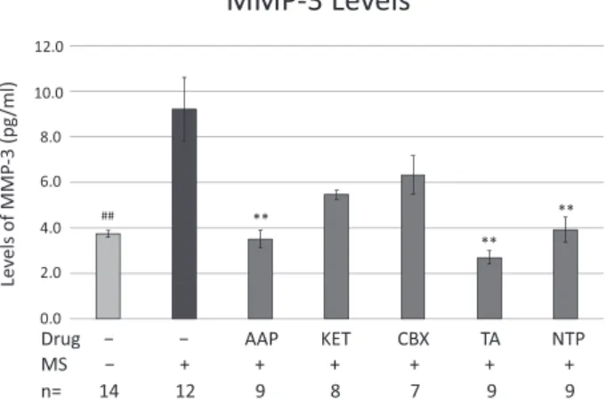
2, which is associated with pain and arachidonate cascade product levels, were measured by ELISA. The expression of integrin α 5 β 1 was significantly increased by mechanical stress. Activation of nuclear factor- κ B by mechanical stress was significantly suppressed by celecoxib only. Mechanical stress-induced increases in tumor necrosis factor-α and matrix metalloproteinase-3 levels were significantly suppressed by acetaminophen, triamcinolone acetonide, and neurotrophin. The mechanical stress-induced increase in prostaglandin E
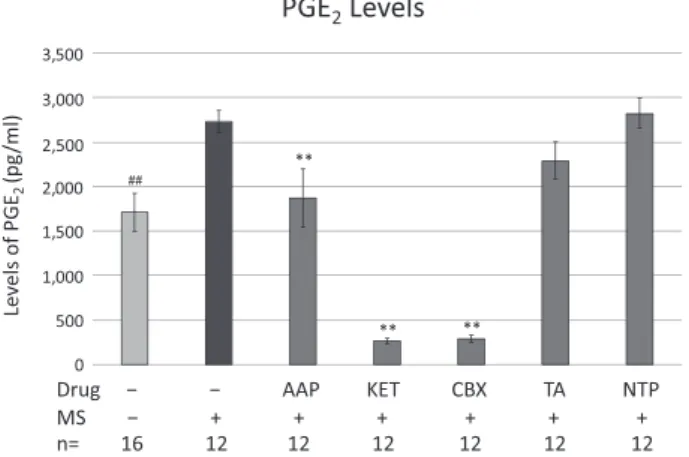
2levels was significantly suppressed by acetaminophen, ketoprofen, and celecoxib. SW982 exposed to mechanical stress is proposed as a model for arthritis, and indeed, the expression of integrin α 5β 1, a membrane receptor protein that binds to fibronectin and the extracellular matrix, and is involved in cell proliferation, differentiation, and neovascularization in osteoarthritis, was significantly upregulated. Following evaluation using this model, acetaminophen was found to possess anti-inflammatory, analgesic, and joint-destruction suppression properties. This drug may, therefore, have applications in the treatment of mechanical stress-induced inflammation.
Key words : osteoarthritis, anti-inflammatory agents, pain, inflammation
Introduction
Osteoarthritis (OA) is an age-related degenerative disease that affects various joints of the whole body, including knee and hip joints. In this disease, synovitis occurs at an early stage Original
1)
Department of Pharmacology, Showa University School of Medicine, 1-5-8 Hatanodai, Shinagawaku, Tokyo 142-8555, Japan.
2)
Department of Orthopedic Surgery, Showa University School of Medicine.
*