Introduction
Low-grade myofibroblastic sarcoma(LGMS)represents a distinct atypical myofibroblastic tumor with fibromatosis-like features and a predilection for the head and neck1). LGMS occurs primarily in adult patients with a slight male predominance ; children are rarely affected1). Although LGMS shows a wide anatomical distribution, it is most common in the head and neck regions, especially in the tongue and oral cavity2, 3). Rare cases involving the salivary gland and the nasal cavity / paranasal sinus have been reported. Here we report a case of LGMS at the base of the tongue. The clinical, histological, and immunohistochemi- cal features are described.
Case Report
A 56-year-old man presented at our hospital with a painless mass in the base of the atypical myofibroblastic tumor that occurs at several sites, primarily within the head and neck regions. A painless, enlarged mass is the most common clinical presentation, but a definitive diagnosis requires both histopathological and immunohistochemical analyses. Histologically, LGMS commonly presents as a cellular lesion composed of spindle-shaped tumor cells arranged primarily in fascicles with a diffusely infiltrative pattern. Immunohistochemically, LGMS shows positive staining for at least one myogenic marker, such as desmin or muscle actin. Here we report a case of LGMS in the base of the tongue.
Our case showed positive immunostaining for desmin and vimentin, and was thus diagnosed as LGMS. The patient received surgery, but no chemotherapy or radiotherapy, and was completely without evidence of the disease 38 months after the surgery.
Key words : low-grade myofibroblastic sarcoma, myofibrosarcoma, myogenic marker
Department of Otolaryngology, Showa University Fujigaoka Hospital, 1-30 Fujigaoka, Aoba-ku, Yokohama 227-8501, Japan.
tongue and difficulty in swallowing. Laryngeal fiberoscopy revealed a tumor at the base of the tongue(Fig. 1).
Magnetic resonance imaging(MRI)revealed a tumor mass that was well-demarcated and myxomatous without calcification. The airway space was stenosed by the tumor. Other analyses were negative for malignancies or tumor metastasis(Fig. 2).
Exfoliative cytology as well as forceps and fine needle aspiration cytology revealed no malignant cells. We therefore diagnosed a benign tumor of the base of the tongue, and sur- gery was performed accordingly. The tumor was covered by a capsule and was not adhered to the surrounding muscles and pharyngeal mucus, allowing resection with the capsule intact.
The patient received no chemotherapy or radiotherapy and was completely without evi- dence of disease 38 months after the surgery(Fig. 3).
Fig. 3. Postoperative MRI revealed no residual tumor, recurrence, or metastasis.
Fig. 2. Preoperative MRI revealed a tumor at the base of the tongue. The mass was well- demarcated and myxomatous without calcifi- cation. MRI confirmed stenosis of the airway due to the tumor.
Fig. 1. Tumor at the base of the tongue. The airway space was stenosed by the tumor.
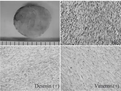
Histopathological findings(Fig. 4)
The resected tissue mass was 51 49 44 mm3. Sectioning revealed a well-demarcated nodular tumor measuring 42 mm. The tumor was solid, myxomatous, and appeared xant- hous without necrotic foci or hemorrhage.
Microscopic examination of the resected specimen showed spindle-shaped, elongated and stellate tumor cells embedded in a myxoid matrix.
Immunohistochemical examination of the tumor sections showed positive staining for desmin and vimentin. Immunostaining was negative for smooth muscle actin(SMA), muscle-specific actin(MSA), epithelial membrane antigen(EMA), S-100 protein, AE1 / AE3, CD34, and anaplastic lymphoma kinase(ALK). The patient was diagnosed as having LGMS.
Discussion
LGMS represents a distinct atypical myofibroblastic tumor with fibromatosis-like features and a predilection for the head and neck1). In most cases, patients present with a painless swelling or an enlarged mass, and pain or related symptoms are rarely reported. In general, the mass involved is firm with pale and fibrous cut surfaces and primarily indistinct margins, although the minority of these neoplasms is well circumscribed with well-defined margins.
Fig. 4. On sectioning, a well-demarcated nodular tumor, measuring 42 mm was observed. The tumor was solid, myxomatous, and appeared xanthous without necrotic foci or hemorrhage. The tumor was immunopositive for desmin and vimentin, but negative for SMA, MSA, EMA S-100 protein, AE1 / AE3, CD34, and ALK.
Radiologically, these lesions have a destructive growth pattern2).
Histologically, most cases of LGMS are characterized by a diffusely infiltrative growth pattern, and by deeply located neoplasms ; tumor cells may grow between individual skeletal muscle fibers. Most cases are composed of cellular fascicles or show a storiform growth pattern of spindle-shaped tumor cells. Importantly, neoplastic cells show at least focally moderate nuclear atypia with enlarged, hyperchromatic, and irregular nuclei and they show slightly increased proliferative activity.
The reported immunohistochemical findings vary for myofibroblast sarcoma(MS). Ment- zel et al2) reported that MS was positive for vimentin, SMA, calponin, and fibronectin, rarely positive for desmin, and negative for laminin and type IV collagen, while Montgom- ery et al4), Eyden et al5, 6)and Coffin et al7)reported SMA immunopositivity in 13 of 15 tumors, MSA staining in 7 of 9, and desmin in 6 of 14, with all of these cases negative for all cytokeratins, S-100 protein, and CD34. In a recent study, Meng et al8) reported that MS tumor cells strongly and extensively expressed vimentin(20 / 20), SMA(18 / 20), MSA
(16 / 20), and fibronectin(20 / 20), while only 2 cases(2 / 20)focally expressed desmin and type IV collagen, h-caldesmon, laminin, ALK1, and CD68 showed uniformly immunonegative staining. Based on these and our results, we conclude that myogenic markers are useful for the diagnosis of LGMS.
Clinically, local MS recurrences are common, whereas metastasis occurs only rarely and then only after a prolonged period. Although LGMSs are low-grade tumors, they exhibit a pattern of aggressive local spread with eventual metastatic dissemination1). The treatment for MS, as for other types of sarcomas, is surgical excision with wide margins. Adjuvant therapies such as chemotherapy and radiotherapy have also been used in some cases8-10), although the optimal treatment of LGMS remains ill-defined.
In our case, surgery alone was considered to be adequate and the patient was disease-free at the most recent oral examination, 38 months postoperatively. Such patients should be carefully observed at follow-ups.
References
1) Keller C, Gibbs CN, Kelly SM, Haller JR, White KS, Coffin CM and Lemors RS : Low-grade myofibrosarcoma of the head and neck : importance of surgical therapy. J Pediatr Hematol Oncol 26:119-120(2004)
2) Mentzel T, Dry S, Katenkamp D and Fletcher C : Low-grade myofibroblastic sarcoma : analysis of 18 cases in the spectrum of myofibroblastic tumors. Am J Surg Pathol 22:1228-1238(1998)
3) Jay A, Piper K, Farthing PM, Carter J and Diwakar A : Low-grade myofibroblastic sarcoma of the tongue. Oral Surg Oral Med Oral Pathol Oral Radiol Endod 104:e52-e58(2007)
4) Montgomery E, Goldblum JR and Fisher C : Myofibrosarcoma : a clinicopathologic study. Am J Surg Pathol 25:219-228(2001)
5) Eyden BP, Banerjee SS, Harris M and Mene A : A study of spindle cell sarcomas showing myofibroblastic dif- ferentiation. Ultrastruct Pathol 15:367-378(1991)
6) Eyden BP and Christensen L : Leiomyosarcoma versus myofibrosarcoma : observations and terminology. Ultra- struct Pathol 17:231-239(1993)
[Received October 14, 2010 : Accepted November 30, 2010]