緒 言
18-fluoro-2-deoxy-glucose positron emission tomogra- phy/computed tomography(FDG-PET/CT) 検査は,
肺癌の肺門・縦隔リンパ節転移の評価に広く用いられて いる.Billéら
1)の成績では,FDG-PET/CTで80.5%の肺 癌症例が正しく病期診断されたが,5.7%が過大評価され
(overstaging),13.8%が過小評価された(understaging).
その場合の転移リンパ節を検出するFDG-PET/CT の感 度・特異度・正診率はそれぞれ54.2%・91.9%・80.5%と なり,必ずしも満足できるものではなかった.
MR拡散強調画像(diffusion-weighted magnetic reso- nance imaging:DWI)は,拡散現象(水分子のブラウ ン運動)の制限領域を描出する撮影法で,みかけの拡散 係数(apparent diffusion coefficient:ADC)がその定量 評価に用いられている.正常組織の水分子の動きに対し,
細胞密度の高い悪性腫瘍等ではかなり拡散が制限された 動きになることを利用し,悪性腫瘍の検出,鑑別,治療 効果判定などにも応用されている.MR拡散強調画像で
は,癌および転移陽性のリンパ節は拡散が低下した領域 として描出されるのに対し,正常組織および転移陰性の リンパ節は描出されない.MR 拡散強調画像は,FDG- PETと違って被曝の問題がなく安価で,MR装置があれ ば施設を選ばない利点がある.MR拡散強調画像では拡 散能の低下した領域は明瞭に描出されるため,ADCの至 適カットオフ値が設定できれば,読影は比較的容易とい える.
著者らは,臨床研究 「肺癌および胸部病変に対するMR 拡散強調画像の有効性の検討」(金沢医科大学倫理委員 会承認番号189)で,MR拡散強調画像の有効性を検討し てきた.そして,肺癌例の転移リンパ節を検出するMR 拡散強調画像の感度・ 特異度・ 正診率は, それぞれ 63.9%・98.4%・90.6%であり,FDG-PET/CT のそれと 同等以上であることを報告した
2).
今回,術前検索でFDG-PET/CTよりもMR拡散強調画 像がリンパ節転移の有無をより正確に診断できた症例を 経験したので報告する.
MR拡散強調画像およびFDG-PET/CT
MR拡散強調画像は,SIEMENS MAGNETOM Avanto 1.5Tを用いて,撮像シーケンスはspin echo-EPI法に,脂 肪抑制にはspectral attenuated inversion recovery(SPAIR)
法を併用し,呼吸同期下に撮影した.ADC 値は,ADC map上でリンパ節に可能な限り最大の関心領域を設定し 測定した.b factorを0と800(sec/mm
2)とした.ADC 値は×10
−3mm
2/secで示した.
●画像診断
MR拡散強調画像が術前肺門・縦隔リンパ節転移の評価に有益であった肺癌の2症例
薄田 勝男a 松井 琢真a 本野 望a 的場 宗孝b 湊 宏c 浦本 秀隆a
要旨:PET-CTより,MR拡散強調画像が肺癌のリンパ節評価に有効であった症例を報告する.症例1は右下 葉の2.9cm大の肺癌例で,PET-CTでcN0,拡散強調画像でcN2,切除によりpN2と診断された.症例2は 右上葉の2.0cm大の肺癌例で,PET-CTで#4R・#4Lのリンパ節にFDGの集積があったが,拡散強調画像で はそれらに拡散の低下はなかった.肺切除および#4Rのリンパ節を切除し,リンパ節は炭珪肺症と診断され た.肺癌のリンパ節の評価では,拡散強調画像が有益な症例がある.
キーワード:MR拡散強調画像,FDG-PET/CT,肺癌,リンパ節転移 Diffusion-weighted magnetic resonance imaging (DWI),
18-fluoro-2-deoxy-glucose positron emission tomography/computed tomography (FDG-PET/CT), Lung cancer, Lymph node metastases
連絡先:薄田 勝男
〒920
‒
0293 石川県河北郡内灘町大学1‒
1a金沢医科大学呼吸器外科学
b同 放射線医学
c同 臨床病理学(現 石川県立中央病院病理診断科)
(E-mail: [email protected])
(Received 19 Jun 2017/Accepted 24 Oct 2017)
Fig. 1 Case 1. (a, b) The non-contrast CT showed a nodule of 29mm in the right lower lobe. (c) FDG-PET/CT showed a
positive accumulation of SUVmax(6.0) in the nodule. (d) DWI showed decreased diffusion (ADC: 0.888×10
−3mm2/sec). (e) Non-contrast CT (#4R lymph node). (f) FDG-PET/CT (#4R lymph node negative). (g) DWI (#4R lymph node, ADC 1.688×10⁻³mm2/sec
)
.(
h)
Non-contrast CT(
#11 lymph node)
.(
i)
FDG-PET/CT(
#11 lymph node negative)
.(
j)
DWI(
#11 lymph node positive, ADC 1.432×10⁻³mm2/sec)
.(
k)
Hematoxylin-eosin(
HE)
staining(
×100)
of a main tumor; lung cancer(
squa- mous cell carcinoma)
.(
l)
HE staining(
×200)
of a #4R lymph node; a metastatic lesion from the lung cancer.a
c
b
d
e
h
f
i
g
j
k l
FDG-PET/CT は,SIEMENS Biograph Sensation 16 を用いて,maximum standardized uptake value(SUV- max)値も同様にリンパ節に可能な限り最大の関心領域 を設定し測定した.
症例1・2とも,FDG-PET/CT とMR 拡散強調画像の 撮像間隔は1ヶ月以内であった.以前の報告
2)に従って,
転移性リンパ節を診断するためのADCのカットオフ値を 1.70×10
−3mm
2/sec,SUVmax のカットオフ値を4.45と した.
症 例
【症例1】
患者:71歳,男性.
生活歴:粉塵曝露歴はなく,喫煙指数は750であった.
現病歴:感冒症状を呈し,近医で胸部X線写真上,浸 潤影を指摘され,肺炎として治療を受けた.浸潤影は改 善したが,右肺下葉に2.9cm大の結節陰影が残存しその 後増大したため,当科に紹介となった.FDG-PET/CTに て,リンパ節にはFDGの有意な集積はなかった(Fig. 1).
一方,MR拡散強調画像では,肺門および縦隔リンパ節 に拡散の低下した制限領域が描出された.右肺下葉切除
およびリンパ節郭清術が施行され,肺癌(扁平上皮癌)
でリンパ節#4R(8個中3個),#10(11個中6個),#11i
(3個中3個)に肺癌の転移を認め,pT1cN2M0(pStage
ⅢA)と病理診断された.
【症例2】
患者:71歳,男性.
生活歴: 解体業に 45 年従事し, 喫煙指数は 1,000 で あった.
現病歴:高血圧・糖尿病・高脂血症で経過観察中に,
胸部単純CT上,右肺上葉に2.0cm大の腫瘤影を認めた.
気管支鏡検査で診断がつかず,胸腔鏡下の診断治療のた め当科に紹介となった.FDG-PET/CTにて,右肺上葉の 原発巣以外に#4R・#4Lのリンパ節にはFDGの有意な集 積を認めた(Fig. 2).一方,MR拡散強調画像では,肺 門および縦隔リンパ節に拡散の低下した制限領域の描出 はなかった.胸腔鏡下に右肺上葉部分切除術および#4R リンパ節切除術が施行され,肺癌(扁平上皮癌)で#4R リンパ節は炭珪肺症と診断された.
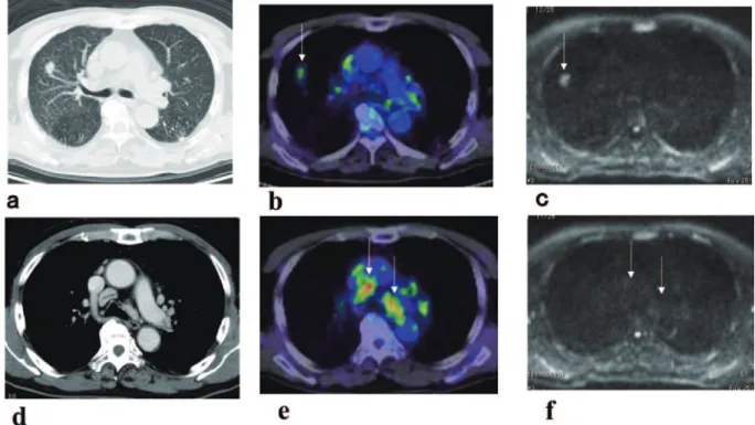
Fig. 2 Case 2. (
a‒
c)
Comparison of a primary lung cancer among non-contrast CT(
a)
, FDG-PET/CT(
b)
and DWI(
c)
.(
a)
The non-contrast CT showed a nodule of 20mm in the right upper lobe.(
b)
FDG-PET/CT showed an FDG accumulation(
SUVmax 4.18)
in the lung cancer(
arrow)
.(
c)
DWI showed decreased diffusion(
ADC: 1.45×10⁻³mm2/sec)
(
arrow)
.(
d‒
f)
Comparison of #4R and #4L lymph nodes among contrast-enhanced CT (d), FDG-PET/CT (e) and DWI (f). (e, f) FDG- PET/CT (e) showed an FDG accumulation (SUVmax 7.57) in the #4R and #4L lymph nodes (arrows). DWI (f) did not show decreased diffusion in the #4R and #4L lymph nodes(arrows).
考 察
著者らは,すでにMR拡散強調画像の有用性を原著で 報告しているが
2),症例報告は行っておらず,日本であ まり知られていないことを考えると,本症例を報告する 意義はあると考え報告した.今回の症例は2例とも扁平 上皮癌であるが,腺癌等の他の組織型でもFDG-PET よ り正確な診断に至った症例の経験を報告している
2)3).
FDG-PET/CT は種々の悪性腫瘍の評価に用いられ,
CT に比較しリンパ節転移の評価に優れていることが報 告されているが,ブドウ糖の代謝を反映した検査のため 偽陽性が少なくないことが指摘されている
4).
MRIの診断技術が進歩し,複数のmeta-analysisによっ て肺癌のリンパ節転移の評価により有効と報告されてい る
5)6).MR拡散強調画像のmeta-analysisの結果,非小細 胞肺癌における肺門および縦隔リンパ節の病期診断の感 度は87%,特異度は88%と高い診断能力を示している
5).
FDG-PET/CTに比較してMR拡散強調画像の長所の一 つとして,MR拡散強調画像による転移性リンパ節の感 度および正診率はFDG-PET/CT のそれより有意に高い と報告されている
2)7).MR拡散強調画像による肺癌88例 の正診率は89%と,FDG-PET/CT(78%)に比較し,有 意に高い
7).
MR拡散強調画像がFDG-PET/CTより正診率が高い理 由として,FDG-PET/CTではMR拡散強調画像に比較し て肺癌のN因子診断が過大評価されること(overstaging)
が少なくないことが挙げられる.実際,炭鉱労働者に対 するFDG-PET/CT でのリンパ節評価はじん肺症のため の高い偽陽性率となり適切ではないと報告されている
8). 珪肺に侵されたリンパ節は,FDG-PET/CT で中等度の FDGの集積を有し偽陽性となりやすい
2).FDG-PET/CT では,肺癌症例に合併した多発肺門・縦隔リンパ節の FDG集積についてその診断に難渋することが多い.FDG- PET/CTで肺門・縦隔リンパ節に多発FDGの集積を認め る症例では,FDG-PET/CTとMR拡散強調画像を比較す ると,MR拡散強調画像の特異度・正診率は,FDG-PET/
CT のそれに比較し有意に良好であった
9).FDG-PET/
CT でリンパ節にFDG の集積がみられた100例の病因の 検討では,癌の転移が11例,炭粉沈着症が40例,反応性 リンパ節炎が39例,肉芽腫が4例,珪肺症が3例であり,
必ずしも悪性病変は多いわけではない
10).
MR拡散強調画像がFDG-PET/CTより正診率が高いも う一つの理由として,FDG-PET/CTでは,肺癌例のN因 子診断が過小評価され(understaging),偽陰性が稀では ないことも報告されている
11).FDG-PET/CTで臨床的Ⅰ A期と診断された非小細胞肺癌例で,14.3%(21/147)が 病理学的N1ないしN2症例であった
11).著者らの160例
の肺癌例の解析によると,FDG-PET/CTで検出できた転 移リンパ節内の腫瘍最大径は11.9±4.1mm と,MR 拡散 強調画像の7.2±4.1mm に比較し,有意に大であった
2). MR拡散強調画像では,FDG-PET/CTよりも小型のリン パ節転移を検出でき,FDG-PET/CTに比較しN因子の偽 陰性が少なかった.しかし,より小さいリンパ節転移巣 は,FDG-PET/CTでもMR拡散強調画像でも検出できな かった.このことは,肺癌のリンパ節評価におけるFDG- PET/CTおよびMR拡散強調画像の診断には限界があり,
偽陰性例がなくならないことを示している.
FDG-PET/CT はN 因子に加え,M 因子が同時に評価 できる点など有用性がある.MR拡散強調画像でも,全 身拡散強調画像を撮影することは容易で,FDG-PET/CT と同様に遠隔転移の情報も提供可能である
12).
日本の「肺癌診療ガイドライン2016年版
13)」で,初め てMRIに関してMR拡散強調画像が小型肺野孤立陰影の 良悪性の鑑別や精査の必要性の判断に有用であることが 引用された.著者らは,いずれ,肺癌診療ガイドライン でMR 拡散強調画像もFDG-PET/CT と同程度の推奨グ レードで採用すべきと考えているが,MR検査が胸部疾 患で一般化していない現状であっても,MR拡散強調画 像 は FDG-PET/CT 検 査 が 施 行 で き な い 施 設 で FDG- PET/CT検査の代用として利用でき,またFDG-PET/CT 検査で判断が難しい症例に適応があると考えている.
MR拡散強調画像の短所として,浸潤性粘液性腺癌の MR拡散強調画像は,もともと細胞成分が少なく水分子 の拡散能が抑制されていないため,ADC値は高値となり 偽陰性となりやすい点が挙げられる
3).また,壊死組織 を有する良性病変は水分子の拡散能が抑制されるため,
ADC値が低値となり偽陽性となりやすい
3).
近年,脂肪抑制方法のSTIR(short TI inversion re- covery)像の有用性も報告されている
14).これは組織の 信号の縦磁化成分が0になるタイミング(null point)の 標準を脂肪に合わせ, 脂肪を抑制した画像であるが,
STIR 法は拡散強調画像よりも非小細胞肺癌のN 因子の 評価に有用と報告されており検討が必要である
14).
肺のMR拡散強調画像については,肺組織が空気と血 管による構造が主体であり,血流による灌流の影響およ び呼吸運動や心拍による影響を受け,他臓器と比較して 磁化率アーチファクトの発生やMRI信号獲得の困難さが 指摘されている.今後検査方法等の標準化が必要とされ ている.
肺門・縦隔リンパ節転移の有無について,FDG-PET/
CT検査よりMR拡散強調画像が有効であった2肺癌切除
例を報告した.肺癌例において,MR拡散強調画像によ
る肺門・縦隔リンパ節転移の評価は,FDG-PET/CTより
有益である可能性がある.
謝辞:本研究は,文部科学省の科学研究費(16K10694)の 助成で行われた.
著者のCOI(conflicts of interest)開示:本論文発表内容に 関して特に申告なし.
引用文献
1) Billé A, et al. Preoperative intrathoracic lymph node staging in patients with non-small-cell lung cancer: Accuracy of integrated positron emission tomography and computed tomography. Eur J Car- diothorac Surg 2009; 36: 440
‒
5.2) Usuda K, et al. Advantages of diffusion-weighted imaging over positron emission tomography-com- puted tomography in assessment of hilar and medi- astinal lymph node in lung cancer. Ann Surg Oncol 2013; 20: 1676
‒
83.3) Usuda K, et al. Diagnostic performance of diffusion weighted imaging of malignant and benign pulmo- nary nodules and masses: Comparison with posi- tron emission tomography. Asian Pac J Cancer Prev 2014; 15: 4629
‒
35.4) Roberts PF, et al. Factors associated with false-posi- tive staging of lung cancer by positron emission to- mography. Ann Thorac Surg 2000; 70: 1154‒9.
5) Peerlings J, et al. The diagnostic value of MR imag- ing in determining the lymph node status of pa- tients with non-small cell lung cancer: A meta-anal- ysis. Radiology 2016; 281: 86
‒
98.6) Shen G, et al. Performance of DWI in the nodal characterization and assessment of lung cancer: A meta-analysis. AJR Am J Roentgenol 2016; 206: 283
‒
90.7) Nomori H, et al. Diffusion-weighted magnetic reso- nance imaging can be used in place of positron emission tomography for N staging of non-small cell lung cancer with fewer false-positive results. J Tho- rac Cardiovasc Surg 2008; 135: 816
‒
22.8) Saydam O, et al. Accuracy of positron emission to- mography in mediastinal node assessment in coal workers with lung cancer. Med Oncol 2012; 29: 589‒
94.
9) Usuda K, et al. Diagnostic performance of diffusion- weighted imaging for multiple hilar and mediasti- nal lymph nodes with FDG accumulation. Asian Pac J Cancer Prev 2015; 16, 6401‒6.
10) Koksal D, et al. The correlation of SUVmax with pathological characteristics of primary tumor and the value of Tumor/Lymph node SUVmax ratio for predicting metastasis to lymph nodes in resected NSCLC patients. J Cardiothorac Surg 2013; 8: 63.
doi: 10.1186/1749
‒
8090‒
8‒
63.11) Park HK, et al. Occult nodal metastasis in patients with non-small cell lung cancer at clinical stage IA by PET/CT. Respirology 2010; 15: 1179‒84.
12) Usuda K, et al. Diagnostic performance of whole- body diffusion-weighted imaging compared to PET- CT plus brain MRI in staging clinically resectable lung cancer. Asian Pac J Cancer Prev 2016; 17:
2775‒80.
13) 日本肺癌学会編.質的画像診断.EBM の手法によ る肺癌診療ガイドライン2016年版.2016;29‒32.
14) Ohno Y, et al. N stage disease in patients with non
‒
small cell lung cancer: Efficacy of quantitative and qualitative assessment with STIR turbo spin-echo imaging, diffusion-weighted MR imaging, and Fluoro- deoxyglucose PET/CT. Radiology 2011; 261: 605‒15.Abstract
Two cases of lung cancer where diffusion-weighted magnetic resonance imaging was useful in the assessment of hilar and mediastinal lymph node metastases
Katsuo Usudaa, Takuma Matsuia, Nozomu Motonoa, Munetaka Matobab, Hiroshi Minatoc and Hidetaka Uramotoa
a
Department of Thoracic Surgery, Kanazawa Medical University
b
Department of Radiology, Kanazawa Medical University
c