4`•. Acta Med Kinki Univ
• Vol. 36, No. 1 33-36, 2011
Primary umbilical malignant melanoma : report of a
caseMasuki Yoshida, Shigeru Kawara and Akira Kawada
Department of Dermatology, Osakasayama,
Kinki Osaka
University Faculty of Medicine, 589-8511, Japan
Abstract
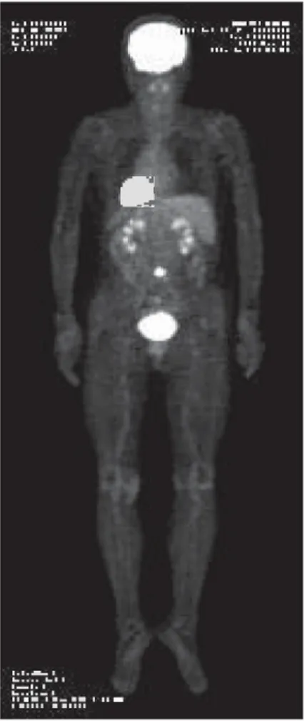
We report a 77-year-old female who showed a primary malignant melanoma on the umbilicus, with 3 years' duration. Physical examination showed a red, hard, and ulcerated tumor (2 cm) arising from the umbilicus, and there was no evidence of node metastases in axillary or groin nodes on either side. Preoperative dermoscopic examination showed an atypical pigment net- work and blue-grey background at the periphery of the tumor. Preoperative positron emission tomographic scan of her body showed hyper- metabolic tumor foci in the umbilicus, but no
distant metastases. Since there was a strong suspicion that this was a malignant melanoma, the patient underwent wide local excision of the primary tumor, including its attachment to the peritoneum. The excisied specimen was examined and reported as a malignant melanoma. The tumor thickness was 17 mm.
The patient underwent postoperative chemo- therapy. A case of umbilical malignant melanoma is reported. Moreover, previously reported cases are described.
Key words : malignant melanoma, umbilicus
Introduction
Neoplastic nodules localized to the umbilicus are relatively uncommon. Malignant tumors account for approximately 43% of all umbilical tumors, and are classified as primary or metas- tatic.' In particular, primary malignant umbili- cal tumors are extremely rare, comprising only 20% of all malignancies in this area, and may be melanomas, squamous or basal cell carcinomas, myosarcomas, or adenocarcinomas.''2
We report a case of primary umbilical malig- nant melanoma, and describe previously report- ed cases.
Case Report
A 77-year-old female exhibited a reddish nod- ule on the umbilicus, which had been present for approximately 3 years and showed umbilical discharge. On physical examination, the tumor was a granulating lesion, partially ulcerated, hard, measured approximately 2.0 X 2.0 cm in diameter, and based on irregular pigmented skin
in the umbilicus (Fig. 1). No axillary or in- guinal lymphadenopathy was noted. Dermos- copic examination showed a homogeneous blue- grey background with a superimposed atypical pigment network (Fig. 2). A positron emission tomographic scan of the body showed hyper- metabolic tumor foci in the umbilicus, but no distant metastases (Fig. 3). Hematological and radiographic data did not suggest any metastases.
Since there was a strong suspicion that this was a malignant melanoma, the tumor was resected with margin of 2 cm and down to the peritoneum. The resected area was closured simply.
A histologic specimen showed marked the following : the tumor was eosinophilic-stained and solid (Fig. 4a). The tumor showed variation in arrangement, with some areas consisting of eosinophil-staining cells and other areas present- ing a more whorled or spindle cell arrangement, and alveolar groups of cells were evident (Fig.
4b). In the tumor, irregularly sized and pigment- ed atypical cells were seen (Fig. 4c). In the dermis, melanophages and lymphocytes were
Received September 17, 2010 ; Accepted September 29, 2010
33
M. Yoshida et al.
detected (Fig. 4d). Moreover, a small aggregate consisting of irregularly arranged cells in the deep dermis was observed. These cells contained a melanin-like pigment. The pigmented atypical cells were positive for S-100 protein, HMB-45, and Melan-A.
Higher magnification of the upper dermis revealed these cells to be composed of hyper- chromatic atypical nuclei and partly of melanin with a vacuolated polygonal cytoplasm. Moder- ate numbers of mitotic figures were present (Fig.
4e). These histologic findings led us to diagnose the lesion as a malignant melanoma. Breslow's tumor thickness was 17 mm. The staging classifi- cation of AUCC was pT4bNOMO, stage IIc.
Because no palpable cutaneous lymph nodes were found, and radiographic data did not sug- gest metastases, the patient did not undergo wider local excision or regional lymph node dissection. Four weeks after excision, the patient underwent chemotherapy (DTIC, ACNU, VCR, and interferon-g). There was no evidence of
local gery.
recurrence or metastases 1
Discussion
year after sur-
It is important to distinguish between primary and metastatic tumors of the umbilicus. Metas- tatic tumors account for 80% of malignancies occurring in or on the umbilicus. The classic
MMI
11.1
•
1-1 In
n n'n
Fig. 1 Clinical view
The tumor was a granulating lesion, partially ulcerat- ed, hard, and measured approximately 2.0 X 2.0 cm in diameter, being based on irregular pigmented skin in the umbilicus.
Fig. 3
n:712"
11.1N'' .:;AN
Positron emission tomography (PET) of the body A PET scan showed hypermetabolic tumor foci in the umbilicus, but no distant metastases.
Fig. 2 Dermoscopic findings
Dermoscopic examination showed a homogeneous blue-grey background with a superimposed atypical pigment network.
34
Primary umbilical malignant melanoma : report of a case
a
•
• • .. +
.• . •
• - . . . .
. . 1.! •
•
• • • . • •
•
„4". ••• _ • - • • • • 2
1.•
• • •
.• •
. . ' •-• '.!
- ;1 •
: - • ".--7;:: •••
C
- • • . ••
b
• -
1111 --., •- ,....-
-
P.i' " ir, .. .'
.. .-. if e / ... i . . ,... --- ,--'..dr - „, ..,," i .1:
7 '(-, • '''' ____. _ . . . d ... , • f., lig .--,..00` 14 16 'Yrer.. •-..I... 1 -,.- .1.(11, 1... '1.1 ;y;rr1r \5+1 - - ,44 „, -: ‘• . ''''•• (.c.r.: i
•- '` -;Jt.---• ‘-1-. - t.•
Li ,"' ... O. ' '11. ..iro ., 42,...,1- „„--,....,.
,.1 ,,..., ,,...,- ...„.. .,, ..., #._ •,,,, :,.!.' ...:"'" ,',..' p-r-,-,- ;-. ---• 1-
1,.. ... -i'd*„'t --..: ...: ..., '..`.7...? a ..._ -.. • - g. A . . _Si.,..:1..'" \-.:.c.-
,....,-.)'•-,A:-.:).'T ti...- :•.--1..,. . IF --,---_`-, •• ar . ..-4, , ).,..--.•,-r.
r
- ' - '',„,,L,..)04,-:-.'.,,,--21.1-it.••..,--.6.4411.'.,.,,4*...:...-.•ii...0.-•it-
..;: .-ti•i'''..''•ii•ji
.i:'j ',:.-A•r..-; 4. ••••,_ ' : .• , 40.-4-/ ‘ki•-`-:-.1,,.1'1,• -..\:.:k '' .•• .... ••.'" • ::-•
e
Fig. 4 Histopathological findings
a : The tumor was eosinophilic and solid (H•E X 41 arrangement (H• E X 40). c : In the tumor, irregular (H• E X 100). d : In the dermis, melanophages am
d
w. - • •
-
e ,
% • -
10.
% r?:
ge 'CI;
a : The tumor was eosinophilic and solid (H•E X 40). b : The tumor showed marked variation in arrangement (H•E X 40). c : In the tumor, irregularly sized and pigmented atypical cells were seen (H•E X 100). d : In the dermis, melanophages and lymphocytes were detected (H•E X 100). e : Higher magnification of the upper dermis showed these cells to be composed of hyperchromatic atypical nuclei and partially of melanin, with a vacuolated polygonal cytoplasm (H•E X 400).
example is the "Sister Mary Joseph nodule,"
representing a metastasis from the gastrointesti- nal tract. Primary malignant tumors occurring on the umbilicus are much less common, accounting for approximately 20% of umbilical malignancies.
In a large series of 112 umbilical tumors,' 48 lesions were malignant and 64 were benign. The 48 patients with malignant tumors were adults
ranging from 21 to 79 years of age, with an average of 57 years. There were 32 male and 16 female patients. Of umbilical malignancies, eight were primary and 40 were metastatic tumors. Primary umbilical malignant melanomas accounted for four of eight primary malignancies.' Umbilical primary malignant tumors included malignant melanoma in 4 patients, basal cell carcinoma in 2, adenocar-
35
M. Yoshida et al.
cinoma in 1, and myosarcoma in 1. Each of the 4 malignant melanomas arose in a pre-existing nevus. The mean age of these patients was 61 years (range : 45-77 years) and the incidence was the same in both sexes. In addition, three patients developed regional or distant metastases over a 5-year follow-up period.'
We have found only 12 cases of primary umbilical malignant melanoma reported in the literature.1-8 Of the 12 cases, 5 were male and 7 were male. The average age of these patients was 53.2 years (range : 30-77 years). Clinically, the skin lesions were black nodules, irregularly shaped brownish or black macules, irregularly pigmented brown and black patches, granula- tion, and ulcerated swelling. The size of skin lesions ranged from 6 to 50 mm. The average tumor thickness of these patients was 1.75 mm (range : 0.5-4.5 mm). Clark's level was mostly III or IV.9 The depth of excision was from the fascia to peritoneum in almost all of these patients. Of these, 5 cases developed metastases after excision of the umbilical tumors. In approximately 60% of these cases, malignant melanomas developed in a pre-existing melanocytic nevus.1'5'6 Therefore, an accurate surveillance of these umbilical nevi, including the excision of suspicious lesions, is recommend- ed. In addition, the peculiar anatomy of the umbilicus can create difficulties in detecting signs of malignancy at this level, delaying inves- tigations. In a recent case of umbilical malig- nant melanoma, a diagnosis of omphalitis was initially made because of the presence of promi- nent inflammatory features that disguised the pigmented lesion.'
The presence of relevant vascular and em- bryological connections of the umbilicus with the peritoneum and other intra-abdominal vis- ceral organs has important implications regard- ing the surgical approach for these patients.2'7 It is known that vestigial remnants of several em-
bryonal structures, including the urachus, viteline artery, vitello-intestinal duct, and round ligament of the liver, pass to the umbilicus.2'6 Therefore, the extension of a tumor developing in the umbilicus along these fibrous remnants has been suggested as a possible risk of incom- plete excision and recurrence or metastatic spread.2'7 Consequently, malignant umbilical tumors, including primary malignant melanomas, should be managed by wide excision to the entire umbilical structure, including its attachment to the underlying peritoneum.
References
1. Steck WD, Helwig EB (1965) Tumors of the um- bilicus. Cancer 18 : 907-915
2. Meine JG, Bailin PL (2003) Primary melanoma of the umbilicus : report of a case and reviewof the rele- vant anatomy. Dermmatol Surg 29 : 404-407 3. Hughes J (1963) Melanoma of the umbilicus : a case
report. J Ir Med Assoc 53 : 94-98
4. Miller MJ, Charles MB (1993) "Iris" technique for immediate umbilical reconstruction. Plast Reconstr Surg 92 : 754-756
5. Hashiro M, Miyamoto T, Sonoda S, Okumura M (1997) Malignant melanoma developing from an intradermal nevus. Dermatology 196: 425-426 6. Colonna MR, Giovannini UM, Sturniolo G, Colon-
na U (1999) The umbilicus : arare site for melanoma.
Clinical conciderations in two cases. Scand J Plast Reconstr Hand Surg 33 : 449-452
7. Campos-Munoz I, Quesada-Cortes A, Ruiz e, Casado M, Pizarro A (2007) Primary melanoma of the umbilicus appearing as omphalitis. Clin Exp Dermatol 52 : 522-524
8. Cecchi R, Pavesi M, Buralli L, Rapicano V, Caudio CD (2009) Primary umbilical melanoma. Austrarias J Dermtol 50 : 220-222
9. Clark WH JR (1966) A classification of malignant melanoma in man correlated with histogenesis and biologic behavior. In Montagna W, Hu F (eds) : Advances in Biology of Skin, Vol 8, The Pigmentary System, New York, pergamon Press, pp 621-647
36