As s oc i at i ons of c ent r al aor t i c pr es s ur e and
br ac hi al bl ood pr es s ur e w
i t h f l ow
m
edi at ed
di l at at i on i n appar ent l y heal t hy J apanes e m
en:
The Ci r c ul at or y Ri s k i n Com
m
uni t i es St udy
( CI RCS) .
著者
Li u Keyang, Cui Renz he, Es hak Ehab S, Cui
M
ei s han, D
ong J i a- Yi , Ki yam
a M
as ahi ko, O
kada
Takeo, Ki t am
ur a Aki hi ko, U
m
es aw
a M
i t s um
as a,
Yam
agi s hi Kaz um
as a, I m
ano H
i r onor i , O
hi r a
Tet s uya, I s o H
i r oyas u
j our nal or
publ i c at i on t i t l e
At her os c l er os i s
vol um
e
259
page r ange
46- 50
year
2017- 04
権利
( C) 2017. Thi s m
anus c r i pt ver s i on i s m
ade
avai l abl e under t he CC- BY- N
C- N
D
4. 0 l i c ens e
ht t p: / / c r eat i vec om
m
ons . or g/ l i c ens es / by- nc - nd/ 4
. 0/
U
RL
ht t p: / / hdl . handl e. net / 2241/ 00146799
ASSOCIATIONS OF CENTRAL AORTIC PRESSURE AND BRACHIAL 1
BLOOD PRESSURE WITH FLOW MEDIATED DILATATION IN 2
APPARENTLY HEALTHY JAPANESE MEN: THE CIRCULATORY 3
RISK IN COMMUNITIES STUDY (CIRCS) 4
5
Keyang Liu1, Renzhe Cui1, Ehab S Eshak1, 2, Meishan Cui1, Jia-Yi Dong1, 6
Masahiko Kiyama3, Takeo Okada3, Akihiko Kitamura3, 4, Mitsumasa Umesawa5,
7
Kazumasa Yamagishi6, Hironori Imano1, Tetsuya Ohira7, Hiroyasu Iso1 8
1. Public Health, Department of Social Medicine, Osaka University Graduate
9
School of Medicine
10
2. Department of Public Health and Preventive Medicine, Minia University,
11
Egypt
12
3. Osaka Center for Cancer and Cardiovascular Disease Prevention
13
4. Tokyo Metropolitan Institute of Gerontology
14
5. Department of Public Health, Dokkyo Medical University, School of Medicine
15
6. Departments of Public Health Medicine, Faculty of Medicine, University of
16
Tsukuba
17
7. Radiation Medical Science Center for the Fukushima Health Management
18
Survey, Fukushima Medical University
19
Number of tables:2. Number of figures:0. 20
21
22
Correspondence to:
23
Professor Hiroyasu Iso, M.D, Ph.D.
24
Public Health, Department of Social Medicine,
25
Osaka University Graduate School of Medicine
26
2-2 Yamadaoka, Suita-shi, Osaka, 565-0871 Japan.
27
Tel: +81-6-6879-3911; Fax; +81-6-6879-3919
28
E-mail: [email protected]
Highlights 30
・We examined the associations of central systolic aortic pressure and brachial
31
systolic pressure with flow mediated dilatation in apparently healthy Japanese
32
men.
33
・Higher central aortic pressure rather than higher brachial blood pressure was
34
associated with lower flow mediated dilatation; the association was evident for
35
men without antihypertensive medication.
36
・Our finding suggests that central systolic aortic pressure, rather than brachial
37
systolic blood pressure, is a useful marker for endothelial dysfunction in men.
38
39
40
41
42
43
44
45
46
47
48
49
50
51
52
53
54
Background and aims. Endothelial dysfunction is considered the first stage in 56
the development of atherosclerosis and cardiovascular disease, and brachial
flow-57
mediated dilation (FMD) is a measure of endothelial function. It is uncertain
58
which of central systolic aortic pressure (CAP) or brachial systolic blood
59
pressure (SBP) is more strongly associated with FMD. Therefore, we examined
60
the correlations of CAP and SBP with FMD in Japanese men.
61
Methods. The study subjects comprised 507 male volunteers aged 30–79 years 62
that were residents in two communities under the Circulatory Risk in
63
Communities Study (CIRCS) between 2013 and 2015. The low percent change
64
of FMD (%FMD) ≤5.0% after 5 minutes of reactive hyperemia evaluated by the
65
brachial artery was used to assess endothelial dysfunction. Values of CAP and
66
SBP were divided into tertiles, with the lowest tertile used as a reference.
67
Results. After adjustment for cardiovascular risk factors, the multivariable odds 68
ratio (95% CI) of low FMD for the highest versus the lowest tertile of CAP was
69
1.5(0.9–2.6) for total subjects and 1.3(0.7–2.5) for those with and 2.4(1.2–4.8)
70
for those without antihypertensive medication use. The corresponding odd ratios
71
for the highest versus lowest tertile of SBP were 0.9(0.5–1.5), 0.8(0.3–2.2), and
72
1.3(0.7–2.5).
73
Conclusions. Higher CAP levels were associated with low FMD for men without 74
antihypertensive medication, but such an association was not found for SBP
75
levels.(word count: 227)
76
Key words: Central aortic pressure ■ Endothelial function ■ Japanese men ■
Cross sectional study
78
79
80
81
82
83
84
85
86
87
88
89
90
91
92
93
94
95
96
97
98
1. Introduction 100
Cardiovascular diseases remain the major cause of morbidity and mortality in
101
developed countries, with atherosclerosis being the leading underlying cause (1).
102
Endothelial dysfunction is considered the first stage in the development of
103
atherosclerosis and cardiovascular disease (2, 3). Endothelial cells form the inner
104
lining of all blood vessels and play a central role in vascular homeostasis; they
105
respond to stimuli, such as hemodynamic changes or blood-borne signals by
106
releasing vasoactive substances (4). Brachial flow-mediated dilation (FMD) is a
107
measure of the release of nitric oxide by the endothelium due to a transient flow
108
stimulus (5) and low brachial FMD was regarded as a cardiovascular disease risk
109
factor (4, 5).
110
Hypertension is a recognized risk factor for the development of atherosclerosis
111
and cardiovascular disease (6-8). Central systolic aortic pressure (CAP) has been
112
reliably determined by mathematically transforming the radial artery pulse
113
waveform to the aortic pulse waveform (9-10). Several studies have also reported
114
that CAP levels were strongly associated with risk of mortality from
115
cardiovascular disease (11, 12). The Circulatory Risk in Communities Study
116
(CIRCS) of 3,002 Japanese men and women reported that CAP levels were 117
correlated more strongly with cardiovascular risk factors than brachial systolic
118
blood pressure (SBP) levels (13). However, evidence for the correlation of CAP
119
with FMD is limited. Additionally, to date, it is unclear which of CAP or SBP is
120
more strongly associated with flow-mediated dilatation. In this study, we
investigated the relationship of CAP and SBP with FMD in the general
122
population.
123
124
125
126
127
128
129
130
131
132
133
134
135
136
137
138
139
140
141
142
2.Materials and Methods
144
2.1. Subjects
145
FMD and CAP measurements were conducted in two communities of the CIRCS,
146
a dynamic cohort study of the Japanese population: Yao City, Osaka Prefecture
147
and Ikawa town, Akita Prefecture under a nationwide study. We recruited 507
148
men aged 30–79 years one by one from January 2013 to May 2015 from
149
participants who underwent annual cardiovascular risk surveys. Informed consent
150
was obtained from community representatives based- on guidelines of the
151
Council for International Organizations of Medical Science to perform an
152
epidemiological study (14). The study protocol was approved by the Ethics
153
Committee of the Osaka University.
154
2.2. Measurement of FMD and CAP
155
All participants had five minutes of rest in the seated posture, using a standard
156
protocol (15). FMD was measured with high-resolution ultrasonography and
157
forearm occlusive cuff by technicians. High-resolution ultrasound with a
10-158
MHz linear array transducer was used to record longitudinal images of the right
159
brachial artery. This transducer system can accurately capture and track the edge
160
of target artery automatically once the probe is placed at the proper position. To
161
standardize the position of the probe, we used a specially designed arm-rest and
162
probe holder. The brachial artery diameter at baseline was measured by this
163
system and then the brachial cuff was inflated to 50mm Hg above SBP for 5
minutes and deflated. Computer-assisted analysis software (UNEX Co. Ltd.,
165
Nagoya, Japan) was used to determine brachial artery diameter
semi-166
automatically, as previously described (16).
167
The baseline longitudinal image of the artery was acquired for 30 seconds, after
168
which the blood pressure cuff was inflated to 50 mmHg above systolic pressure
169
for 5 minutes. FMD change (%FMD) was defined by the following
170
formula: %FMD = ((maximal artery lumen diameter after cuff release-artery
171
lumen diameter at baseline)/artery-lumen diameter at baseline) ×100, according
172
to published guidelines for determining endothelial function (17). The coefficient
173
of inter-observer variability for FMD measurements in our laboratory was 5.7 %,
174
while that of intra-observer variability were 11.1% apart from 2 months and
175
10.8% apart from 4 months. In previous studies, the coefficient of inter-observer
176
variability for FMD measurement was 1.3% to 3.5% (18, 19), and that for
intra-177
observer variability was 15.6% apart from 48 hours and 18.3% apart from 3
178
months (20).
179
CAP was measured by technicians with an automated tonometer, HEM-9000AI
180
(Omron, Healthcare Co., Kyoto, Japan). A previous clinical study used both
181
HEM-9000AI and standard cardiac catheterization to examine the validity and
182
reproducibility of CAP levels among 18 hypertension patients aged 47–78 years.
183
The correlation coefficient was 0.95 (p < 0.001) between CAP levels by the two
184
measurement systems, and 0.93 (p < 0.001) between the repeated CAP
185
measurement by HEM-9000AI (6).
2.3. Measurement of cardiovascular risk factors
187
We previously reported the protocols for measuring cardiovascular risk factors,
188
such as blood pressure, serum lipids, body mass index (BMI), assessment of
189
smoking and drinking habits, hypertension, and diabetes mellitus (10, 21, 22).
190
Height in stocking feet and weight in light clothing were measured. Body mass
191
index (BMI) (kg/m2) was calculated as weight in kilograms divided by height in
192
square meters. Trainedobservers measured SBP and diastolic blood pressure
193
(DBP) using a standard mercury sphygmomanometer on the right arm after
194
participants had rested for 5 minutes (23). An interview was conducted to
195
confirm information on habits, including drinking status, tobacco status,
196
hypertension, and diabetes mellitus medication use. For drinking status, persons
197
who reported consuming 0.3 gō (equivalent to 7 grams of ethanol) or more per
198
week were regarded as current drinkers. Former drinkers were defined as
199
abstainers for the previous 3 months or more. Trained interviewers also
200
determined information on smoking status, use of antihypertensive agents, and
201
medical history. Persons who smoked ≥1 cigarette per day were defined as
202
current smokers.
203
Blood samples were obtained on the same day as annual cardiovascular risk
204
surveys from participants and the serum was separated immediately.
205
Measurements of serum triglycerides were performed using a fluorometric
206
method by an Autoanalyzer II (Technicon, Tarrytown, NY, U.S.A.), while total
207
cholesterol and high density lipoprotein (HDL)-cholesterol measurements were
performed at the Osaka Medical Center for Health Science and Promotion lipid
209
reference laboratory, a certified member of the US National Cholesterol
210
Reference Method Laboratory Network (CRMLN), using enzymatic methods by
211
an auto analyzer Olympus AU 2700 (24). Serum glucose measurements were
212
performed by the hexokinase method, using the same instrument. Diabetes
213
mellitus was defined as a fasting glucose level of ≥7.8 mmol/L, a non-fasting
214
glucose level of ≥11.1 mmol/L, or use of medication for diabetes mellitus (25).
215
Hypertension was defined as SBP ≥140 mmHg, DBP ≥90 mmHg, or use of
216
antihypertensive medication (26).
217
2.4. Statistical analysis
218
We defined the low FMD as %FMD≤5.0 (lowest 30 percentile) based on
219
previous reports that used the receiver-operating characteristic analysis (27, 28).
220
Values of cardiovascular risk factors in subjects with %FMD≤5.0 and >5.0 are
221
presented as mean ± standard deviation (SD) or proportions (%). The odd ratios
222
(OR) with the respective 95% confidence intervals (CIs) of the low FMD were
223
calculated according to tertiles of and 1-SD increment of CAP and SBP levels, by
224
using logistic regression analysis, after adjusting for age in one model, and
225
further adjustment for potential confounding factors including area of residence,
226
heart rate, brachial artery baseline diameter, total serum cholesterol, serum
227
triglycerides, history of diabetes mellitus, drinking status and smoking status.
228
The analyses were repeated by stratifying antihypertensive medication use.
229
All statistical analyses were performed with SAS version 9.4 software (SAS
Institute Inc., Cary, NC, USA). All probability values for statistical tests were
231
two-tailed and values of p <0.05 were regarded as statistically significant.
232
233
234
235
236
237
238
239
240
241
242
243
244
245
246
247
248
249
250
251
3.Results
253
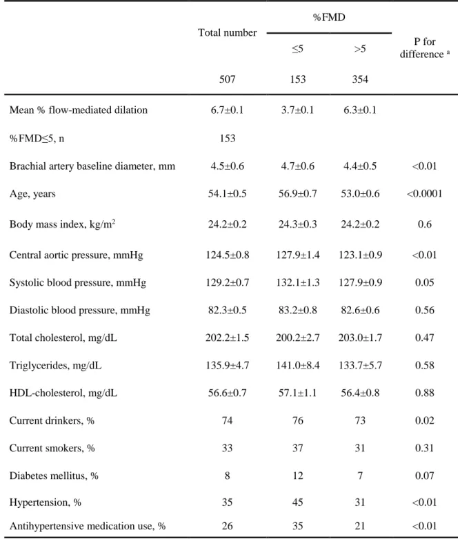
The characteristics of 507 Japanese men are summarized in Table 1. The mean
254
values of %FMD, age and BMI were 6.7, 54.1 years and 24.2kg/m2, respectively.
255
Compared with participants in the group of %FMD≤5, those in the group
256
of %FMD>5 had lower CAP, lower SBP levels and smaller brachial artery
257
baseline diameter, and were less likely to be drinkers, diabetics and hypertensive.
258
The ORs (95% CI) of the low FMD according to tertiles and 1-SD increment
259
for CAP and SBP levels are given in Table 2. Among total 507 subjects, the
260
multivariable ORs (95% CI) of the low FMD was 1.5(0.9–2.6) for the highest
261
versus lowest tertiles of CAP, and 1.2(1.0-1.5) for 1-SD increment (16.3 mmHg)
262
of CAP levels; while were 0.9(0.5–1.5) for the highest versus lowest tertiles of
263
SBP, and 1.0(0.8-1.3) for 1-SD increment (13.9 mmHg) of SBP levels.
264
When these associations were stratified by antihypertensive medication use,
265
significant positive associations between CAP and the low FMD were observed
266
primarily in subjects without antihypertensive medication use; the multivariable
267
ORs (95%CI) of the low FMD was 2.4(1.2–4.8) for the lowest versus highest
268
tertiles and 1.3(1.0-1.7) for 1-SD increment of CAP levels. There were no
269
difference in the associations between SBP and low FMD in participants with
270
and without the use of antihypertensive medication.
271
272
273
4.Discussion
275
In the present community-based study of 507 Japanese men aged 30–79 years,
276
CAP, but not brachial SBP, levels were correlated with the low FMD. The
277
association between CAP and low FMD levels was evident for men who did not
278
use antihypertensive medications.
279
Low FMD is a surrogate marker of early atherosclerosis in Japanese (29),
280
American (30), and European subjects (31). In a clinical study of 384 patients
281
with suspected cardio metabolic disorders, %FMD was significantly reduced in
282
patients with nonalcoholic fatty liver disease, diabetes, history of coronary heart
283
disease, metabolic syndrome, and in those taking antihypertensive drugs (31).
284
The Multi-Ethnic Study of Atherosclerosis for 2,936 men and women (mean age
285
61 years) showed that a 1-SD (2.8%) increase in %FMD values was associated
286
with lower risk of incident auricular fibrillation [Hazard ratio (HR) =0.84,
287
95%CI=0.70, 0.99], suggesting that markers of endothelial dysfunction
288
contributes to the pathogenesis of auricular fibrillation (30).
289
To our knowledge our study is the first to show that CAP levels were associated
290
more with reduced %FMD than SBP levels in men without use of
291
antihypertensive medication. Lind L has reported that CAP measurement was not
292
superior over traditional blood measurements regarding its relation to
293
endothelium-dependent vasodilatation or FMD (32). However, that study was
294
conducted only among elder participates over 70 years old. It was previously
295
shown that the absolute difference between aortic and brachial systolic pressures
declined with age (<20y up to 69 years) and then the difference plateaued after
297
ages≥70 years (33). Our study supports the previous finding from a clinical study
298
of 201 type 2 diabetes patients that an ankle-brachial index, a surrogate marker of
299
atherosclerosis, was more strongly correlated with CAP than SBP levels (CAP:
300
r=0.162, p=0.04, SBP: r=0.083, p=0.30)(34). Compared with a 10 mmHg
301
increment of SBP, the same increment of CAP was more strongly associated with
302
mortality risk from cardiovascular disease in a cohort study of normotensive and
303
untreated hypertensive Taiwanese; the multivariable HR (95% CI) of
304
cardiovascular mortality was 1.34(1.10–1.49) for CAP and 0.96(0.79–1.16) for
305
SBP (12).Our previous study found that CAP levels were associated with
306
subclinical damage expressed by minor ST-T ECG abnormalities (8).
307
Furthermore, a clinical study of 146 hypertensive patients reported that left
308
ventricular mass change was more strongly correlated with CAP than SBP (35).
309
These findings support CAP as a more sensitive marker for the loading
310
conditions on the heart and coronary arteries than SBP.
311
In the current study, the lack of association between CAP levels with %FMD
312
among subjects with antihypertensive medication use may be due to the dilution
313
of association after lowering CAP levels by various amounts. On the other hand,
314
the lack of association between SBP levels and %FMD in total subjects
315
regardless of antihypertensive medication use might probably due to the small
316
number of severe high blood pressure patients (SBP≥160 mmHg, n=21).
317
The strengths of the present study include the use of a noninvasive technique
for measuring CAP and FMD and the standardized measurements of other
319
cardiovascular risk factors in community population-based samples (22).
320
However, this study has several limitations as follows: first, details of
321
antihypertensive drug treatment were not available. Antihypertensive drugs such
322
as calcium channel antagonist, ACE-inhibitors and AT1-receptor antagonists can
323
improve endothelial function (36). However, we could not investigate whether
324
the lack of association between CAP levels and reduced %FMD in men with
325
antihypertensive medication use might be attributable to the effects of those
326
drugs or not. Second, reduced %FMD was the only indicator for endothelial
327
dysfunction in the current study; no data were available for nitrate-induced
328
vasodilatation. Last, our subjects were not recruited randomly, but they were
329
selected consecutively, and thus generalizability of our findings is limited. We
330
also were unable to include women in the current analysis because of very small
331
sample size, and further investigation will be necessary.
332
In conclusion, higher CAP levels were associated with low FMD for men
333
without antihypertensive medication, but such an association was not found for
334
SBP levels.
335
336
Conflict of interest 337
None declared.
338
Financial support 339
This study was supported by Grant-in-Aid for Scientific Research C (No.
25490790 in 2012-2014) from the Ministry of Health, Education, Culture, Sports,
341
Science and Technology, Japan.
342
Author contributions 343
Keyang Liu, Renzhe Cui, Ehab S. Eshak, Jia-Yi Dong, Meishan Cui and
344
Masahiko Kiyama participated in the study design and data collection; Keyang
345
Liu, Renzhe Cui and Ehab S. Eshak analyzed the data; Keyang Liu, Renzhe Cui,
346
Akihiko Kitamura and Hiroyasu iso participated in interpretation of data and
347
drafting of the manuscript; Keyang Liu, Renzhe Cui and Ehab S. Eshak provided
348
statistical expertise. Takeo Okada, Akihiko Kitamura, Mitsumasa Umesawa,
349
Kazumasa Yamagishi, Hironori Imano, Tetsuya Ohira and Hiroyasu Iso
350
participated in the study concept and design, acquisition of data and
351
interpretation of data, and critical revision of the manuscript.
352
Acknowledgements 353
The authors are grateful to Haytham A. Sheerah, Osaka University for his
354
contribution to this study. The full member list of the CIRCS Investigators is
355
presented in Appendix.
356
Appendix 357
The CIRCS study is a collaborative study managed by the Osaka Center for
358
Cancer and Cardiovascular Disease Prevention, University of Tsukuba, Osaka
359
University and Ehime University. The authors thank the CIRCS investigators
360
who contributed to this study: Professor Emeritus Yoshio Komachi (University of
361
Tsukuba), Professor Emeritus Hideki Ozawa (Medical College of Oita), Former
professor Minoru Iida (Kansai University of Welfare Sciences), Professor
363
Emeritus Takashi Shimamoto (University of Tsukuba), Dr. Yoshinori Ishikawa
364
(Consultant of Osaka Center for Cancer and Cardiovascular Disease Prevention),
365
Professor Yoshihiko Naito (Mukogawa Women’s University), and Professor
366
Tomonori Okamura (Keio University). 367
368
369
370
371
372
373
374
375
376
377
378
379
380
381
382
383
References 385
1. Fuster V, Kelly BB, Vedanthan R. Global cardiovascular health: urgent need
386
for an intersectoral approach. J Am Coll Cardiol. 2011; 58:1208-1210
387
2. Simsek H, Sahin M, Gunes Y, Akdag S, Akil MA, et al. A novel
388
echocardiographic method as an indicator of endothelial dysfunction in
389
patients with coronary slow flow. Eur Rev Med Pharmacol Sci. 2013;
17:689-390
693
391
3. Yeboah J, Folsom AR, Burke GL, Johnson C, Polak JF, et al. Predictive value
392
of brachial flow-mediated dilation for incident cardiovascular events in a
393
population-based study: the multi-ethnic study of atherosclerosis. Circulation.
394
2009; 120:502-509
395
4. Widlansky ME, Gokce N, Keaney Jr JF, Vita JA. The clinical implications of
396
endothelial dysfunction. J Am Coll Cardiol. 2003; 42:1149-1160
397
5. Yeboah J, Folsom AR, Burke GL, Johnson C, Polak JF, et al. Predictive Value
398
of Brachial Flow-Mediated Dilation for Incident Cardiovascular Events in a
399
Population-Based Study. Circulation. 2009; 120: 502-509
400
6. Obuchowicz A, Kniażewska M, Zmudzińska-Kitczak J, Urban K,
Gonciarz-401
Majda A. Concentrations of tumour necrosis factor-α and its soluble receptors
402
in the serum of teenagers with atherosclerosis risk factors: obesity or obesity
403
combined with hypertension. J Pediatr Endocrinol Metab. 2014; 27:1209-1212
404
7. Whelton PK. Sodium, potassium, blood pressure, and cardiovascular disease
405
in humans. Curr Hypertens Rep. 2014;16:465
8. Cui R, Li Y, Krisztina G, Yamagishi K, Umesawa M, et al. An association
407
between central aortic pressure and subclinical organ damage of the heart
408
among a general Japanese cohort: Circulatory Risk in Communities Study
409
(CIRCS). Atherosclerosis. 2014; 232:94-98
410
9. Takazawa K, Kobayashi H, Shindo N, Tanaka N, Yamashina A. Relationship
411
between radial and central arterial pulse wave and evaluation of central aortic
412
pressure using the radial arterial pulse wave. Hypertens Res 2007; 30:219-228
413
10. Pauca AL, O’Rourke MF, Kon ND. Prospective evaluation of a method for
414
estimating ascending aortic pressure from the radial artery pressure waveform.
415
Hypertension. 2001; 38:932–937
416
11. Roman MJ, Devereux RB, Kizer JR, Lee ET, Galloway JM, et al. Central
417
pressure more strongly relates to vascular disease and outcome than does
418
brachial pressure: the Strong Heart Study. Hypertension. 2007; 50:197-203
419
12. Wang KL, Cheng HM, Chuang SY, Spurgeon HA, Ting CT, et al. Central or
420
peripheral systolic or pulse pressure: which best relates to target organs and
421
future mortality? J Hypertens. 2009; 27:461-7
422
13. Cui R, Li Y, Krisztina G, Yamagishi K, Umesawa M, et al. An association
423
between central aortic pressure and subclinical organ damage of the heart
424
among a general Japanese cohort: Circulatory Risk in Communities Study
425
(CIRCS). Atherosclerosis. 2014; 232:94-8
426
14. International guidelines for ethical review of epidemiological studies. Law
427
Med Health Care. 1991; 19: 247-258
15. Kohara K, Tabara Y, Oshiumi A, Miyawaki Y, Kobayashi T, et al. Radial
429
augmentation index: a useful and easily obtainable parameter for vascular
430
aging. Am J Hypertens. 2005; 18:11S-14S
431
16. Maruhashi T, Soga J, Fujimura N, Idei N, Mikami S, et al. Relationship
432
between flow-mediated vasodilation and cardiovascular risk factors in a large
433
community-based study. Heart. 2013; 99: 1837-1842
434
17. Corretti MC, Anderson TJ, Benjamin EJ, Celermajer D, Charbonneau F, et
435
al. International Brachial Artery Reactivity Task Force. Guidelines for the
436
ultrasound assessment of endothelial-dependent flow-mediated vasodilation of
437
the brachial artery: a report of the International Brachial Artery Reactivity
438
Task Force. J Am Coll Cardiol. 2002; 39: 257-265
439
18. Furumoto T, Saito N, Dong J, Mikami T, Fujii S, et al. Association of
440
cardiovascular risk factors and endothelial dysfunction in Japanese
441
hypertensive patients: implications for early atherosclerosis. Hypertens Res.
442
2002; 25: 475-480
443
19. Suzuki K, Elkind MS, Boden-Albala B, Jin Z, Berry G, et al. Moderate
444
alcohol consumption is associated with better endothelial function: a cross
445
sectional study. BMC Cardiovasc Disord. 2009, 9: 8.doi:
10.1186/1471-2261-446
9-8
447
20.Charakida M, de Groot E, Loukogeorgakis SP, Khan T, Lüscher T,et al.
448
Variability and reproducibility of flow-mediated dilatation in a multicentre
449
clinical trial. Eur Heart J. 2013; 34:3501-7
21. Cui R, Iso H, Yamagishi K,Tanigawa T, Imano H, et al. Ankle-arm blood
451
pressure index and cardiovascular risk factors in elderly Japanese men.
452
Hypertens Res. 2003; 26:377-382
453
22. Imano H, Kitamura A, Sato S, Kiyama M, Ohira T, et al. Trends for blood
454
pressure and its contribution to stroke incidence in the middle-aged Japanese
455
population: the Circulatory Risk in Communities Study (CIRCS). Stroke.
456
2009; 40:1571-1577
457
23. Kirkendall WM, Feinleib M, Freis ED,Mark AL. Recommendations for
458
human blood pressure determination by sphygmomanometers. Subcommittee
459
of the AHA Postgraduate Education Committee. Circulation. 1980;
62:1146A-460
1155A
461
24. Nakamura M, Morita M, Yabuuchi E, Yukami M, Kuruma S, et al. The
462
evaluation and the results of cooperative cholesterol and triglyceride
463
standardization program by WHO-CDC. Risho Byori.1982; 30:325–332 [in
464
Japanese]
465
25. American Diabetes Association. Diagnosis and Classification of Diabetes
466
Mellitus. Diabetes Care. 2010 Jan; 33(Suppl 1): S62–S69
467
26. Whitworth JA, World Health Organization, International Society of
468
Hypertension Writing Group. 2003 World Health Organization
469
(WHO)/International Society of Hypertension (ISH) statement on management
470
of hypertension. J Hypertens. 2003; 21:1983-92
471
27. Maruhashi T, Nakashima A, Soga J, Fujimura N, Idei N, et al. Hyperuricemia
is independently associated with endothelial dysfunction in postmenopausal
473
women but not in premenopausal women. BMJ Open. 2013; 3: e003659.
474
28. Teragawa H, Kato M, Kurokawa J, Yamagata T, Matsuura H, et al.
475
Usefulness of flow-mediated dilation of the brachial artery and/or the
intima-476
media thickness of the carotid artery in predicting coronary narrowing in
477
patients suspected of having coronary artery disease. Am J Cardiol. 2001,
478
15;88:1147-51.
479
29. Maruhashi T, Soga J, Fujimura N, Idei N, Mikami S, et al. Relationship
480
between flow-mediated vasodilation and cardiovascular risk factors in a large
481
community-based study. Heart. 2013; 99:1837-1842
482
30. O'Neal WT, Efird JT, Yeboah J, Nazarian S, Alonso A, et al. Brachial
Flow-483
Mediated Dilation and Incident Atrial Fibrillation The Multi-Ethnic Study of
484
Atherosclerosis. Arterioscler Thromb Vasc Biol. 2014; 34:2717-2720
485
31. Pastori D, Loffredo L, Perri L, Baratta F, Scardella L, et al. Relation of
486
nonalcoholic fatty liver disease and Framingham Risk Score to flow-mediated
487
dilation in patients with cardiometabolic risk factors. Am J Cardiol. 2015;
488
115:1402-1406
489
32. Lind L. Endothelium-dependent vasodilation in relation to different
490
measurements of blood pressure in the elderly: the prospective investigation of
491
the vasculature in Uppsala Seniors study. Blood Press Monit. 2008; 13:245-50
492
33. McEniery CM, Yasmin, McDonnell B, Munnery M, Wallace SM, et al.
493
Central pressure: variability and impact of cardiovascular risk factors: the
Anglo-Cardiff Collaborative Trial II. Hypertension. 2008; 51:1476-1482
495
34. Jung CH, Jung SH, Kim KJ, Kim BY, Kim CH, et al. Differential
496
associations of central and brachial blood pressure with carotid atherosclerosis
497
and microvascular complications in patients with type 2 diabetes. BMC
498
Cardiovasc Disord. 2014; 14:23
499
35. de Luca N, Asmar RG, London GM, O'Rourke MF, Safar ME. Selective
500
reduction of cardiac mass and central blood pressure on low-dose combination
501
perindopril/indapamide in hypertensive subjects. J Hypertens. 2004;
22:1623-502
1630
503
36. Schiffrin EL. Circulatory therapeutics: use of antihypertensive agents and
504
their effects on the vasculature. J Cell Mol Med. 2010; 14: 1018–1029
505
37. Bots ML, Westerink J, Rabelink TJ, de Koning EJ. Assessment of
flow-506
mediated vasodilatation (FMD) of the brachial artery: effects of technical
507
aspects of the FMD measurement on the FMD response. Eur Heart J.
508
2005;26:363-8
509
510
511
512
513
514
515
Table 1. Mean values ± standard deviations and proportions of cardiovascular risk factors among 507 Japanese men.
Total number
%FMD
P for difference a
≤5 >5
507 153 354
Mean % flow-mediated dilation 6.7±0.1 3.7±0.1 6.3±0.1
%FMD≤5, n 153
Brachial artery baseline diameter, mm 4.5±0.6 4.7±0.6 4.4±0.5 <0.01
Age, years 54.1±0.5 56.9±0.7 53.0±0.6 <0.0001
Body mass index, kg/m2 24.2±0.2 24.3±0.3 24.2±0.2 0.6
Central aortic pressure, mmHg 124.5±0.8 127.9±1.4 123.1±0.9 <0.01
Systolic blood pressure, mmHg 129.2±0.7 132.1±1.3 127.9±0.9 0.05
Diastolic blood pressure, mmHg 82.3±0.5 83.2±0.8 82.6±0.6 0.56
Total cholesterol, mg/dL 202.2±1.5 200.2±2.7 203.0±1.7 0.47
Triglycerides, mg/dL 135.9±4.7 141.0±8.4 133.7±5.7 0.58
HDL-cholesterol, mg/dL 56.6±0.7 57.1±1.1 56.4±0.8 0.88
Current drinkers, % 74 76 73 0.02
Current smokers, % 33 37 31 0.31
Diabetes mellitus, % 8 12 7 0.07
Hypertension, % 35 45 31 <0.01
Antihypertensive medication use, % 26 35 21 <0.01
Table 2. Age- and multivariable-adjusted odds ratio (95% CI) of low FMD according to tertiles of central aortic pressure and
systolic blood pressure in Japanese men.
Tertiles of central systolic aortic pressure
(mmHg) OR per 1-SD increment b
Tertiles of brachial systolic blood pressure
(mmHg) OR per 1-SD increment b T1 (Low) T2 T3 (High) T1 (Low) T2 T3 (High)
Total subjects, No. 169 173 165 165 171 171
Range of pressure ≤115 116-130 ≥131 ≤122 123-135 ≥136
Mean %FMD ± SD
Age-adjusted %FMD 6.9±0.2 6.7±0.2 6.4±0.2 6.8±0.2 6.6±0.2 6.6±0.2 Multivariable-adjusted %FMD a 6.8±0.2 6.8±0.2 6.4±0.2 6.6±0.2 6.6±0.2 6.7±0.2
Low FMD, No. 41 51 61 44 53 56
Age-adjusted OR 1 1.1(0.7-1.8) 1.5(0.9-2.4) 1.2(1.0-1.4) 1 1.0(0.6-1.7) 1.0(0.6-1.7) 1.2(0.9-1.4) Multivariable-adjusted OR a 1 1.1(0.6-1.9) 1.5(0.9-2.6) 1.2(1.0-1.5) 1 0.9(0.5-1.6) 0.9(0.5-1.5) 1.0(0.8-1.3)
Subjects without antihypertensive medication use 142 124 111 145 127 105
Range of pressure ≤113 114-128 ≥129 ≤118 119-132 ≥133
Mean %FMD ± SD
Age-adjusted %FMD ± SD 7.3±0.3 6.9±0.3 6.6±0.3 7.2±0.3 6.8±0.3 6.8±0.3
Multivariable-adjusted %FMD ± SD a 7.1±0.3 7.0±0.3 6.6±0.3 7.1±0.3 6.8±0.3 6.9±0.3
Low FMD, No 29 32 38 30 38 31
Age-adjusted OR 1 1.5(0.8-2.7) 2.0(1.1-3.7) 1.2(1.0-1.5) 1 1.3(0.7-2.3) 1.4(0.8-2.5) 1.1(0.9-1.4) Multivariable-adjusted OR a 1 1.9(0.9-3.9) 2.4(1.2-4.8) 1.3(1.0-1.7) 1 1.2(0.7-2.3) 1.3(0.7-2.5) 1.1(0.8-1.4) Subjects using antihypertensive medication 27 49 54 20 44 66
Range of pressure ≤123 124-137 ≥138 ≤128 129-140 ≥141
Mean %FMD ± SD
Multivariable-adjusted %FMD ± SD a 5.5±0.5 6.2±0.4 5.7±0.4 4.5±0.6 6.2±4 6.0±0.3
Low FMD, No. 12 19 23 14 15 25
Age-adjusted OR 1 1.2(0.5-2.8) 1.3(0.6-3.1) 1.1(0.8-1.5) 1 0.5(0.2-1.1) 0.8(0.3-1.8) 1.0(0.7-1.4) Multivariable-adjusted OR a 1 1.1(0.4-3.0) 1.4(0.5-3.8) 1.2(0.8-1.8) 1 0.5(0.2-1.4) 0.8(0.3-2.2) 0.9(0.5-1.4)
1-SD for CAP= 16.3 mmHg, and 1-SD for SBP= 13.9 mmHg.
a Adjusted for age, area of residence, heart rate, brachial artery baseline diameter, total serum cholesterol, serum triglycerides, history of diabetes mellitus, drinking
status, and smoking status.