Zinc-pyrithione exerts diverse actions on human leukemia K562 cells:
A preliminary study on hormetic action
Yosuke Endo* • Zhe-Hao Han*
• Jun Kurochi* • Masahiro Sakata* • Akinori Maetaka* •
Takayasu Yonezawa* • Kazuki Koizumi • Yasuo Oyama
Division of Environmental Symbiosis Studies, Graduate School of Integrated Arts and Sciences, The University of Tokushima, Tokushima 770-8502, Japan
* 2011 Class for Chemicals Evaluation Corresponding author:
Yasuo Oyama, Ph.D.
Email: [email protected]
_________________________________________________________________________________________ Abstract Zinc-pyrithione is widely used as a topical agent. It may be likely that zinc-pyrithione possesses unique cytotoxic action because zinc exerts diverse actions on process of cell death. In this study, we examined the cytotoxicity of zinc-pyrithione on human leukemia K562 cells in order to examine the possibility of hormetic action. Zinc-pyrithione at concentrations ranging from 0.3 µM to 3 µM induced diverse actions on K562 cells. The agent at 0.3 and 1 µM significantly inhibited spontaneous occurrence of cell death during 72 h incubation while it was not the case for 3 µM. Significant decrease in population of shrunken cells was observed when the concentrations of zinc-pyrithione were 0.3 and 1 µM. Cell growth was significantly inhibited by 1 and 3 µM zinc-pyrithione, but not by 0.3 µM. Although the inhibition of cell growth by zinc-pyrithione was concentration-dependent, the inhibition of spontaneous cell death was observed only at lower concentrations (0.3 and 1 µM) of zinc-pyrithione. Results suggest that zinc-pyrithione possesses diverse actions, including hormetic action, on proliferative cells.
Keywords: Zinc-pyrithione • Cytotoxicity • K562 cells
_________________________________________________________________________________________
1. Introduction
Zinc-pyrithione is a topical agent in a range of medicinal and cosmetic applications (Fredriksson and Faergemann, 1983; Marks et al., 1985; Guthery et al, 2005). Despite its extensive use, the information concerning potential toxicity to mammalian cells is very limited. Zinc-pyrithione increases intracellular Zn2+ concentration in several types of cells (Kim et al., 1999; Rudolf and Cervinka, 2010; Lamore and Wondrak, 2011). Intracellular Zn2+ inhibits and promotes the process of cell death (Fraker and Telford, 1997; Truong-Tran et al., 2000, 2001). Therefore, it is presumably suggested that zinc-pyrithione exerts unique cytotoxic action such as hormetic action. In this study, we examined the cytotoxicity of zinc-pyrithione on human leukemia K562 cells in order to examine the possibility of hormetic action.
2. Materials and methods 2.1. Chemicals
Zinc-pyrithione was purchased from Wako Pure Chemicals (Osaka, Japan). Propidium iodide was a product of Molecular Probes Inc. (Eugene, USA). Other agents were obtained from Sigma Chemical Co. (St. Louis, USA).
2.2. Cell preparation and experimental protocol Human leukemia K562 cells were cultured in the 24 wells of Falcon tissue culture plates placed in a CO2 incubator (Sanyo, Tokyo) at a temperature of
37°C. Each well contained 2 mL of RPMI 1640 medium with glutamine (300 mg/L) and 10 % fetal bovine serum (Dainippon Pharmaceutical Co. Ltd., Osaka).
Zinc-pyrithione was initially dissolved in dimethyl sulfoxide (DMSO). The DMSO solution containing zinc-pyrithione at different concentrations
was added to the cell suspensions (2 mL) at 1–2 h after the commencement of incubation, and the incubation with the agent was continued for next 72 h. The final concentration of DMSO was 0.1 % in the cell suspension.
2.3. Analysis on cytotoxicity
Cytograms (forward scatter intensity versus side scatter intensity) were obtained from K562 cells by flow cytometer (Cyto-ACE150, JASCO, Tokyo). Cell growth (cell proliferation) and lethality were simultaneously estimated (Chikahisa et al., 1998). The number of cells counted during a programmed time of 30 sec by a flow cytometer indicates cell density (cell growth) since the number is correlated to that estimated by an erythrocytometer with a coefficient of 0.98. Cell lethality was estimated from the percentage population of cells stained with 5 µM propidium iodide to total cells. Since propidium iodide, a dye highly impermeant to intact plasma membranes, is unable to stain living cells, K562 cells exerting propidium fluorescence are presumably dead and/or have compromised membranes. The fluorescence was measured at 2 min after adding propidium iodide to the cell suspension. The excitation wavelength for propidium was 488 nm. Emission was monitored at wavelength of 600 ± 20 nm.
2.4. Statistical analysis
Statistical analysis was performed by using Tukey’s multivariate method. A P value of < 0.05 was considered significant. Values were expressed as mean ± standard deviation (S.D.) of four experiments.
3. Results
3.1. Effect of zinc-pyrithione on growth of K562 cells The number of K562 cells counted during a programmed time by a flow cytometer after 72 h incubation was 3668 ± 141 under control condition while it was 182 ± 22 before the start of incubation. In the presence of 0.1 % DMSO (a solvent for zinc-pyrithione), the number was 3743 ± 231 that was similar to the control (without DMSO). The result indicates no effect of 0.1 % DMSO on the cell growth. The 72 h incubation of cells with zinc-pyrithione at concentrations ranging from 0.03 µM to 0.3 µM did not significantly affect the number of cells. However, in the cases of 1 µM and 3 µM zinc-pyrithione, the numbers of cells were
respectively 1399 ± 203 and 493 ± 85, indicating the inhibition of cell growth by zinc-pyrithione. Results are summarized in Fig. 1.
Figure 1. Effect of zinc-pyrithione on cell growth of K562 cells during 72 h incubation. The values at abscissa are relative to the control. Column and bar indicate mean and S.D. of four experiments, respectively. Symbol (**) indicates significant difference (P < 0.01) against both CONTROL and DMSO.
3.2. Change in cell lethality of K562 cells by zinc-pyrithione
The cells counted by flow cytometer consisted of living and dead cells. The percentage population of cells stained with propidium iodide indicates cell lethality. As shown in Fig. 2, 19.7 ± 0.4 % of cells exerted propidium fluorescence under control condition. In the case of incubation with 0.3 µM zinc-pyrithione, the population was 6.1 ± 0.3 % although the cell growth in the presence of 0.3 µM zinc-pyrithione was similar to the control (Fig. 1). However, the population was 19.7 ± 3.9 % after 72 h incubation with 3 µM zinc-pyrithione that greatly inhibited cell growth (Fig. 2). Therefore, zinc-pyrithione at low concentrations (0.3 and 1 µM) seems to inhibit cell death that spontaneously occurs during the control incubation. The results are summarized in Fig. 3. Zinc-pyrithione at 0.3 and 1 µM significantly reduced the lethality while it was not the case for other concentrations.
Figure 2. Change in population of cells exerting propidium fluorescence by zinc-pyrithione. The line under each histogram indicates the population of cells exerting the fluorescence, presumably the population of dead cells. Each histogram was constructed with cells (3699, 3467, or 619) counted for a programmed time.
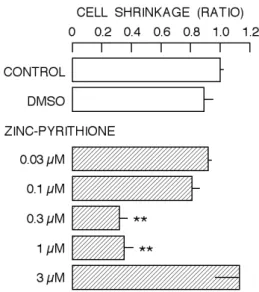
3.3. Change in cell population of shrunken cells by zinc-pyrithione.
Zinc modifies the process of cell death (Fraker and Telford, 1997; Truong-Tran et al., 2000, 2001). Thus, zinc can promotes and inhibits cell death. As shown in Fig. 4, the population of shrunken cells (area S of cytogram), one of common features in apoptotic cells, was 26.1 ± 0.7 % under control condition. On the other hand, in the presence of 0.3 µM zinc-pyrithione, it was 8.2 ± 1.3 %. However, the population after the incubation with 3 µM zinc-pyrithione was 29.5 ± 5.3 %. Thus, 0.3 µM zinc-pyrithione suppressed the increase in population of shrunken cells while it was not the case for 3 µM zinc-pyrithione. The results are summarized in Fig. 5. Zinc-pyrithione at 0.3 and 1 µM significantly reduced the increase in population of shrunken cells while it was also not the case for other concentrations.
Figure 3. Dose-dependent change in cell lethality by zinc-pyrithione. The value of abscissa is relative to the control. Column and bar indicate mean and S.D. of four experiments, respectively. Symbol (**) indicates significant difference (P < 0.01) against both CONTROL and DMSO.
Figure 4. Change in population of shrunken cells by zinc-pyrithione. The line divides cells into two populations (normal cell population and shrunken cell population, N and S areas). Shrunken cells are belonged to area S. CONT: Control, ZP: Zinc-pyrithione.
Figure 5. Dose-dependent change in population of shrunken cells by zinc-pyrithione. The value of abscissa is relative to the control. Column and bar indicate mean and S.D. of four experiments, respectively. Symbol (**) indicates significant difference (P < 0.01) against both CONTROL and DMSO.
4. Discussion
Zinc-pyrithione at 0.3 and 1 µM seems to suppress cell death that spontaneously occurs during 72 h incubation (Figs. 2 and 3). However, the cell lethality of cells incubated with 3 µM zinc-pyrithione was slightly high than that of control cells (Fig. 3).
Thus, the agent at 3 µM may start to increase cell lethality. The modification of process of cell death by zinc-pyrithione presumably suggests its hormetic action. Zinc exerts diverse actions on the process of cell death (Fraker and Telford, 1997; Truong-Tran et al., 2000, 2001). Therefore, it is not surprising that the compounds containing zinc induce such hormetic action. Zinc-pyrithione at 0.3 and 1 µM reduced the population of shrunken cells (Figs. 4 and 5), one of parameters for apoptotic cells (Bortner and Cidlowski, 1998, 2007). Thus, zinc-pyrithione at 0.3 and 1 µM may inhibit the process of apoptosis in some cells, resulting in the decrease in cell lethality.
Zinc-pyrithione at 1 and 3 µM significantly suppressed the cell growth (Fig. 1). In the case of 1 µM, the inhibition by zinc-pyrithione was associated with the decrease in both cell lethality and population of shrunken cells (Figs. 2-5). On the other hand, at 3 µM, the cell lethality and population of shrunken cells were significantly higher than those at 0.3 and 1 µM (Figs. 2-5). This observation indicates that zinc-pyrithione possesses diverse actions with different concentration-dependent manners on proliferative cells.
Conflict of interest statement
Authors declare that there are no conflicts of interest in this study.
References
Bortner CD, Cidlowski JA (1998) A necessary role for cell shrinkage in apoptosis. Biochem Pharmacol 56:1549-1559.
Bortner CD, Cidlowski JA (2007) Cell shrinkage and monovalent cation fluxes: role in apoptosis. Arch Biochem Biophys 462:176-188.
Chikahisa L, Oyama Y, Okazaki E, Noda K (1996) Fluorescent estimation of H2O2-induced changes
in cell viability and cellular nonprotein thiol level of dissociated rat thymocytes. Jpn J Pharmacol 71:299-305.
Fraker PJ, Telford WG (1997) A reappraisal of the role of zinc in life and death decisions of cells. Proc Soc Exp Biol Med 215: 229-236.
Fredriksson T, Faergemann J (1983) Double-blind comparison of a zinc pyrithione shampoo and its shampoo base in the treatment of tinea versicolor. Cutis 31:436-437.
Guthery E, Seal LA, Anderson EL (2005) Zinc
pyrithione in alcohol-based products for skin antisepsis: persistence of antimicrobial effects. Am J Infect Control 33:15-22.
Kim CH, Kim JH, Moon SJ, Chung KC, Hsu CY, Seo JT, Ahn YS (1999) Pyrithione, a zinc ionophore, inhibits NF-kappaB activation. Biochem Biophys Res Commun 259:505-509.
Lamore SD, Wondrak GT (2011) Zinc pyrithione impairs zinc homeostasis and upregulates stress response gene expression in reconstructed human epidermis. Biometals (In Press)
Marks R, Pearse AD, Walker AP (1985) The effects of a shampoo containing zinc pyrithione on the control of dandruff. Br J Dermatol 112:415-422. Rudolf E, Cervinka M (2010) Zinc pyrithione induces
cellular stress signaling and apoptosis in Hep-2 cervical tumor cells: the role of mitochondria and lysosomes. Biometals. 23:339-354.
(2001) The role of zinc in caspase activation and apoptotic cell death. Biometals 14:315-330. Truong-Tran AQ, Ho LH, Chai F, Zalewski PD
(2000) Cellular zinc fluxes and the regulation of apoptosis/gene-directed cell death. J Nutr 130:1459S-1466S.
Article History:
Received MS – 22 July 2011
Received revised MS – 30 July 2011 Accepted MS – 30 July 2011