Original Article
INTRODUCTION
A conjoined gene (CG), a read-through transcript, or cis-splicing between adjacent genes (cis-SAGe) is defined as a fusion gene resulting from the splicing of two neighboring
genes in the same coding orientation.1,2 A CG is a novel
gene expression mechanism and interest in its presence in both normal and cancer cells has been on the increase. Many CGs have been discovered only recently and listed on a database (ConjoinG, http://metasystems.riken.jp/conjoing/). Some CGs have been identified in several types of cancers and their tumorigenesis of them has been studied intensively.
However, few reports have dealt with CGs in lymphoma.3
The genetic pathogenesis of non-Hodgkin B-cell lym-phoma (B-NHL) is highly heterogeneous. By and large, two types of genetic pathogenesis of B-NHL have been researched. One is dysregulated expression of oncogenes, cyclins, transcription factors and other entities resulting from
the translocation of immunoglobulin heavy and light chain
genes on chromosomes 14q32, 2p12, and 22q11.4-7 The
other is a number of genetic mutations, which lead to dysreg-ulation of various signaling pathways including NFκB, PI3K/ MTOR, JAK-STAT, B-cell differentiation and focal adhesion, as well as of epigenetic modification and other
conse-quences.8 Furthermore, though in rare cases, fusion
tran-scripts resulted from chromosomal rearrangements in B-NHL have been reported, as exemplified by API2-MALT1 or
TBL1XR1-TP63.9,10 In addition to researching these patho-geneses, we also considered the possibility that the expres-sion of lymphoma-specific CGs might create a novel genetic pathogenesis and be useful as a genetic biomarker of lymphoma.
In this study, we used paired-end RNA sequencing (RNA-Seq) to reveal the comprehensive expression of CGs in the diffuse large B-cell lymphoma (DLBCL) cell line KPUM-UH1. Furthermore, we analyzed by means of
Detection of novel and recurrent conjoined genes in
non-Hodgkin B-cell lymphoma
Yosuke Matsumoto,
1)Taku Tsukamoto,
2)Yoshiaki Chinen,
2,3)Yuji Shimura,
2)Nana Sasaki,
4)Hisao Nagoshi,
5)Ryuichi Sato,
6)Hiroko Adachi,
6)Masakazu Nakano,
6)Shigeo Horiike,
2)Junya Kuroda,
2)Tomohiko Taki,
7)Kei Tashiro,
6)Masafumi Taniwaki
8)For this study, we investigated comprehensive expression of conjoined genes (CGs) in non-Hodgkin B-cell lymphoma (B-NHL) cell line KPUM-UH1 by using paired-end RNA sequencing. Furthermore, we analyzed the expression of these transcripts in an additional 21 cell lines, 37 primary samples of various malignancies and peripheral blood mononuclear cells of four normal individuals. Seventeen CGs were detected in KPUM-UH1: CTBS-GNG5, SRP9-EPHX1, RMND5A-ANAPC, OTX1-EHBP1,
ATF2-CHN1, PRKAA1-TTC33, LARP1-MRPL22, LOC105379697-BAK1, TIAM2-SCAF8, SPAG1-VPS13B, WBP1L-CNNM2, NARS2-GAB2, CTSC-RAB38, VAMP1-CD27-AS1, LRRC37A2-NSF, UBA2-WTIP and ZNF600-ZNF611. To our knowledge,
10 of these genes have not been previously reported. The various characteristics of the CGs included in- and out-of-frame fusions, chimeras involving non-coding RNA and transcript variants. A finding of note was that LARP1-MRPL2 was character-ized as in-frame fusion and was recurrently expressed in B-NHL samples. In this study, variety of CGs was expressed both in malignant and normal cells, some of which might be specific to lymphoma.
Keywords: Conjoined genes, LARP1, MRPL2, non-Hodgkin B-cell lymphoma
Received: July 31, 2020. Revised: October 21, 2020. Accepted: January 27, 2021. J-STAGE Advance Published: April 20, 2021 DOI: 10.3960/jslrt.20033
1)Department of Hematology, Japanese Red Cross Kyoto Daiichi Hospital, Kyoto, Japan, 2)Division of Hematology and Oncology, Kyoto Prefectural University of Medicine, Kyoto,
Japan, 3)Department of Hematology, Fukuchiyama City Hospital, Fukuchiyama, Japan, 4)Department of Hematology, Japanese Red Cross Kyoto Daini Hospital, Kyoto, Japan, 5)Department of Hematology and Oncology, Hiroshima University, Hiroshima, Japan, 6)Department of Genomic Medical Sciences, Kyoto Prefectural University of Medicine,
Kyoto, Japan, 7)Department of Medical Technology, Kyorin University Faculty of Health Science, Tokyo, Japan, 8)Center for Molecular Diagnostics and Therapeutics, Kyoto
Prefectural University of Medicine, Kyoto, Japan.
Corresponding author: Yosuke Matsumoto, MD, PhD, Department of Hematology, Japanese Red Cross Kyoto Daiichi Hospital, 15-749, Honmachi, Higashiyama-ku, Kyoto 605-8981, Japan. E-mail: [email protected]
Copyright © 2021 The Japanese Society for Lymphoreticular Tissue Research
This work is licensed under a Creative Commons Attribution-NonCommercial-ShareAlike 4.0 International License.
reverse transcription polymerase chain reaction (RT-PCR) the expression of these transcripts in additional 21 cell lines, 37 primary samples of various malignancies and peripheral blood mononuclear cells (PBMCs) of four healthy individuals.
MATERIALS AND METHODS Cell lines and primary samples
The study population included two B-NHL cell lines
(KPUM-UH1 and -MS3) established at our institute,11 20
purchased cell lines (B-NHL: JeKo-1, Mino, JVM, Z-138, MLMA, P32/ISH, Namalwa, A3/KAW and RAJI; hemato-logical malignancies other than B-NHL: DND-41, YNH-1, MOLM-13, MOLM-1 and K562; solid tumors: GOTO, NB-1, COLO-320, MeWo, MKN45 and KATO-III), and pri-mary samples from 37 Japanese patients with various malig-nancies who were diagnosed at the KPUM hospital between
May 2005 and October 2014 and were previously described12
(Supplementary Table 1). PBMCs of four healthy individu-als were individu-also studied. Inforrmed consent was obtained from each patient and healthy individual in accordance with our institutional guidelines.
To detect recurrent CGs specific to the histological sub-type of lymphoma we applied DLBCL cell line to paired-end RNA sequencing because DLBCL is the most common sub-type of B-NHL. KPUM-UH1 was established from pleural effusion lymphoma cells of 77 years old female patient with DLBCL. Cytogenetic features of this cell line were
previ-ously reported.11 Flow cytometric analysis of the lymphoma
cells immunophenotype showed positivity for CD10, CD19, CD20, CD25, and negativity for CD5 and CD23. Light chain restriction was detected (κ:λ=0.5%:95.4%).
DNA and RNA extraction
Genomic DNA was extracted using the Wizard Genomic DNA Purification kit (Promega Corporation, Madison, WI, USA). Total RNA was extracted using the AllPrep DNA/ RNA/Protein Mini Kit (QIAGEN, Venlo, The Netherlands), and reverse transcribed into cDNA with the aid of QuantiTect Reverse Transcription (QIAGEN).
Sample collection, library preparation and RNA-Seq
Total RNA was extracted from KPUM-UH1. The qual-ity and quantqual-ity of total RNA were evaluated by using RNA 6000 Nano Kit of Agilent 2100 Bioanalyzer (Agilent Technologies, Santa Clara, CA, USA). The library for next generation sequencing (NGS) was prepared with the Ribo-Zero Gold LT Sample Prep Kit (Illumina, San Diego, CA, USA) from 1.0 µg of total RNA using TruSeq Stranded Total RNA. In brief, after depletion of ribosomal RNA, the remaining RNA was purified, fragmented, and subjected to first and second strand cDNA synthesis. The cDNA was then end-repaired, adenylated at the 3’ end, and ligated with paired-end sequencing adaptors. After amplification by polymerase chain reaction (PCR) and removal of unligated
adaptors using Agencourt AMPure XP beads (Beckman Coulter, Tokyo, Japan), the quality and quantity of the gener-ated library was assessed by using both the Agilent DNA 1000 Kit of the Agilent 2100 Bioanalyzer (Agilent Technologies) and the KAPA NGS qPCR Kit (Kapa Biosystems, Wilmington, MA). Finally, sequencing was performed in the NGS Core Facility at Kyoto Prefectural University of Medicine with the HiScanSQ instrument (Illumina) by the 100-bp paired-end method.
Detection of fusion transcripts with the RNA-Seq data
NGS raw data was converted into FASTQ files using CASAVA software (version 1.8.2). For quality control, we removed the low quality reads if 1) over 80% of the sequences consisted of bases with a quality score below 20 and 2) lengths were shorter than 30 bps. In addition, 3’ end bases with a quality score less than 20 were trimmed with the aid of PRINSEQ (version 0.20.4) and FASTX (version 0.0.13). For the alignment to the human genome reference (UCSC hg19) and detection of the fusion transcripts in silico, the following programs were used: deFuse (version 0.6.1), FusionHunter (version 1.4), and TopHat-Fusion (version 2.0.9).
Genomic copy number analysis
Genomic copy number in KPUM-UH1 was analyzed by means of GeneChip Human mapping SNP 6.0 array (Affymetrix, Santa Clara, CA, USA) according to the manu-facturer’s instructions. Chromosome copy numbers were calculated using the Affymetrix Genotyping Console Browser.
RT-PCR and primers
To confirm the presence of CGs detected by RNA-Seq and to detect these genes in the cell lines and the primary samples listed above, RT-PCR was performed and followed by nucleotide sequencing. To show conserved and constant expression of CGs, mRNA was extracted from KPUM-UH1 for RNA-Seq and for RT-PCR independently and at different times. For conventional 35-cycle RT-PCR analysis (30 s at 94 °C, 30 s at 55 °C, 30 s at 72 °C), the AmpliTaq Gold 360 Master Mix (Applied Biosystems; Thermo Fisher Scientific, Inc., Waltham, MA, USA) was used, and 5 μl of the RT-PCR product was electrophoresed in a 3% agarose gel. The primer sets were designed to detect each of the CGs as shown in Supplementary Table 2.
Nucleotide sequencing
After purification of the above mentioned RT-PCR prod-ucts and cycle sequencing, capillary electrophoresis was per-formed on an Applied Biosystems 3130 Genetic Analyser. The RT-PCR products were then sequenced in both directions by using forward and reverse specific primers.
RESULTS
revealed 2,929 fusion gene transcripts, of which only 24 (0.8%) genes between coding regions. Of these 24, 17 (71%) were CGs: CTBS-GNG5, SRP9-EPHX1,
RMND5A-ANAPC, OTX1-EHBP1, ATF2-CHN1, PRKAA1-TTC33, LARP1-MRPL22, LOC105379697-BAK1, TIAM2-SCAF8, SPAG1-VPS13B, WBP1L-CNNM2, NARS2-GAB2, CTSC-RAB38, VAMP1-CD27-AS1, LRRC37A2-NSF, UBA2-WTIP
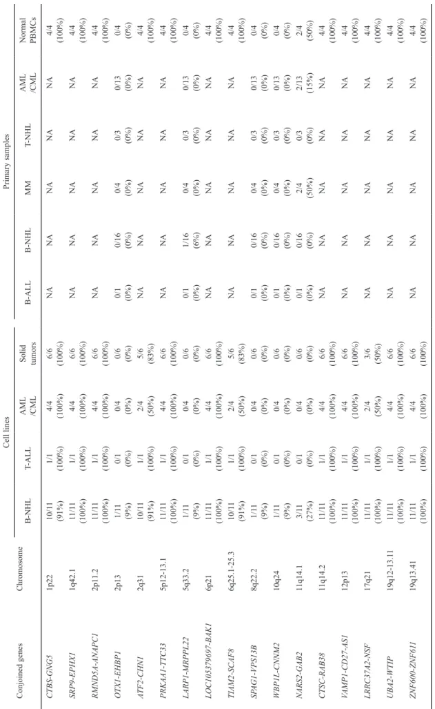
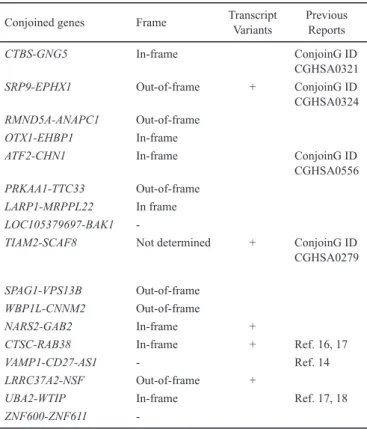
and ZNF600-ZNF611 (Fig. 1-3, Supplementary Fig. 1, Supplementary Table 3). DNA copy number SNP arrays confirmed that none of these genes in KPUM-UH1 had a genomic deletion between the two adjacent genes (Fig. 1b). The base sequences of these chimeric transcripts were veri-fied by RT-PCR and the subsequent sequencing analysis. The expression of these chimeric transcripts in various tumor cell lines, primary samples and normal PBMCs are listed in Table 1. Of the search results listed by ConjoinG and in pre-vious reports, 10 CGs were novel. The CGs showed in- and out-of-frame fusions, chimeras involving non-coding RNA and transcript variants. These representative characteristics verified by RT-PCR and the subsequent sequence analysis are listed in Table 2.
DISCUSSION
In this study, fusion transcripts in the DLBCL cell line KPUM-UH1 were comprehensively analyzed by means of RNA-Seq. While numerous fusion transcripts were detected, very few were found between coding regions (24/2929, 0.8%). In addition, most of these fusion tran-scripts were CGs (17/24, 71%). We detected ten novel CGs in a lymphoma cell line KPUM-UH1. These results suggest
that many novel CGs and their functions are still unknown and may be discovered in the future.
The 12 CGs (CTBS-GNG5, SRP9-EPHX1,
RMND5A-ANAPC, ATF2-CHN1, PRKAA1-TTC33, LOC105379697-BAK1, TIAM2-SCAF8, CTSC-RAB38, VAMP1-CD27-AS1, LRRC37A2-NSF, UBA2-WTIP and ZNF600-ZNF611) were
detected in both various tumor cell lines and PBMCs of all four normal individuals (Table 1). The expression of these CGs was not analyzed in the primary samples in this study because these samples might have been contaminated by
nor-mal PBMCs. Of these 12 CGs, CTBS-GNG5,13-15
CTSC-RAB38,16,17 VAMP1-CD27-AS114 and UBA2-WTIP17,18 were previously reported to be expressed in several cancer types and normal tissue, and might therefore be associated with cell functions common to both various tumor populations and in normal cells. On the other hand, OTX1-EHBP1,
SPAG1-VPS13B and WBP1L-CNNM2 were detected only in
KPUM-UH1, and LARP1-MRPL22 in KPUM-UH1 and one primary DLBCL sample (L-1840) (Fig. 1). In contrast to the CGs widely expressed in both normal and tumor cells, the expression of these CGs in the limited number of lymphoma samples points to the heterogeneity and specificity of each malignant tumor.
In KPUM-UH1, seven CGs (Table 2) were found to be formed by in-frame fusion, which might be translated to the chimeric proteins and have a certain function. Although we did not analyze the expression of chimeric proteins and func-tion of the CGs in this study, other studies have investigated the functions of some in-frame fusion genes. For example,
CTBS-GNG5 was reported to show a growth inhibitory
func-tion in some cancer cell lines through the analysis of
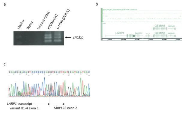
Fig. 1. Expression and base sequence of MRPL22. a Expression of a chimeric transcript of LARP1-MRPL22 shown by RT-PCR. b High-density SNP genotyping arrays showed no genomic deletion between LARP1 and MRPL22. The base sequences of LARP1 transcript variant X1-4 are listed on https://www.ncbi.
nlm.nih.gov/gene/23367. c Sequencing analysis of 241bp RT-PCR product of LARP1-MRPL22. PBMCs, peripheral blood mononuclear cells; DLBCL, diffuse large B-cell lymphoma.
Fig. 2. Expression shown by RT-PCR (a) and subsequent sequencing analysis (b and c) of con-joined genes involving non-coding RNA, LOC105379697 and CD27-AS1. LOC105379697 is listed on https://www.ncbi.nlm.nih.gov/gene/105379697, and https://www.ncbi.nlm.nih.gov/ nuccore/XR_954461.1?report=genbank. MCL, mantle cell lymphoma; HCL, hairy cell leuke-mia; BL, Burkitt lymphoma.
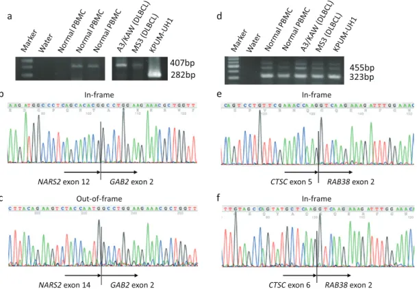
Fig. 3. Expression verified by RT-PCR and subsequent sequencing analysis of transcript variants of conjoined genes. a PCR products of NARS2-GAB2. b Sequencing analysis of 282bp RT-PCR product of NARS2-GAB2. c Sequencing analysis of 407bp RT-PCR product of NARS2-GAB2. d PCR products of CTSC-RAB38. e Sequencing analysis of 323bp RT-PCR product of CTSC-RAB38. f Sequencing analysis of 425bp RT-PCR product of CTSC-RAB38. MM, multiple myeloma; APL, acute promyelocytic leukemia.
Conjoined genes Chromosome Cell lines Primary samples B-NHL T-ALL AML /CML Solid tumors B-ALL B-NHL MM T-NHL AML /CML Normal PBMCs CTBS-GNG5 1p22 10/1 1 (91%) 1/1 (100%) 4/4 (100%) 6/6 (100%) NA NA NA NA NA 4/4 (100%) SRP9-EPHX1 1q42.1 11 /11 (100%) 1/1 (100%) 4/4 (100%) 6/6 (100%) NA NA NA NA NA 4/4 (100%) RMND5A-ANAPC1 2p1 1.2 11 /11 (100%) 1/1 (100%) 4/4 (100%) 6/6 (100%) NA NA NA NA NA 4/4 (100%) OTX1-EHBP1 2p13 1/1 1 (9%) 0/1 (0%) 0/4 (0%) 0/6 (0%) 0/1 (0%) 0/16 (0%) 0/4 (0%) 0/3 (0%) 0/13 (0%) 0/4 (0%) ATF2-CHN1 2q31 10/1 1 (91%) 1/1 (100%) 2/4 (50%) 5/6 (83%) NA NA NA NA NA 4/4 (100%) PRKAA1-TTC33 5p12-13.1 11 /11 (100%) 1/1 (100%) 4/4 (100%) 6/6 (100%) NA NA NA NA NA 4/4 (100%) LARP1-MRPPL22 5q33.2 1/1 1 (9%) 0/1 (0%) 0/4 (0%) 0/6 (0%) 0/1 (0%) 1/16 (6%) 0/4 (0%) 0/3 (0%) 0/13 (0%) 0/4 (0%) LOC105379697-BAK1 6p21 11 /11 (100%) 1/1 (100%) 4/4 (100%) 6/6 (100%) NA NA NA NA NA 4/4 (100%) TIAM2-SCAF8 6q25.1-25.3 10/1 1 (91%) 1/1 (100%) 2/4 (50%) 5/6 (83%) NA NA NA NA NA 4/4 (100%) SP AG1-VPS13B 8q22.2 1/1 1 (9%) 0/1 (0%) 0/4 (0%) 0/6 (0%) 0/1 (0%) 0/16 (0%) 0/4 (0%) 0/3 (0%) 0/13 (0%) 0/4 (0%) WBP1L-CNNM2 10q24 1/1 1 (9%) 0/1 (0%) 0/4 (0%) 0/6 (0%) 0/1 (0%) 0/16 (0%) 0/4 (0%) 0/3 (0%) 0/13 (0%) 0/4 (0%) NARS2-GAB2 11q14.1 3/1 1 (27%) 0/1 (0%) 0/4 (0%) 0/6 (0%) 0/1 (0%) 0/16 (0%) 2/4 (50%) 0/3 (0%) 2/13 (15%) 2/4 (50%) CTSC-RAB38 11q14.2 11 /11 (100%) 1/1 (100%) 4/4 (100%) 6/6 (100%) NA NA NA NA NA 4/4 (100%) VAMP1-CD27-AS1 12p13 11 /11 (100%) 1/1 (100%) 4/4 (100%) 6/6 (100%) NA NA NA NA NA 4/4 (100%) LRRC37A2-NSF 17q21 11 /11 (100%) 1/1 (100%) 2/4 (50%) 3/6 (50%) NA NA NA NA NA 4/4 (100%) UBA2-WTIP 19q12-13.1 1 11 /11 (100%) 1/1 (100%) 4/4 (100%) 6/6 (100%) NA NA NA NA NA 4/4 (100%) ZNF600-ZNF61 1 19q13.41 11 /11 (100%) 1/1 (100%) 4/4 (100%) 6/6 (100%) NA NA NA NA NA 4/4 (100%) Table 1.
Expression of the conjoined genes in various tumor cell lines, primary samples and normal peripheral blood mononuclear cells.
B-NHL, B-cell non-Hodgkin lymphoma;
T-ALL,
T-cell acute lymphoblastic leukemia;
AML, acute myeloid leukemia; CML, chronic myelogeneous leukemia
; B-ALL,
T-cell acute lympho
-blastic leukemia; MM, multiple myeloma;
T-NHL,
chimera-targeting small inhibitory RNA.13 Moreover, UBA2-WTIP chimeric protein reportedly could alter
SUMOylation activity.18 A noteworthy finding of our study
was that LARP1-MRPL22 is an in-frame fusion gene and recurrently expressed in DLBCL samples (Fig. 1). LARP1 plays a role in ribosomal translation through interaction with
mammalian target of rapamycin complex 1 (mTORC1),19 and
is highly expressed in various epithelial cancers.20 MRPL22
gene encodes 39S mitochondrial ribosomal protein (MRP) L22. MRPs regulate not only cellular respiration but also apoptosis as well as modify tumorigenesis and metastasis in
numerous cancers.21 Because LARP1-MRPL22 fusion
tran-script did not contain the base sequences translated into the functional domains of LARP1, this fusion gene might have lymphomagenic function mainly originated from the MRP. To detect recurrent CGs specific to each histological subtype of lymphoma, analysis of further number of patient speci-mens is needed.
LOC105379697-BAK1 and VAMP1-CD27-AS1 were
identified as CGs associated with non-coding RNA (Fig. 2). Although discoveries of chimeric transcripts involving non-coding RNA have been on the increase, their function in nor-mal and nor-malignant cells remains unknown. Recently, we reported finding novel chimeric transcripts involving non-coding RNA, PVT1 in multiple myeloma (MM) and acute myeloid leukemia (AML), which could lead to dysregulation
of the expression of fusion partner genes.22,23
CGs were found to express transcript variants as well as
conventional genes (Fig. 3). In this study, the presence of transcript variants in SRP9-EPHX1, TIAM2-SCAF8,
NARS2-GAB2, CTSC-RAB38, and LRRC37A2-NSF were confirmed
by means of sequencing analysis. We also established there were two types of the transcript variant expression in the CGs. Expression of the transcripts of NARS2-GAB2 was seen to vary among the samples including normal PBMCs. While an out-of-frame chimeric transcript between NARS2 exon 14 and GAB2 exon 2 was detected in two normal PBMCs, two DLBCL cell lines (A3/KAW and KPUM-MS3), two primary MM samples, and two primary acute promyelo-cytic leukemia samples, an in-frame chimeric transcript between NARS2 exon 12 and GAB2 exon 2 was detected only in two DLBCL cell lines KPUM-UH1 and A3/KAW. The transcript variants of the other CGs, on the other hand, were detected concurrently in the same samples.
In conclusion, we detected 17 CGs in the DLBCL cell line KPUM-UH1, 10 of which were novel. These genes show various characteristics including in- and out-of-frame fusions, chimeras involving non-coding RNA and transcript variants. In our study, while most CGs were detected both in normal PBMCs and malignant cells, in-frame fusion CG,
LARP1-MRPL2 was specifically and recurrently expressed in
B-NHL samples. CGs are one of gene expression mecha-nisms commonly found in normal and malignant cells, some of which might be specific to malignancies. Clarification of oncogenesis associated with lymphoma-specific CGs and establishment of novel genetic biomarkers using CGs are the next challenges.
CONFLICT OF INTEREST
The authors have no conflict of interest.
REFERENCES
1 Akiva P, Toporik A, Edelheit S, et al. Transcription-mediated gene fusion in the human genome. Genome Res. 2006; 16 : 30-36.
2 Parra G, Reymond A, Dabbouseh N, et al. Tandem chimerism as a means to increase protein complexity in the human genome. Genome Res. 2006; 16 : 37-44.
3 López-Nieva P, Fernández-Navarro P, Graña-Castro O, et al. Detection of novel fusion-transcripts by RNA-Seq in T-cell lymphoblastic lymphoma. Sci Rep. 2019; 9 : 5179.
4 Dalla-Favera R, Bregni M, Erikson J, et al. Human c-myc onc gene is located on the region of chromosome 8 that is translo-cated in Burkitt lymphoma cells. Proc Natl Acad Sci USA. 1982; 79 : 7824-7827.
5 Taub R, Kirsch I, Morton C, et al. Translocation of the c-myc gene into the immunoglobulin heavy chain locus in human Burkitt lymphoma and murine plasmacytoma cells. Proc Natl Acad Sci USA. 1982; 79 : 7837-7841.
6 Taniwaki M, Nishida K, Ueda Y, et al. Interphase and meta-phase detection of the breakpoint of 14q32 translocations in B-cell malignancies by double-color fluorescence in situ hybrid-ization. Blood. 1995; 85 : 3223-3228.
Conjoined genes Frame TranscriptVariants Previous Reports
CTBS-GNG5 In-frame ConjoinG ID
CGHSA0321
SRP9-EPHX1 Out-of-frame + ConjoinG ID
CGHSA0324 RMND5A-ANAPC1 Out-of-frame
OTX1-EHBP1 In-frame
ATF2-CHN1 In-frame ConjoinG ID
CGHSA0556
PRKAA1-TTC33 Out-of-frame
LARP1-MRPPL22 In frame
LOC105379697-BAK1
-TIAM2-SCAF8 Not determined + ConjoinG ID
CGHSA0279
SPAG1-VPS13B Out-of-frame
WBP1L-CNNM2 Out-of-frame
NARS2-GAB2 In-frame +
CTSC-RAB38 In-frame + Ref. 16, 17
VAMP1-CD27-AS1 - Ref. 14
LRRC37A2-NSF Out-of-frame +
UBA2-WTIP In-frame Ref. 17, 18
ZNF600-ZNF611
-Table 2. Characteristics of the conjoined genes detected in KPUM-UH1.
Only transcript variants confirmed by means of sequencing analysis are listed in this table.
7 Taniwaki M, Sliverman GA, Nishida K, et al. Translocations and amplification of the BCL2 gene are detected in interphase nuclei of non-Hodgkin’s lymphoma by in situ hybridization with yeast artificial chromosome clones. Blood. 1995; 86 : 1481-1486.
8 Moffitt AB, Dave SS. Clinical Applications of the Genomic Landscape of Aggressive Non-Hodgkin Lymphoma. J Clin Oncol. 2017; 35 : 955-962.
9 Akagi T, Motegi M, Tamura A, et al. A novel gene, MALT1 at 18q21, is involved in t(11;18) (q21;q21) found in low-grade B-cell lymphoma of mucosa-associated lymphoid tissue. Oncogene. 1999; 18 : 5785-5794.
10 Scott DW, Mungall KL, Ben-Neriah S, et al. TBL1XR1/TP63: a novel recurrent gene fusion in B-cell non-Hodgkin lymphoma. Blood. 2012; 119 : 4949-4952.
11 Sasaki N, Kuroda J, Nagoshi H, et al. Bcl-2 is a better therapeu-tic target than c-Myc, but attacking both could be a more effec-tive treatment strategy for B-cell lymphoma with concurrent Bcl-2 and c-Myc overexpression. Exp Hematol. 2011; 39 : 817-828.e1.
12 Matsumoto Y, Chinen Y, Shimura Y, et al. Recurrent intragenic exon rearrangements of SOBP and AUTS2 in non-Hodgkin B-cell lymphoma. Int J Hematol. 2020; 111 : 75-83.
13 Plebani R, Oliver GR, Trerotola M, et al. Long-range transcrip-tome sequencing reveals cancer cell growth regulatory chimeric mRNA. Neoplasia. 2012; 14 : 1087-1096.
14 Qin F, Song Z, Babiceanu M, et al. Discovery of CTCF-sensitive Cis-spliced fusion RNAs between adjacent genes in human prostate cells. PLoS Genet. 2015; 11 : e1005001.
15 Lee Y, Park S, Lee SH, Lee H. Characterization of genetic aber-rations in a single case of metastatic thymic adenocarcinoma. BMC Cancer. 2017; 17 : 330.
16 Grosso AR, Leite AP, Carvalho S, et al. Pervasive transcription read-through promotes aberrant expression of oncogenes and RNA chimeras in renal carcinoma. eLife. 2015; 4 : e09214. 17 Chwalenia K, Qin F, Singh S, Tangtrongstittikul P, Li H.
Connections between Transcription Downstream of Genes and cis-SAGe Chimeric RNA. Genes. 2017; 8 : 338.
18 Rodríguez-Martín B, Palumbo E, Marco-Sola S, et al. ChimPipe: accurate detection of fusion genes and transcription-induced chimeras from RNA-seq data. BMC Genomics. 2017; 18 : 7.
19 Fonseca BD, Lahr RM, Damgaard CK, Alain T, Berman AJ. LARP1 on TOP of ribosome production. Wiley Interdiscip Rev RNA. 2018; e1480.
20 Stavraka C, Blagden S. The La-Related Proteins, a Family with Connections to Cancer. Biomolecules. 2015; 5 : 2701-2722. 21 Kim HJ, Maiti P, Barrientos A. Mitochondrial ribosomes in
can-cer. Semin Cancer Biol. 2017; 47 : 67-81.
22 Nagoshi H, Taki T, Hanamura I, et al. Frequent PVT1 rear-rangement and novel chimeric genes NBEA and PVT1-WWOX occur in multiple myeloma with 8q24 abnormality. Cancer Res. 2012; 72 : 4954-4962.
23 Chinen Y, Sakamoto N, Nagoshi H, et al. 8q24 amplified seg-ments involve novel fusion genes between NSMCE2 and long noncoding RNAs in acute myelogenous leukemia. J Hematol Oncol. 2014; 7 : 68.