Review J Tokyo Wom Med Univ 89(Extra1): E100-E109, 2019 Jul
Does Anti-Vascular Therapy Reprogram the Immunosuppressive
Microenvironment in Gastric Cancer?
Shuji Kitahara1
and Yasuhiro Suzuki2 1
Department of Anatomy and Developmental Biology, School of Medicine, Tokyo Women s Medical University, Tokyo, Japan 2
Department of Vascular Biology, Institute of Development, Aging and Cancer, Tohoku University, Sendai, Japan (Accepted August 24, 2018)
Abnormal cell proliferation, a characteristic of malignant tumors, is accompanied by angiogenesis, through a com-plex molecular pathway involving various vascular growth factor groups. The abnormal blood vessels formed by the cancer establish a hypoxic and low pH environment in the vicinity of the tumor, causing a decrease in immu-nogenicity. Since the cancer cells produce immunosuppressive factors that further induce the related cells, an im-munosuppressive environment optimal for the growth of the tumor is established. Although the gastrointestinal tract maintains homeostasis by specific immune surveillance mechanisms involving the lymph nodes and Peyer s patch, once a malignant tumor develops, a system that escapes the monitoring mechanism by taking advantage of the immune-checkpoint route is newly generated. Although normalization of the tumor microenvironment us-ing molecular target drugs has attracted attention in recent years as the next generation cancer treatment, it is reported that this method also affects the host immune mechanism simultaneously. However, since many aspects of this intervention including the detailed mechanism of the immune mechanism for tumor microenvironment normalization, the prognosis, and the side-effects are still unclear, the clinical applications are not feasible yet. We believe that normalization of the tumor environment and tackling its mechanism, can construct a host environ-ment that maximizes the therapeutic effect of anti-cancer therapies including chemotherapy, immunotherapy, and radiation therapy, in addition to restoring the host immunity of the gastrointestinal tract. In other words, this could become the next generation cancer treatment. This review article focuses on tumor angiogenesis, tumor microenvironment, and the specific immune surveillance mechanism of the gastrointestinal tract, and finally pro-poses what is necessary for the“reprogramming of tumor microenvironment”.
Key Words: tumor blood vessel, tumor microenvironment, immunotherapy, gastric cancer
Introduction
Gastric cancer (GC) is the third most common can-cer with the second highest mortality rate world-wide, with one million new patients diagnosed every year.1
The vast majority of GC are adenocar-cinomas, which can be subdivided into intestinal
and diffuse types according to the Lauren classifica-tion.2
Tumor microenvironment processes such as angiogenesis, fibrosis, and inflammation are critical for the local progression and metastasis of solid tu-mors, including those occurring in gastrointestinal cancer.3
These processes create a tumor
microenvi-Corresponding Author: Shuji Kitahara, Department of Anatomy and Developmental Biology, School of Medicine, Tokyo Women s Medical University, 8-1 Kawada-cho, Shinjuku-ku, Tokyo 162-8666, Japan. [email protected]
doi: 10.24488/jtwmu.89.Extra1_E100
Copyright Ⓒ 2019 Society of Tokyo Women s Medical University. This is an open access article distributed under the terms of Creative Commons Attribution License (CC BY), which permits unrestricted use, distribution, and reproduction in any medium, provided the original source is properly credited.
ronment characterized by hypoxia, which sup-presses the immune system s ability to fight the cancer. As a consequence, no single chemotherapy agent or combination regimen consistently leads to objective tumor shrinkage, and novel treatment strategies for GC are desperately needed.3
The tumor vasculature is an essential component of the tumor microenvironment, influencing tumor behavior and treatment response. It can be tar-geted specifically using anti-angiogenic drugs.4-6
Tu-mor blood vessels are histopathologically different from normal blood vessels. They have irregular shape, diameter, and branching patterns and can-not be classified as arterioles, venules, or capillaries.7
Their endothelial cells are interconnected loosely with abnormal pericytes (decreased pericyte cover-age ) that are responsible for their leaky nature. When compared to normal blood vessels, tumor vessels appear immature. Angiogenesis is not just dependent on endothelial cell invasion and prolifera-tion, but also requires the pericyte coverage of vas-cular sprouts for stabilization and maturation of vascular walls. This phenotype might be associated with structural aberrations in their basement mem-brane.8
Vascular abnormalities associated with the tumor may be attributed to the tumor microenvironment.9
These abnormalities may contribute to the develop-ment of tumor resistance to conventional chemo- , radio-, and immune-based therapies. Dr. Jain10
pro-posed that an appropriate anti-angiogenic treat-ment can lead to normalization of the tumor vascu-lature by reducing vascular permeability and inter-stitial fluid pressure, thus improving blood flow and tumor perfusion. A normalized vasculature can re-duce hypoxia and enhance the delivery of oxygen and cytotoxic agents for radiation therapy, but also for anti-tumor immune response.11
Preclinical and clinical studies have supported the hypothesis that anti-angiogenic therapy can normalize the tumor vasculature, at least transiently.4
On the other hand, tumor cells do not act alone. Malignant tumors develop a complex multifaceted relationship with the environment, and simply mak-ing them accessible may not be sufficient to
pro-duce vulnerability to treatment. The immune re-sponse of the host is critical to the success of an im-munotherapy regimen, such as immuno-checkpoint inhibition.4
However, the determinants of the re-sponse are not completely understood. Tumor infil-tration by immune cells such as cytotoxic T-lymphocytes varies widely in density, composition, and clinical significance.12
Blood vascular and lymphatic endothelial cells play important roles in trafficking immune cells, controlling the microenvironment, and modulating the immune response. Improving access to the ma-lignant tumor through vascular alteration with anti-angiogenic drugs may provide an effective combi-natorial strategy for immunotherapy, and might be widely applicable to various tumor types, especially GC.13
Moving forward, these insights may be useful for designing new approaches, for example by com-bining anti-angiogenic agents with immune-checkpoint inhibitors that will produce a substantial improvement in the overall survival of malignant GC patients. This review article focuses on the po-tential new approach to GC therapy.
Tumor Blood Vessels Can Modulate the Tumor Microenvironment
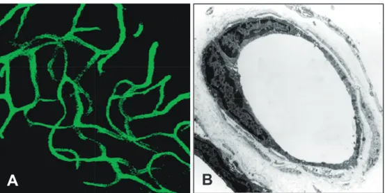
Tumor angiogenesis is a vital process in the pro-gression and metastasis of solid malignant tumors. Angiogenesis within the tumor develops abnormal leaky blood vessels. It has been reported that tumor vessels can create microenvironments that are histopathologically distinct from normal vessels (Figure 1, 2).14
Electron microscopy revealed that most tumor vessels in lung adenocarcinoma have deformed diameters (Figure 2A). In addition, the tu-mor endothelial cells have loose interconnections, intercellular openings, and abnormal pericytes, which are likely to be responsible for vessel leaki-ness. Moreover, the structural abnormalities in the basement membrane of tumor blood vessels are also responsible for their relative immaturity com-pared to normal blood vessels (Figure 2B-E). Ac-cordingly, a tumor blood vessel has an abnormal blood flow and is excessively leaky. Insufficient blood flow to the tumor tissue leads to hypovascu-lar areas, severe hypoxia, and necrosis.
Figure 1 Normal blood vessels in mouse subcutaneous tissue have smooth endothelial
cells.
A: Tomato lectin-labeled vascular architecture in normal mouse subcutaneous region. B: Ultra-thin sections showing normal blood vessel in mouse subcutaneous tissue.
B
A
Although GC is a highly angiogenic cancer, it is characterized by hypoxia.3
Hypoxia may promote GC growth and progression, and promote resis-tance to existing therapies. Conversely, inducing vessel normalization and alleviating hypoxia might delay GC progression and metastasis. Vascular en-dothelial growth factor (VEGF) is the key factor for the abnormal structure and function of tumor ves-sels.14
One of the cues driving evasive resistance is increased hypoxia induced by anti-VEGF treat-ment. Strong inhibition of the VEGF pathway re-sults in pruning of the tumor vasculature, which in-duces hypoxia in malignant tumors.2
Tumor hy-poxia leads to hyhy-poxia-inducible factor 1 alpha subunit ( HIF 1α ) stabilization, which induces hy-poxia responsive genes including VEGF. Thus, VEGF treatment imposes a vicious cycle resulting in evasive resistance. We recently identified that the current treatment dose of sorafenib (anti-VEGF) can induce hypoxia in liver cancer, which resulted in infiltration by immune-suppressive leukocytes.2,15
Based on these results, we hypothesize that dose ti-tration of selective anti-angiogenic agents is war-ranted to optimize treatment and elicit anti-tumor immunity. Founded on the above considerations, if we can control tumor angiogenesis and normalize tumor blood vessels, the tumor microenvironment in GC may improve to allow anti-tumor immunity.
Why Target the Immune Checkpoints in GC?
Cancer immunotherapy has shown great promise to transform clinical oncology in a radical way by substantially improving outcomes in certain ad-vanced malignant cases. However, in most patients, the immune-suppressive microenvironment inter-feres with the development of an appropriate anti-tumor immune response. Co-inhibitory antigen presentation signals, called immune checkpoints, are often activated in the malignant tumor tissue, which results in the evasion of host immunity.16
Based on the success of blocking programmed cell death 1 (PD-1)/programmed cell death 1 ligand 1 ( PD-L 1 ) in melanoma, targeting an immune-checkpoint is an emerging strategy that is gaining significant interest for malignant cancer therapy.17
Anti-immune-checkpoint therapy is ideal for GC pa-tients for at least three reasons. First, GC (especially virus-induced GC) is immunogenic, but the immune response is suppressed by multiple mechanisms. This suggests an anti-immune-checkpoint blockade could be effective.1
Second, gastric cancers have vascular abnormalities that lead to hypoxia, fibrosis, and immune suppression. Modulating these proc-esses with anti-angiogenic therapy could potentially shift the tumor microenvironment toward promo-tion of an anti-tumor response.18
Finally, gastrointes-tinal cancer is particularly prone to chronic
inflam-Figure 2 Electron micrograph of metastatic adenocarcinoma showing the morphological
changes to tumor blood vessels in gastrointestinal cancer.
A: Ultra-thin sections showing tumor blood vessel abnormalities in mouse intestinal tumor. Tumor vessel irregularities included basement membranes.
B-E: Higher magnifications of (A). Abnormal endothelial cells with long protuberances (B, double arrowheads). Note the multi-layered basement membranes (C, arrows). Abnormal endothelial cells are loosely interconnected (D, arrowheads) and have intercellular open-ings (E, arrowhead). Asterisks, endothelial cell. Arrows indicate basement membrane. Scale bars: 10 μm (A), 2.5 μm (B-E).
Lumen RBC RBC RBC WBC B Pc 䙈 Tumor Tumor 䙈 䙈 䙈 䙈 Tumor Lumen Lumen C D E A B C D E
mation, which can create an immune-suppressive microenvironment.
Despite its wide use in the clinic, chemotherapy has limited efficacy and high toxicity. Clearly, new systemic treatment approaches that are more effi-cacious and less toxic ― such as low dose anti-angiogenic therapy combined with immunotherapy ― are desperately needed. Our preliminary data
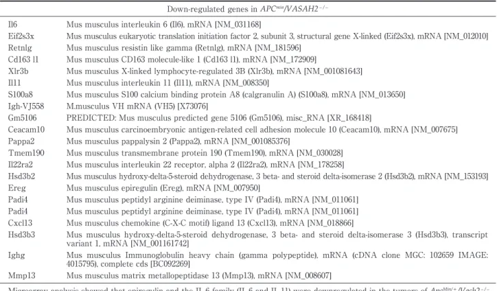
showed that the inhibition of tumor angiogenesis factor ( vasohibin 2 ) could improve the chronic in-flammation in gastrointestinal cancer (Table 1,
Fig-ure 3: The ApcMin /+
mouse spontaneously develops multiple intestinal adenomas that clinically mimic those observed in patients with familial adenoma-tous polyposis and undergo early transformation into adenocarcinomas. In addition, ApcMin/+
Table 1 Microarray analysis showing downregulation of genes in ApcMin/+/Vash2−/− mice compared with ApcMin/+mice.
Down-regulated genes in APCmin/VASAH2−/−
Il6 Mus musculus interleukin 6 (Il6), mRNA [NM_031168]
Eif2s3x Mus musculus eukaryotic translation initiation factor 2, subunit 3, structural gene X-linked (Eif2s3x), mRNA [NM_012010] Retnlg Mus musculus resistin like gamma (Retnlg), mRNA [NM_181596]
Cd163 l1 Mus musculus CD163 molecule-like 1 (Cd163 l1), mRNA [NM_172909]
Xlr3b Mus musculus X-linked lymphocyte-regulated 3B (Xlr3b), mRNA [NM_001081643] Il11 Mus musculus interleukin 11 (Il11), mRNA [NM_008350]
S100a8 Mus musculus S100 calcium binding protein A8 (calgranulin A) (S100a8), mRNA [NM_013650] Igh-VJ558 M.musculus VH mRNA (VH5) [X73076]
Gm5106 PREDICTED: Mus musculus predicted gene 5106 (Gm5106), misc_RNA [XR_168418]
Ceacam10 Mus musculus carcinoembryonic antigen-related cell adhesion molecule 10 (Ceacam10), mRNA [NM_007675] Pappa2 Mus musculus pappalysin 2 (Pappa2), mRNA [NM_001085376]
Tmem190 Mus musculus transmembrane protein 190 (Tmem190), mRNA [NM_030028] Il22ra2 Mus musculus interleukin 22 receptor, alpha 2 (Il22ra2), mRNA [NM_178258]
Hsd3b2 Mus musculus hydroxy-delta-5-steroid dehydrogenase, 3 beta- and steroid delta-isomerase 2 (Hsd3b2), mRNA [NM_153193] Ereg Mus musculus epiregulin (Ereg), mRNA [NM_007950]
Padi4 Mus musculus peptidyl arginine deiminase, type IV (Padi4), mRNA [NM_011061] Padi4 Mus musculus peptidyl arginine deiminase, type IV (Padi4), mRNA [NM_011061] Cxcl13 Mus musculus chemokine (C-X-C motif) ligand 13 (Cxcl13), mRNA [NM_018866]
Hsd3b3 Mus musculus hydroxy-delta-5-steroid dehydrogenase, 3 beta- and steroid delta-isomerase 3 (Hsd3b3), transcript variant 1, mRNA [NM_001161742]
Ighg Mus musculus Immunoglobulin heavy chain (gamma polypeptide), mRNA (cDNA clone MGC: 102659 IMAGE: 4015795), complete cds [BC092269]
Mmp13 Mus musculus matrix metallopeptidase 13 (Mmp13), mRNA [NM_008607]
Microarray analysis showed that epiregulin and the IL-6 family (IL-6 and IL-11) were downregulated in the tumors of ApcMin/+/Vash2−/−
mice compared with control mice.
pure C57BL/6 background were mated to Vash2− / − mice of a mixed C57BL/6 background, and the re-sulting pups were screened for the Min mutation and for the Vash2−/−
gene by PCR). These results in-dicate that the combination of anti-angiogenic ther-apy and immunotherther-apy ( such as immune-checkpoint inhibition) is an effective treatment for GC.
How Does Gastrointestinal Cancer Evade Host Immunity?
The majority of gastric cancers are associated with infectious agents, including the Helicobacter pylori bacterium and Epstein-Barr virus (EBV).19
Recently, The Cancer Genome Atlas (TCGA)project reported that PD-L1 expression is elevated by 15% in EBV-positive gastric cancers. An evaluation of the mes-senger RNA (mRNA) revealed elevated expressions of Janus kinase 2 (JAK2), PD-L1, and programmed cell death 1 ligand 2 (PD-L2).1
In addition, Lin et al.20
reported that non-Asian GC was significantly en-riched in signatures related to T-cell biology, includ-ing cytotoxic T-lymphocyte antigen 4 (CTLA-4) sig-naling. Similarly, in the tissue microarray
(TMA)co-horts, non-Asian GC showed significantly higher ex-pression of T-cell markers and lower exex-pression of the immunosuppressive T-regulatory (Treg) cell marker FoxP3. In lymphocyte-rich gastric carcino-mas the stroma has even been termed a tertiary lymphoid tissue .21
However, GC usually evade im-mune surveillance. Multiple imim-mune-suppressive mechanisms have been proposed.
The stomach is inherently“tolerogenic” to pre-vent aberrant immunity in response to potential an-tigens absorbed by the epithelium.22
On the other hand, GC are inflammation-induced malignancies because they often occur in a diseased stomach with a background of gastritis.23
The underlying chronic inflammation and viral infection create an immune-suppressive environment in the stomach through the production of cytokines including inter-leukin (IL)-6, IL-11, tumor necrosis factor-alpha ( TNF-α ) , and transforming growth factor-beta (TGF-β).24,25
Our data also showed that the regulation of vasohibin2, an angiogenesis factor, could down-regulate IL-6 and IL-11 (Table 1).
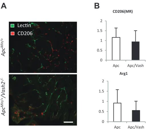
eva-Figure 3 Inhibition of pro-tumor angiogenesis factor vasohibin2 changes the tumor
mi-croenvironment.
A: Double-immunostaining of tomato lectin and CD206 detecting M2 macrophage in a tu-mor of ApcMin/+ mice and ApcMin/+/Vash2−/− mice. The number of M2 macrophages trend toward an increase in response to vasohibin2 inhibition. Scale bar: 100 μm.
B: Gastrointestinal tumors of ApcMin/+ mice and ApcMin/+/Vash2−/− mice were examined for M1 and M2 like macrophage factor (CD206, Arg1) by qRT-PCR. The expression of Arg1 trend toward a decrease in ApcMin/+/Vash2−/− mice.
Apc
Min/ +Apc
Min/ +/Vash2
-/-LecƟn
CD206
0 0.5 1 1.5 2 Apc Apc/Vash CD206(MR) 0 0.5 1 1.5 2 Apc Apc/Vash Arg1A
B
sion in cancer involves infiltration of tumors by immune-suppressive leukocytes such as Tregs and myeloid-derived suppressor cells (MDSCs). Exhaus-tion of CD4+T cells has also been reported as a mechanism of immune evasion in advanced cancer patients.26,27
While the immune response to specific antigens is recognized by major histocompatibility receptors, co-stimulatory and co-inhibitory mole-cules regulate the intensity of the response. Im-mune checkpoints are co-inhibitory molecules that are physiologically expressed for the maintenance of self-tolerance.28
In the GC microenvironment, immune-checkpoint molecules such as CTLA-4 and PD-L1 are overexpressed and broadly induce the evasive mechanism.
Anti-vascular Therapy Can Modulate the Immune Response
Reactivation of the immune response is key to over-coming treatment-resistant GC. Growing evidence is showing that combining anti-angiogenic therapy with immunotherapy in certain contexts may im-prove the immune response to solid cancers.11,29
Sev-eral other studies have evaluated the change in im-mune response after anti-angiogenic therapy. For example, bevacizumab has been shown to enhance the proportion and function of dendritic cells (DC) in solid cancer patients30
. In mouse models, it has been reported that an anti-VEGF antibody can enhance the number and function of DC.31
Previously, Huang et al. 32
reported an interesting finding regarding the treatment dose of anti-angiogenic therapy in a mouse model of breast cancer. When anti-vascular
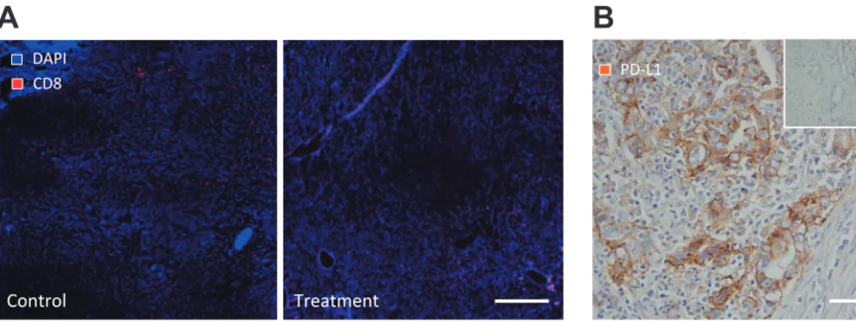
Figure 4 Anti-vascular therapy can modulate digestive tumor immune response.
A: Double-immunostaining of 4',6-diamidino-2-phenylindole (DAPI) and CD8. Lymphocytes infiltrated to the center of the malignant tumor. Right image - anti-angiogenic treatment model of mouse liver cancer. In the treatment model, CD8 positive lymphocytes infiltrated to the tumor center. Scale bar: 100 μm.
B: Immunostaining of PD-L1 in a human pathological gastrointestinal cancer sample. A substantial number of tumor cells expressed PD-L1 in the tumor area. The inset indicates a PD-L1 negative human sample. Scale bar: 100 μm.
B
A
DAPI CD8 Control Treatment PD-L1endothelial growth factor receptor 2 (VEGFR2) neu-tralizing antibody was administered at a low dose, the structure of the tumor vasculature was normal-ized and anti-tumor immunity was promoted. Con-versely, a high dose of anti-VEGFR 2 neutralizing antibody induced vascular pruning and increased tissue hypoxia within the tumor. When they looked at the infiltrating leukocytes, the number of tumor-infiltrating macrophages increased while glucocorti-coid receptor (Gr1)+cells decreased. Interestingly, the macrophages showed a decrease in M2 pheno-type, suggesting an improvement in anti-tumor im-mune activity. Treg activity was also decreased in the low dose anti-VEGFR 2 neutralizing antibody group compared to the high dose group. Our results concur with the M 2 phenotype macrophage data (Figure 3A, B). Recently, when we used therapeutic doses of sorafenib in liver cancer models, we de-tected increased hypoxia and an increase in Gr1+ cells, Tregs, and macrophages.12,15
In addition, low dose anti-VEGFR2 neutralizing antibody could in-duce infiltration of cluster of differentiation (CD) 8 positive lymphocytes in liver cancer (Figure 4A). These experiments suggest a potential benefit on the immune response to GC with dose titrations of anti-VEGF therapy.
Immune-checkpoint Targeting Has Potential to Overcome the Immune-suppressive Environment
To overcome the immune-suppressive environment in GC, we have to focus on the immune-checkpoint blockade, especially on anti-PD-1 / PD-L 1 therapy. Numerous agents targeting PD-L 1 / PD-1 check point are in various phases of clinical development. However, the correlation between PD-L 1 expres-sion and the prognosis of solid tumors, such as GC, is controversial. Here, we propose new strategies to investigate the potential value of immune-checkpoint blockade and anti-angiogenic therapy in the prognostic prediction of GC.
PD-1 is a CD28 superfamily member that conveys co-inhibitory signals for T-cell receptors.33
PD-1 is expressed in CD8+T cells, Tregs, and MDSCs.34,35
PD-1 also regulates peripheral tolerance and auto-immunity. Chronic exposure to antigen leads to the over-expression of PD-1 in T cells, which induces anergy or cell exhaustion.36
Cancer cells can evade immune surveillance by hijacking PD-L1/PD-1 sig-naling. By expressing PD-L1 or PD-L2, PD-1 is acti-vated in tumor-infiltrating lymphocytes, shutting down the immune response.37
PD-1/PD-L1 expres-sion can be detected in clinical samples and is sig-nificantly correlated with the stage of human GC (Figure 4B), local recurrence rate, and poor
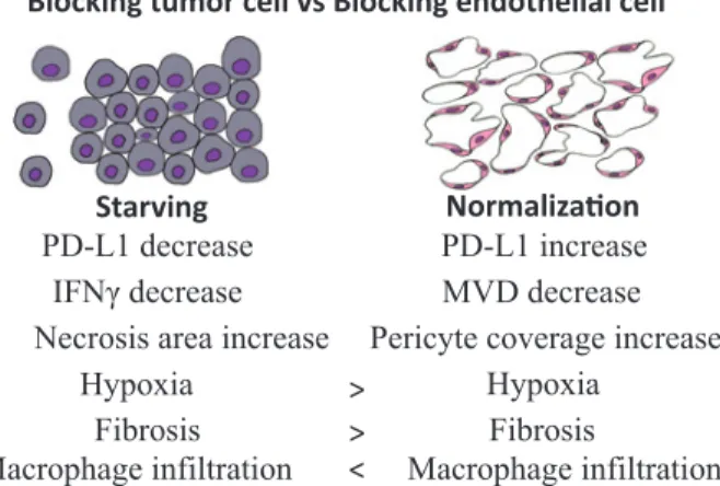
progno-Figure 5 Targeting tumor VEGFR2 vs targeting
en-dothelial cell VEGFR2.
Blocking VGEFR2 in endothelial cells could normalize tumor blood vessels and improve hypoxia and fibrosis compared to blocking VGEFR2 in tumor cells. Note that, single anti-tumor treatments were not efficacious in gastric cancer. Modulating these processes with anti-angiogenic therapy might potentially modify the immune microenvironment and promote anti-tumor response.
Starving NormalizaƟon
MVD decrease Pericyte coverage increase
Hypoxia Fibrosis PD-L1 increase PD-L1 decrease ,)1Ȗ decrease Hypoxia
Necrosis area increase Fibrosis >
>
Macrophage infiltration < Macrophage infiltration
Blocking tumor cell vs Blocking endothelial cell
sis.1
These data support the potential of anti-PD-1/ PD-L1 therapy in GC.
Conclusion
We therefore propose that vascular normalization can improve an immune response against GC. The work to be performed will lay the groundwork for a new paradigm of immunotherapy that modulates the tumor microenvironment (Figure 5), and could rapidly impact clinical practice.32
Acknowledgements
We thank Mrs. Hiromi Sagawa and Mrs. Kae Motomaru of Tokyo Women s Medical University for their techni-cal help. Special thanks go to Dr. Dan G Duda and the gastric cancer research team of Edwin L Steele Labora-tories for Tumor Biology, Department of Radiation On-cology, Massachusetts General Hospital, Harvard Medi-cal School and Yasufumi Sato of the Department of Vas-cular Biology, Institute of Development, Aging and Can-cer, Tohoku University, Sendai, Japan, and my col-leagues from the Department of Anatomy and Develop-mental Biology, Tokyo Women s Medical University, Tokyo, Japan for their advice and expertise.
Conflicts of Interest: The authors declare no
con-flicts of interest associated with this manuscript.
References
1.Cancer Genome Atlas Research Network: Compre-hensive molecular characterization of gastric ade-nocarcinoma. Nature 513: 202―209, 2014
2.Hu B, El Hajj N, Sittler S et al: Gastric cancer: Clas-sification, histology and application of molecular pa-thology. J Gastrointest Oncol 3: 251―261, 2012 3.Park DJ, Thomas NJ, Yoon C et al: Vascular
endo-thelial growth factor a inhibition in gastric cancer. Gastric Cancer 18: 33―42, 2015
4.Ramjiawan RR, Griffioen AW, Duda DG : Anti-angiogenesis for cancer revisited: Is there a role for combinations with immunotherapy? Angiogenesis 20: 185―204, 2017
5.Hato T, Zhu AX, Duda DG : Rationally combining anti-VEGF therapy with checkpoint inhibitors in hepatocellular carcinoma. Immunotherapy 8: 299― 313, 2016
6.Hato T, Goyal L, Greten TF et al: Immune check-point blockade in hepatocellular carcinoma: current progress and future directions. Hepatology 60 : 1776―1782, 2014
7.Kitahara S, Morikawa S, Shimizu K et al: Alteration of angiogenic patterns on B16BL6 melanoma devel-opment promoted in Matrigel. Med Mol Morphol 43: 26―36, 2010
8.Baluk P, Morikawa S, Haskell A et al: Abnormali-ties of basement membrane on blood vessels and endothelial sprouts in tumors. Am J Pathol 163 : 1801―1815, 2003
9.Jain RK: Normalization of tumor vasculature : an emerging concept in antiangiogenic therapy. Sci-ence 307: 58―62, 2005
10.Jain RK: Normalizing tumor microenvironment to treat cancer: bench to bedside to biomarkers. J Clin Oncol 31: 2205―2218, 2013
11.Huang Y, Stylianopoulos T, Duda DG et al: Benefits of vascular normalization are dose and time de-pendent――letter. Cancer Res 73: 7144―7146, 2013 12.Chen Y, Ramjiawan RR, Reiberger T et al: CXCR4
inhibition in tumor microenvironment facilitates anti-programmed death receptor-1 immunotherapy in sorafenib-treated hepatocellular carcinoma in mice. Hepatology 61: 1591―1602, 2015
13.Setia N, Agoston AT, Han HS et al: A protein and mRNA expression-based classification of gastric cancer. Mod Pathol 29: 772―784, 2016
14.Kitahara S, Suzuki Y, Morishima M et al: Vasohibin-2 modulates tumor onset in the gastrointestinal tract by normalizing tumor angiogenesis. Mol Can-cer 13: 99, 2014
15.Chen Y, Huang Y, Reiberger T et al: Differential ef-fects of sorafenib on liver versus tumor fibrosis me-diated by stromal-derived factor 1 alpha/C-X-C re-ceptor type 4 axis and myeloid differentiation antigen-positive myeloid cell infiltration in mice. He-patology 59: 1435―1447, 2014
16.Chen Y, Ramjiawan RR, Reiberger T et al: CXCR4 inhibition in tumor microenvironment facilitates anti-programmed death receptor-1 immunotherapy
in sorafenib-treated hepatocellular carcinoma in mice. Hepatology 61: 1591―1602, 2015
17.Xu C, Fillmore CM, Koyama S et al: Loss of Lkb1 and Pten leads to lung squamous cell carcinoma with elevated PD-L 1 expression. Cancer Cell 25 : 590―604, 2014
18.Hodi FS, O Day SJ, McDermott DF et al: Improved survival with ipilimumab in patients with metas-tatic melanoma. N Engl J Med 363: 711―723, 2010 19.Oh JK, Weiderpass E: Infection and cancer: global
distribution and burden of diseases. Ann Glob Health 80: 384―392, 2014
20.Lin SJ, Gagnon-Bartsch JA, Tan IB et al: Signatures of tumour immunity distinguish Asian and non-Asian gastric adenocarcinomas. Gut 64: 1721―1731, 2015
21.Aloisi F, Pujol-Borrell R : Lymphoid neogenesis in chronic inflammatory diseases. Nat Rev Immunol 6: 205―217, 2006
22.Pelaseyed T, Bergström JH, Gustafsson JK et al: The mucus and mucins of the goblet cells and en-terocytes provide the first defense line of the gas-trointestinal tract and interact with the immune system. Immunol Rev 260: 8―20, 2014
23.Yoon H, Kim N: Diagnosis and management of high risk group for gastric cancer. Gut Liver 9 : 5 ― 17, 2015
24.Putoczki TL, Thiem S, Loving A et al: Interleukin-11 is the dominant IL-6 family cytokine during gas-trointestinal tumorigenesis and can be targeted therapeutically. Cancer Cell 24: 257―271, 2013 25.Zhang S, Desrosiers J, Aponte-Pieras JR et al:
Hu-man immune responses to H. pylori HLA Class II epitopes identified by immunoinformatic methods. PLoS One 9: e94974, 2014
26.Hoechst B, Voigtlaender T, Ormandy L et al: Myeloid derived suppressor cells inhibit natural kil-ler cells in patients with hepatocellular carcinoma via the NKp30 receptor. Hepatology 50 : 799 ― 807, 2009
27.Gabitass RF, Annels NE, Stocken DD et al: Ele-vated myeloid-derived suppressor cells in pancre-atic, esophageal and gastric cancer are an inde-pendent prognostic factor and are associated with significant elevation of the Th 2 cytokine
interleukin-13. Cancer Immunol Immunother 60 : 1419―1430, 2011
28.Okazaki T, Chikuma S, Iwai Y et al: A rheostat for immune responses: the unique properties of PD-1 and their advantages for clinical application. Nat Immunol 14: 1212―1218, 2013
29.Zou Y, Meng J, Chen W et al: Modulation of pheno-typic and functional maturation of murine dendritic cells (DCs) by purified Achyranthes bidentata poly-saccharide (ABP). Int Immunopharmacol 11: 1103― 1108, 2011
30.Osada T, Chong G, Tansik R et al: The effect of anti-VEGF therapy on immature myeloid cell and dendritic cells in cancer patients. Cancer Immunol Immunother 57: 1115―1124, 2008
31.Gabrilovich DI, Ishida T, Nadaf S et al: Antibodies to vascular endothelial growth factor enhance the efficacy of cancer immunotherapy by improving endogenous dendritic cell function. Clin Cancer Res 5: 2963―2970, 1999
32.Huang Y, Yuan J, Righi E et al: Vascular normaliz-ing doses of antiangiogenic treatment reprogram the immunosuppressive tumor microenvironment and enhance immunotherapy. Proc Natl Acad Sci U S A 109: 17561―17566, 2012
33.Freeman GJ, Long AJ, Iwai Y et al: Engagement of the PD-1 immunoinhibitory receptor by a novel B7 family member leads to negative regulation of lym-phocyte activation. J Exp Med 192: 1027―1034, 2000 34.Francisco LM, Salinas VH, Brown KE et al: PD-L1
regulates the development, maintenance, and func-tion of induced regulatory T cells. J Exp Med 206: 3015―3029, 2009
35.Nikolova M, Lelievre JD, Carriere M et al: Regula-tory T cells differentially modulate the maturation and apoptosis of human CD8+ T-cell subsets. Blood 113: 4556―4565, 2009
36.Barber DL, Wherry EJ, Masopust D et al: Restoring function in exhausted CD8 T cells during chronic viral infection. Nature 439: 682―687, 2006
37.Iwai Y, Ishida M, Tanaka Y et al: Involvement of PD-L1 on tumor cells in the escape from host im-mune system and tumor immunotherapy by PD-L1 blockade. Proc Natl Acad Sci U S A 99 : 12293 ― 12297, 2002
抗血管新生療法は胃がんにおける免疫抑制微小環境を改善できるか? 1東京女子医科大学医学部解剖学・発生生物学講座 2東北大学加齢医学研究所腫瘍循環研究分野 キタハラ シュウジ ス ズ キ ヤスヒロ 北原 秀治1 ・鈴木 康弘2 悪性腫瘍の特徴所見である異常細胞増殖には,様々な血管増殖因子群による複雑な分子経路を介した血管新生 が伴う.癌の作る異常血管は,腫瘍周辺の低酸素,低 pH 環境などを作り出し,免疫原性の低下を引き起こす.癌 細胞は免疫抑制因子を産生し,さらに関連細胞が惹起され,癌生育に最適な免疫抑制性環境が構築されていく.一 方,消化管は,リンパ節,パイエル板といった特異的な免疫監視機構を有し恒常性を維持しているが,一旦悪性腫 瘍が発生すると,免疫チェックポイント経路を利用した,監視機構を逃れるシステムが新たに構築される.近年, 次世代の癌治療法として,分子標的治療薬を用いた腫瘍微小環境の正常化が注目されており,同時にこの方法が宿 主免疫機構にも影響を与えることが報告され始めている.しかしながら,こうした免疫機構をはじめ,腫瘍微小環 境正常化に至るまでの詳しいメカニズムや,正常化後の予後,副作用に関しては未だ不明な点が多く,現在も臨床 応用は出来ていない.われわれは上記を背景とし,この「腫瘍微小環境の正常化,およびそのメカニズムへのアプ ローチ」が,結果として,消化管における強力な宿主免疫力を回復させると共に,化学療法,免疫療法,放射線療 法などの治療効果を最大限に発揮させる宿主環境を構築する,つまり次世代の癌治療法としての可能性を確信し ている.本総説は,腫瘍血管新生,腫瘍微小環境,消化管の特異的な免疫監視機構に焦点をあて,最終的に「腫瘍 微小環境のリプログラミング」を起こすためには何が必要かを提案する.