Increase of hsa-mir-144-3p in umbilical cord blood is
associated with the development of
atopic dermatitis in infancy
(臍帯血中の hsa-mir-144-3p の上昇は乳児期のアトピー性皮膚炎の発症に関連する)
Graduate School of Medical and Pharmaceutical Sciences
Chiba University
Field of study: Frontier Medicine and Pharmacy
Academic supervisor: Prof. Naoki Shimojo
CONTENTS
Page Introduction 3 Methods 4 Results 9 Discussion 21 References 23 List of figuresFigure 1. MiR-144 levels are elevated in the cord serum of infants who developed AD at 1 year of age.
14
Figure 2. MiRs in maternal serum (MS) and cord serum (CS) are not correlated.
15
Figure 3. MiR-144 levels in cord serum are not related to the total IgE levels in serum at 1 year of age.
15
Figure 4. MiR-144 transfection decreases ABCA1 expression in HPEKs 17
Figure 5. MiR-144 induces nuclear translocation of NF-κB p65 subunit translocation
18
Figure 6. MiR-144 increases HDM-induced hBD-2 expression in keratinocytes
20
List of tables
Table 1. Baseline characteristics of the infants and their mothers in the cohort population and the nested case-control population defined by propensity score matching
10
Table 2. Baseline characteristics of nested case control population 11
Table 3. MicoRNAs in umbilical cord serum (CS) and maternal serum (MS) associated with the development of AD at 1 year
INTRODUCTION
Atopic dermatitis is a common chronic inflammatory disease of childhood with a significant burden on patients and caregivers (1). It is estimated that the prevalence of AD is around 10 – 20% in the developed where it is believed to have plateaued, whereas in developing countries it is of lower prevalence, but on the rise (2). It is known that most cases (up to 60%) of AD appear during the first year of life (3). Although most cases tend to resolve within the first few years, some may persist into adulthood. In addition even cases that resolve early tend to leave behind hypersensitive skin and may also relapse (4). In addition, the dual exposure to allergen hypothesis suggests that tolerance to antigens occurs in the neonate through high-dose oral exposure and allergen sensitization occurs through low-dose cutaneous exposure (5). This makes AD a risk factor for developing food allergies later on in life. Therefore to prevent early development and sequelae of AD, it is important to understand the triggers that set-off the disease process.
The skin in AD is characterized by several abnormalities in barrier function including altered lipid composition, decreased number of tight junctions, altered protease activity and altered antimicrobial activity, leaving the skin prone to microbial infection and allergen sensitization (6). Nonetheless, little is known about the initial changes that
specifically occur in keratinocytes, which are the key players in barrier function. MicroRNAs (miRs), which are short (~22 nucleotides) non-coding RNAs, are mainly involved in post-transcriptional gene regulation (7). MiRs have been implicated in the pathogenesis of many diseases including allergic diseases (8, 9). Therefore, in this study, we aimed to find the early miR dysregulation seen in infants who later developed AD and look at their effect on keratinocytes.
METHODS
CHIBA (Chiba HIgh risk Birth cohort for Allergy) study
A birth cohort study of newborn infants with a family history of allergic disease was set up in Seikei-kai Chiba Medical Center and Chiba University Hospital in Chiba, one of the prefectures around the Tokyo region, from January 2010 to December 2011. This study protocol was approved by the Bioethics Review Committee, Faculty of Medicine, Chiba University, Japan (approval number 754).
Study participants
The study was advertised by use of a poster at the above hospitals and all women attending the above hospitals who were at 34 weeks of gestation, who, whose partner, and/or their children had any allergic diseases such as atopic dermatitis, asthma, allergic rhinitis, and/or food allergies were invited to participate in the study.
The eligibility criteria were:
a. Japanese ethnicity
b. Parent/s and/or sibling/s of the expected child has one or more allergic diseases
c. Not having or expected to have any severe congenital abnormalities such as
congenital heart disease
The mothers and their expected babies were enrolled in the study after obtaining informed consent. Following the birth of the study subjects, we reconfirmed the subject’s eligibility, and there were no babies excluded due to congenital abnormalities. We did not exclude babies having any perinatal abnormalities. The guardians were requested by letter to take the study subjects to each clinic at 1 month, 6 months, 1 year, and 2 years of age for physical examinations.
Perinatal information
Perinatal information such as gestational age at birth, birth weight, mode of delivery, and APGAR scores was obtained from medical records of the mothers.
Questionnaires
At 1 month, 6 months, 1 year, and 2 years of age, the guardians of the study subjects were interviewed about the health of their children (symptoms or a diagnosis of allergic
diseases), breast-feeding, exposure to second-hand tobacco smoke at home, and exposure to dogs or cats. Diagnosis of wheezing was made by history of 3 or more episodes of
wheezing or by the subject’s family doctor. Diagnosis of both allergic rhinitis and food allergies was made by the subject’s family doctor.
Physical examination
Physical examinations were performed at 1 month and 6 months of age, by Dr. Naoki Shimojo at Chiba University Hospital or by Dr. Hiroko Suzuki at Seikei-kai Chiba Medical Center. Diagnosis of infantile face eczema was made by physical examination and/or by the history of itchy eczema on the face by 6 months of age.
Physical examination of all participants was repeated 1 year and 2 years of age, by Dr. Naoki Shimojo at Chiba University Hospital, and a diagnosis of AD was made using the guidelines for management of AD by Japanese Dermatological Association..
Blood sampling
Blood samples were collected from the mothers at 36 weeks of gestation and cord blood, and blood samples at 1 year and 2 years of age were collected from the infants. The serum samples were stored at -30ºC within 24 h until measurement of allergen-specific IgEs.
Allergen-specific IgE assay
Levels of specific IgEs to Dermatophagoides farinae (Derf), cat dander and Japanese cedar pollen in the mothers’ sera and those to egg white, milk, Derf, cat dander and Japanese cedar pollen in umbilical cord serum and the children’s sera at 1 and 2 years of age were measured by ImmunoCAP®. A specific IgE level > 0.7 UA/mL was considered positive.
Cell culture
Neonatal Human Primary Epithelial Keratinocytes (HPEKs) were obtained from
CELLnTEC Advanced Cell Systems AG (Bern, Switzerland). The cells were cultured in CnT-Prime medium (CELLnTEC #CNT-PR).
All cells tested negative for Mycoplasma.
MiRNA transfection and house dust mite (HDM) stimulation of HPEKs
A human miR-144 mimic (C-300612-05-0002), a miRNA positive control for GAPDH
(CP-001000-02-05)and a miRNA negative control (CN-001000-01-05)were purchased
from Dharmacon (Horizon Discovery, UK). HPEKs were transfected with miRNAs using
the LipofectamineTM RNAiMAX Transfection Reagent (Invitrogen, USA) following the
manufacturer’s instructions. Briefly, HPEKs were seeded to reach 60-80% confluency at transfection. For triplicates on a 24-well plate, the LipofectamineTM RNAiMAX reagent was diluted in 50 µl of Opti-MEM® medium (Gibco) and added to 3 µl of the 10 mM
pmol. Cells were incubated with the miRNAs for 24-48 hours, stimulated with 25 µg/ml of HDM (Torii Pharmaceutical Co. Ltd, Japan) for a further 24 hours and then used for qPCR and immunostaining.
Total RNA extraction and qPCR
Total RNA was extracted from serum using the miRNeasy Serum/Plasma Kit (Qiagen, Hilden, Germany). The initial screening of samples for candidate miRNAs was done using the Exiqon Serum/Plasma Focus microRNA PCR Panel, 96-well (V4.MI) which contains assays for 179 individual microRNAs (Exiqon, Denmark). Total RNA was extracted from all cell types using the miRCURY™ RNA Isolation Kit – Cell & Plant
(Exiqon, Denmark). Complementary DNA was synthesized using SuperScript® III
First-Strand Synthesis SuperMix (Invitrogen, USA) and real-time PCR was performed using the QuantiTect SYBR Green PCR kit (Qiagen, Germany) on a CFX Connect Real-Time System (Bio-Rad).
Fluorescence microscopy and filipin staining for cholesterol
HPEKs were grown to 60-70% confluence on glass coverslips and fixed in 4%
paraformaldehyde for 10 minutes. The cells were permeabilized with 0.3% saponin for 20 minutes, washed and blocked with 3% BSA in PBS for 1 hour. The cells were stained overnight with a 1:200 dilution of rabbit polyclonal antibodies to ABCA1 (ab7360, Abcam, UK). Goat polyclonal Alexa Fluor® 488-conjugated antibodies to rabbit IgG (ab150077, Abcam, UK) were used as secondary antibodies at a 1:300 dilution for 1 hour of staining. Staining for cholesterol was done for a further 30 minutes with a working solution of 0.05 mg/ml Filipin III (CAS 480-49-9, Cayman Chemical, USA) dissolved in DMSO and diluted in PBS. Imaging was done on a Keyence BZ-X700 (Keyence, Japan).
HPEKs were grown on coverslips as for fluorescence microscopy as mentioned above. Forty-eight hours following transfection, the cells were co-incubated for 24 hours with 25 µg/ml of HDM extract and 15 nmol of NK-κB p65 inhibitor JSH-23 (Calbiochem®, Merck, Germany). The cells were stained overnight with a 1:200 dilution of rabbit monoclonal antibodies to the NF- κB p65 subunit (#8242, Cell Signaling Technology, USA). Goat polyclonal Alexa Fluor® 488-conjugated antibodies to rabbit IgG (ab150077, Abcam, UK) were used as secondary antibodies at a 1:300 dilution for 1 hour of staining. Cells were stained with 300 nM solution of DAPI for 5 minutes. Imaging was done on a Keyence BZ-X700 (Keyence, Japan).
Statistical analysis
Statistical analysis was done on GraphPad Prism® 6.0f. PLS analysis was done on JMP®
Pro 12.0.1.All data are shown as mean ± SEM. Mann-Whitney Test and the Wilcoxon test
were used for comparison of groups. P less than 0.05 was considered significant.All results shown are from 3 independent experiments.
RESULTS
Cord blood miR-144 is elevated in children who develop AD at 1 year of age
To identify the miRNA changes that are seen in the maternal serum (MS) and the umbilical cord serum (CS) of children with AD, we selected a nested case-control population of 36 children who did (AD) or did not develop AD (No AD) at 1 year of age and their mothers by propensity score matching. The baseline characteristics of the infants and their mothers in the nested case-control population were similar to those of the cohort population (tables 1 and 2).
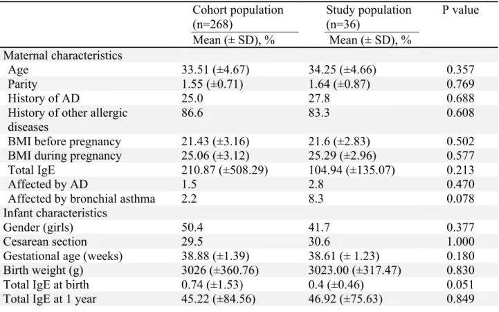
Table 1. Baseline characteristics of the infants and their mothers in the cohort population and the nested case-control population defined by propensity score matching
Maternal age, parity, BMI, gestational age, and birth weight were all compared by Student’s t-test, and serum total IgE levels by Mann-Whitney test. Other variables were compared by Pearson’s χ2 test
Cohort population (n=268) Mean (± SD), % Study population (n=36) Mean (± SD), % P value Maternal characteristics Age 33.51 (±4.67) 34.25 (±4.66) 0.357 Parity 1.55 (±0.71) 1.64 (±0.87) 0.769 History of AD 25.0 27.8 0.688
History of other allergic diseases
86.6 83.3 0.608
BMI before pregnancy 21.43 (±3.16) 21.6 (±2.83) 0.502
BMI during pregnancy 25.06 (±3.12) 25.29 (±2.96) 0.577
Total IgE 210.87 (±508.29) 104.94 (±135.07) 0.213
Affected by AD 1.5 2.8 0.470
Affected by bronchial asthma 2.2 8.3 0.078
Infant characteristics
Gender (girls) 50.4 41.7 0.377
Cesarean section 29.5 30.6 1.000
Gestational age (weeks) 38.88 (±1.39) 38.61 (± 1.23) 0.180
Birth weight (g) 3026 (±360.76) 3023.00 (±317.47) 0.830
Total IgE at birth 0.74 (±1.53) 0.4 (±0.46) 0.051
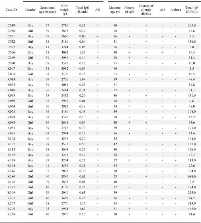
Table 2. Baseline characteristics of nested case control population
Infants Mothers
Case ID Gender age (weeks) Gestational Birth weight (g) Total IgE (IU/mL) AD Maternal age (y) History of AD History of allergic disease
AD Asthma Total IgE (IU/mL)
C019 Boy 37 2710 0.32 + 40 - + - - 305.0 C038 Girl 39 2848 0.19 - 26 - + - - 12.8 C051 Boy 38 2660 0.09 - 36 - + - - 2.5 C052 Girl 38 3180 0.64 + 31 - + - - 136.0 C062 Boy 41 3244 0.08 + 36 - + - - 6.8 C066 Boy 38 3012 1.34 + 30 + + + - 80.4 C069 Girl 39 2956 0.10 + 26 + + - - 11.3 C070 Boy 38 3380 0.13 - 35 - + - - 10.0 K007 Boy 38 2952 0.04 - 40 - - - - 2.5 K008 Girl 38 3190 0.26 + 25 - + - + 62.5 K013 Boy 39 2706 1.98 + 39 - - - - 48.4 K032 Boy 39 3082 0.28 + 31 - - - - 87.8 K040 Boy 36 2464 0.31 - 37 - + - - 31.1 K045 Boy 38 2412 0.29 - 36 - + - - 131.0 K059 Girl 38 2990 0.04 - 38 + + - - 9.6 K074 Girl 40 2912 0.18 + 33 + + - - 48.0 K078 Boy 36 3110 0.45 + 39 - + - - 104.0 K079 Boy 38 3390 0.10 - 38 - + - - 13.2 K083 Girl 39 2642 0.04 - 30 + + - - 13.6 K085 Boy 39 3712 0.70 + 39 - - - - 123.0 K095 Boy 39 2984 0.13 + 36 - + - - 11.4 K102 Boy 40 3508 0.30 - 35 - - - - 144.0 K107 Boy 38 3522 0.50 - 42 + + - - 187.0 K112 Boy 38 2604 0.25 + 30 - + - - 126.0 K132 Boy 40 3202 0.17 + 38 - + - - 42.3 K138 Boy 37 3274 0.22 + 27 + + - - 113.0 K144 Boy 41 3536 0.11 + 34 - + - + 27.6 K146 Girl 37 2802 0.30 - 30 - + - + 304.0 K180 Girl 40 2890 0.62 - 28 - + - - 688.0 K196 Girl 39 2832 0.04 + 40 - - - - 2.5 K197 Girl 40 3190 0.23 + 37 + + - - 104.0 K199 Girl 39 2940 0.65 - 39 - + - - 255.0 K205 Girl 40 3468 0.26 - 36 - + - - 14.2 K207 Girl 38 2770 1.15 - 35 + + - - 313.0 K209 Boy 38 2904 1.67 - 31 + + - - 165.0 K228 Girl 40 2850 0.16 - 30 - + - - 41.4
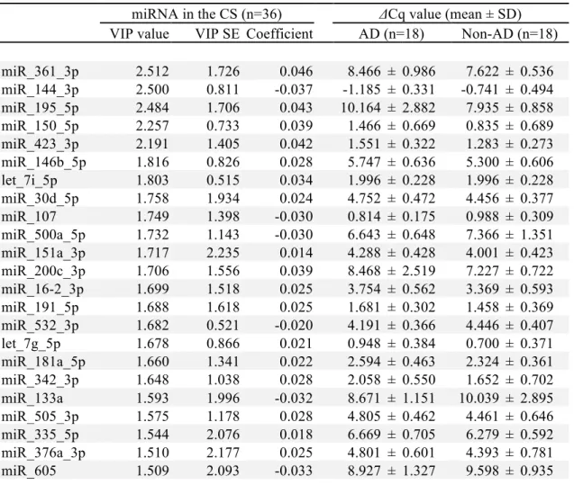
In the 36 selected subjects, we analyzed the expression profile of 179 miRs. By use of partial least squares regression, we identified the miRs that were related to the development of AD at 1 year. Candidate miRs with a VIP score of 1.5 or more were selected for further evaluation (table 3).
Table 3. MicoRNAs in umbilical cord serum (CS) and maternal serum (MS) associated with the development of AD at 1 year. Partial least squares regression was
used to identify miRNAs associated with the development of AD at 1 year of age. The miRs in CS and MS with a VIP score over 1.5 were selected as candidates for further analysis.
miRNA in the CS (n=36) ΔCq value (mean ± SD)
VIP value VIP SE Coefficient AD (n=18) Non-AD (n=18)
miR_361_3p 2.512 1.726 0.046 8.466 ± 0.986 7.622 ± 0.536 miR_144_3p 2.500 0.811 -0.037 -1.185 ± 0.331 -0.741 ± 0.494 miR_195_5p 2.484 1.706 0.043 10.164 ± 2.882 7.935 ± 0.858 miR_150_5p 2.257 0.733 0.039 1.466 ± 0.669 0.835 ± 0.689 miR_423_3p 2.191 1.405 0.042 1.551 ± 0.322 1.283 ± 0.273 miR_146b_5p 1.816 0.826 0.028 5.747 ± 0.636 5.300 ± 0.606 let_7i_5p 1.803 0.515 0.034 1.996 ± 0.228 1.996 ± 0.228 miR_30d_5p 1.758 1.934 0.024 4.752 ± 0.472 4.456 ± 0.377 miR_107 1.749 1.398 -0.030 0.814 ± 0.175 0.988 ± 0.309 miR_500a_5p 1.732 1.143 -0.030 6.643 ± 0.648 7.366 ± 1.351 miR_151a_3p 1.717 2.235 0.014 4.288 ± 0.428 4.001 ± 0.423 miR_200c_3p 1.706 1.556 0.039 8.468 ± 2.519 7.227 ± 0.722 miR_16-2_3p 1.699 1.518 0.025 3.754 ± 0.562 3.369 ± 0.593 miR_191_5p 1.688 1.618 0.025 1.681 ± 0.302 1.458 ± 0.369 miR_532_3p 1.682 0.521 -0.020 4.191 ± 0.366 4.446 ± 0.407 let_7g_5p 1.678 0.866 0.021 0.948 ± 0.384 0.700 ± 0.371 miR_181a_5p 1.660 1.341 0.022 2.594 ± 0.463 2.324 ± 0.361 miR_342_3p 1.648 1.038 0.028 2.058 ± 0.550 1.652 ± 0.702 miR_133a 1.593 1.996 -0.032 8.671 ± 1.151 10.039 ± 2.895 miR_505_3p 1.575 1.178 0.028 4.805 ± 0.462 4.461 ± 0.646 miR_335_5p 1.544 2.076 0.018 6.669 ± 0.705 6.279 ± 0.592 miR_376a_3p 1.510 2.177 0.025 4.801 ± 0.601 4.393 ± 0.781 miR_605 1.509 2.093 -0.033 8.927 ± 1.327 9.598 ± 0.935
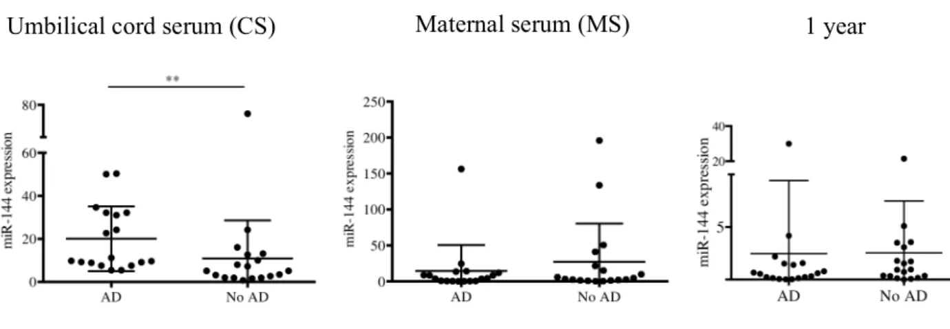
The miR expression analysis of the 36 sera by qPCR showed that miR-144 was significantly higher in the CS of children who developed AD at 1 year of age. This difference was not observed in MS at 36 weeks of gestation or in the children at 1 year of age (Fig 1). We did not find any correlation between miR expression in MS and CS, suggesting that the miRs in MS may not have a direct effect on babies (Fig 2). We found that the miR-144 expression in CS was not associated with total IgE levels at 1 year of age (Fig 3). As this suggests that miR-144 may play a role in the pathogenesis of AD
independent of atopy, we focused on the role of miR-144 in keratinocytes.
miRNA in the MS (n=36) ΔCq value (mean ± SD)
VIP value VIP SE Coefficient AD (n=18) Non-AD (n=18)
miR_301a_3p 2.854 1.594 0.051 3.835 ± 0.602 3.034 ± 0.665 miR_133b 2.732 0.713 -0.054 6.106 ± 0.976 9.384 ± 3.774 miR_133a 2.676 1.019 -0.049 5.572 ± 1.192 9.007 ± 4.028 miR_424_5p 2.407 0.538 0.031 2.421 ± 0.638 1.770 ± 0.656 miR_27b_3p 2.256 1.341 0.030 1.106 ± 0.561 0.590 ± 0.549 miR_485_3p 2.196 0.437 0.020 4.960 ± 0.859 4.149 ± 0.946 miR_148b_3p 2.146 0.760 0.031 1.597 ± 0.418 1.279 ± 0.300 miR_142_3p 1.992 0.749 0.021 -0.009 ± 0.530 -0.471 ± 0.622 miR_101_3p 1.943 0.547 0.017 -0.094 ± 0.693 -0.548 ± 0.449 miR_15a_5p 1.937 0.967 0.020 -2.700 ± 0.375 -2.995 ± 0.386 miR_296_5p 1.933 0.914 -0.025 8.126 ± 3.826 11.298 ± 4.370 miR_128 1.912 1.230 0.018 2.018 ± 0.388 1.706 ± 0.428 miR_21_5p 1.866 0.984 0.010 -3.049 ± 0.615 -3.409 ± 0.306 miR_141_3p 1.854 1.329 0.019 5.498 ± 2.701 3.998 ± 1.006 miR_30d_5p 1.812 1.902 0.028 4.828 ± 0.475 4.463 ± 0.541 miR_191_5p 1.785 0.715 -0.019 1.495 ± 0.564 1.832 ± 0.374 miR_320a 1.780 1.126 0.017 0.221 ± 0.569 -0.160 ± 0.516 miR_154_5p 1.775 0.892 -0.029 8.173 ± 3.880 11.122 ± 4.515 miR_382_5p 1.766 1.037 -0.026 7.050 ± 2.941 9.668 ± 4.429 miR_126_3p 1.654 1.024 0.010 -1.341 ± 0.650 -1.761 ± 0.649 miR_215 1.654 0.630 0.016 2.621 ± 0.761 2.075 ± 0.920 miR_375 1.626 1.334 0.027 5.251 ± 2.724 3.848 ± 1.528 miR_766_3p 1.618 1.255 -0.018 6.211 ± 2.376 8.413 ± 4.318 let_7i_3p 1.539 0.918 0.025 9.650 ± 3.562 7.721 ± 2.850
Figure 1. MiR-144 levels are elevated in the cord serum of infants who developed AD at 1 year of age. MiR-144 levels were estimated in CS, MS and serum at 1 year of age
using quantitative RT-PCR. MiR-144 expression was significantly increased in the CS of the AD group. This difference was not seen in the MS or serum at 1 year of age. **P <
0.005, Mann-Whitney test. AD n = 18; No AD n = 18. CS, cord serum; MS, maternal serum; AD, atopic dermatitis.
Umbilical cord serum (CS) 1 year
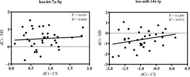
Figure 2. MiRs in maternal serum (MS) and cord serum (CS) are not correlated.
Levels of 179 miRs in MS and CS were measured by qPCR. The levels of each miR did not show any correlation between MS and CS. The data shows 2 representative miRs including miR-144, which was investigated further in this study.
Figure 3. MiR-144 levels in cord serum are not related to the total IgE levels in serum at 1 year of age. The miR-144 levels in cord serum were plotted against the Serum total
IgE levels at 1 year of age. There was no correlation between the levels.
-2.0 -1.5 -1.0 -0.5 0.0 0.5 0 100 200 300 400 500 dCt value of CS miR-144
Serum total IgE at 1 year (IU/ml)
P = 0.5807 R2 = 0.0952
ABCA1 is a target of miR-144
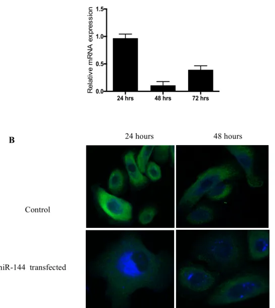
Using the commercially available software microRNA.org (10), TargetScan (11) and miRBase (12), we identified ATP-binding cassette transporter A1 (ABCA1) as a target of miR-144. ABCA1 is involved in cholesterol efflux from cells and miR-144 has been shown downregulate ABCA1 expression leading to cholesterol accumulation within cells (13, 14). To observe the effect of miR-144 on the expression of ABCA1 in keratinocytes, we transfected human epithelial keratinocytes with miR-144 and looked at the expression of ABCA1. MiR-144 transfected cells showed decreased ABCA1 mRNA (Fig 4A) and protein levels (Fig 4B). Keratinocytes also showed the accumulation of cholesterol following miR-144 transfection (Fig 4B).
House dust mite (HDM) stimulation of miR-144 transfected keratinocytes leads to increased translocation of the NF-κB p65 subunit
ABCA1 knockout in macrophages has been shown to cause cholesterol accumulation and exaggerated cytokine response to the TLR agonists (15). This exaggerated response is believed to be due to the enhanced recruitment of all MyD88-dependent TLRs (TLR 2, 4, 7 and 9) into lipid rafts leading to enhanced downstream
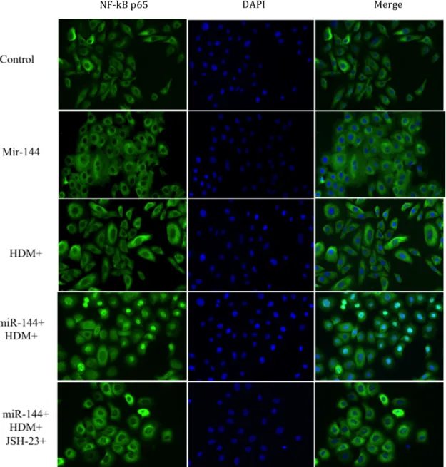
signaling and pro-inflammatory cytokine production through NF-κB and activator protein-1 (AP-1) signaling (15, 16). Therefore, we looked at how miR-144 transfected keratinocytes with decreased ABCA1 expression responded to house dust mite (HDM) stimulation, which is a well know trigger of inflammation in AD. With HDM stimulation miR-144 increased the nuclear translocation of the NF-κB p65 subunit (Fig 5).
A
B
Figure 4. MiR-144 transfection decreases ABCA1 expression in HPEKs. (A) MiR-144
transfection decreased the mRNA levels of ABCA1. (B) HPEKs grown in media only or transfected with miR-144 for 24 and 48 hours. Cells were stained with Alexa Fluor 488 for ABCA1 (green) and filipin for cholesterol (blue). MiR-144 transfection caused a decreased in ABCA1 expression leading to cholesterol accumulation within cells. HPEKs, human primary epithelial keratinocytes.
24 hrs 48 hrs 72 hrs 0.0 0.5 1.0 1.5 Re la tiv e m RNA e x p re s s io n 24 hours 48 hours Control miR-144 transfected
Figure 5. MiR-144 induces nuclear translocation of NF-κB p65 subunit translocation.
HDM induced the nuclear translocation of p65 (green). This was enhanced when miR-144 was introduced into the HPEKs prior to HDM stimulation. The p65 translocation was abrogated by JSH-23 (p65 translocation inhibitor). The nucleus is stained in blue with DAPI.
NF-kB p65 DAPI Merge
HDM stimulation of miR-144 transfected keratinocytes leads to increased mRNA expression of human beta-defensin 2 (hBD-2)
As we did not see any pro-inflammatory cytokine production from keratinocytes in response to NF-κB activation, we looked at other molecules that play a role in the
pathogenesis of AD via NF-κB activation. Anti-microbial peptides (AMPs) are small cationic peptides present in epithelia that show direct anti-microbial activity and have also been shown to play a role in antigen presentation, cytokine release and the recovery of barrier function (17, 18). The AMPs implicated in AD include the inducible beta defensins hBD-2 and 3, and the cathelicidin LL-37. Although some studies show a decreased
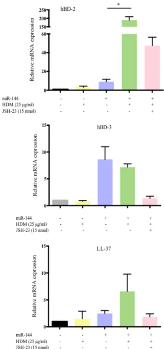
production of AMPs in AD, others show an increased secretion in acute compared to chronic lesions (19, 20). In addition, hBDs are known to induce the production of pro-inflammatory cytokines from keratinocytes and act as chemo-attractants for mast cells, inducing the release of pruritogenic mediators and increasing vascular permeability (21-23). In fact, hBD 2 levels were shown to be significantly higher in the lesional stratum corneum compared to non-lesional skin and that the levels correlate with SCORing of Atopic Dermatitis (SCORAD) scores and trans-epidermal water loss (TEWL) (24). We did observe that miR-144 transfected HPEKs, when stimulated with HDM extract, significantly increased the mRNA expression of hBD-2. The levels decreased when the transfected cells were co-incubated with HDM and the NF-κB inhibitor JSH-23 (Fig 6). Human
beta-defensin 3 and LL-37 mRNA levels did not change significantly in response to HDM stimulation. As NF-κB activation has been shown to increase hBD-2 production, the significant increase in hBD-2 mRNA levels that we observed may be a consequence of the NF- κB activation (25).
Figure 6. MiR-144 increases HDM-induced hBD-2 expression in keratinocytes. HPEKs
were transfected with miR-144 for 48 hours and stimulated with 25 µg/ml of HDM extract for a further 24 hours with or without 15 nmol of p65 translocation inhibitor, JSH-23. The mRNA levels of hBD2, hBD3 and cathelicidin LL-37 were quantified by qPCR. HBD-2 showed a significant increase in expression following HDM stimulation. *P < 0.05, Kruskal-Wallis test. Data shown are from 3 independent experiments. HDM, house dust
DISCUSSION
MiRs are increasingly being investigated for their role in disease pathogenesis and as biomarkers. In spite of this, there is still a dearth of knowledge about the actual roles that miRs may have in allergic diseases. In this study we found that miR-144 is higher in cord blood of infants who developed AD at 1 year of age. MiR-144 targets ABCA1 and miR-144 transfection into keratinocytes decreased the expression of ABCA1 leading to the accumulation of cholesterol. MiR-144-induced cholesterol accumulation has been shown to induce pro-inflammatory cytokine production in THP-1 macrophages (13). However, we did not detect any significant change in TNF-α, TSLP, IL-25, IL-17A, IL-17F or IL-33 levels in serum, or the mRNA levels in transfected keratinocytes (data not shown). In addition, TNF-α was undetectable in the culture supernatants of transfected keratinocytes.
In psoriasis, another chronic inflammatory skin disease, cholesterol accumulation has also been shown to promote IL-17A signaling in keratinocytes (26). IL-17A, the signature cytokine of Th17 cells, is expressed highly in early-onset pediatric AD skin lesions as opposed to normal and adult AD skin, along side the increase in other related molecules including LL-37 (27). Although we did not find any cytokine changes in the serum, the changes may be visible in early AD skin itself. There is also an increasing body of evidence that identifies the role of cationic peptides in non-IgE-mediated stimulation of mast cells (28). Our finding that miR-144 did not correlate with serum IgE levels also supports the fact that the early skin barrier dysfunction may be assisted by mast cell-derived cytokines such as IL-31. This may suggest that the initial activation may come from the resident innate immune cells of the skin.
Our study is limited in several aspects: we conducted this study on the premise that miR-144 targets keratinocytes. We did not investigate any change in miRs or other
Also, we have only looked at the keratinocyte response to HDM: it is worthwhile investigating the response to Staphylococcus aureus, which has a prominent role in the pathogenesis of AD.
Nevertheless, at present there is little knowledge about the earliest triggers that initiate barrier damage in AD. To prevent the onset of this predominantly childhood disease, it is essential to identify and if possible, remedy them. Therefore, we believe that our
primary finding that miR-144 is elevated in the umbilical cord blood provides an important clue to the understanding of what prompts early-life AD and adds a new dimension to the understanding of this disease.
REFERENCES
1. Carroll CL, Balkrishnan R, Feldman SR, Fleischer AB, Manuel JC. The burden of
atopic dermatitis: impact on the patient, family, and society. Pediatr Dermatol. 2005;22(3):192-9.
2. Deckers IA, McLean S, Linssen S, Mommers M, van Schayck CP, Sheikh A.
Investigating international time trends in the incidence and prevalence of atopic eczema 1990-2010: a systematic review of epidemiological studies. PLoS One. 2012;7(7):e39803.
3. Illi S, von Mutius E, Lau S, Nickel R, Grüber C, Niggemann B, et al. The natural
course of atopic dermatitis from birth to age 7 years and the association with asthma. J Allergy Clin Immunol. 2004;113(5):925-31.
4. Garmhausen D, Hagemann T, Bieber T, Dimitriou I, Fimmers R, Diepgen T, et al.
Characterization of different courses of atopic dermatitis in adolescent and adult patients. Allergy. 2013;68(4):498-506.
5. Fox DE, Lack G. Peanut allergy. Lancet. 1998;352(9129):741.
6. Agrawal R, Woodfolk JA. Skin barrier defects in atopic dermatitis. Curr Allergy
Asthma Rep. 2014;14(5):433.
7. He L, Hannon GJ. MicroRNAs: small RNAs with a big role in gene regulation. Nat
Rev Genet. 2004;5(7):522-31.
8. Rebane A. microRNA and Allergy. Adv Exp Med Biol. 2015;888:331-52.
9. Rebane A, Akdis CA. MicroRNAs in allergy and asthma. Curr Allergy Asthma
Rep. 2014;14(4):424.
10. Betel D, Wilson M, Gabow A, Marks DS, Sander C. The microRNA.org resource:
11. Agarwal V, Bell GW, Nam JW, Bartel DP. Predicting effective microRNA target sites in mammalian mRNAs. Elife. 2015;4.
12. Griffiths-Jones S. The microRNA Registry. Nucleic Acids Res. 2004;32(Database
issue):D109-11.
13. Hu YW, Hu YR, Zhao JY, Li SF, Ma X, Wu SG, et al. An agomir of miR-144-3p
accelerates plaque formation through impairing reverse cholesterol transport and promoting pro-inflammatory cytokine production. PLoS One. 2014;9(4):e94997.
14. de Aguiar Vallim TQ, Tarling EJ, Kim T, Civelek M, Baldán Á, Esau C, et al.
MicroRNA-144 regulates hepatic ATP binding cassette transporter A1 and plasma high-density lipoprotein after activation of the nuclear receptor farnesoid X
receptor. Circ Res. 2013;112(12):1602-12.
15. Zhu X, Lee JY, Timmins JM, Brown JM, Boudyguina E, Mulya A, et al. Increased
cellular free cholesterol in macrophage-specific Abca1 knock-out mice enhances pro-inflammatory response of macrophages. J Biol Chem. 2008;283(34):22930-41.
16. Zhu X, Owen JS, Wilson MD, Li H, Griffiths GL, Thomas MJ, et al. Macrophage
ABCA1 reduces MyD88-dependent Toll-like receptor trafficking to lipid rafts by reduction of lipid raft cholesterol. J Lipid Res. 2010;51(11):3196-206.
17. Lai Y, Gallo RL. AMPed up immunity: how antimicrobial peptides have multiple
roles in immune defense. Trends Immunol. 2009;30(3):131-41.
18. Kiatsurayanon C, Niyonsaba F, Smithrithee R, Akiyama T, Ushio H, Hara M, et al.
Host defense (Antimicrobial) peptide, human β-defensin-3, improves the function of the epithelial tight-junction barrier in human keratinocytes. J Invest Dermatol. 2014;134(8):2163-73.
19. Nograles KE, Suárez-Fariñas M, Shemer A, Fuentes-Duculan J, Chiricozzi A,
Cardinale I, et al. Atopic dermatitis keratinocytes exhibit normal T(H)17 cytokine responses. J Allergy Clin Immunol. 2010;125(3):744-6, 6.e1-6.e2.
20. Hata TR, Kotol P, Boguniewicz M, Taylor P, Paik A, Jackson M, et al. History of eczema herpeticum is associated with the inability to induce human β-defensin (HBD)-2, HBD-3 and cathelicidin in the skin of patients with atopic dermatitis. Br J Dermatol. 2010;163(3):659-61.
21. Niyonsaba F, Ushio H, Hara M, Yokoi H, Tominaga M, Takamori K, et al.
Antimicrobial peptides human beta-defensins and cathelicidin LL-37 induce the secretion of a pruritogenic cytokine IL-31 by human mast cells. J Immunol. 2010;184(7):3526-34.
22. Niyonsaba F, Kiatsurayanon C, Ogawa H. The role of human β-defensins in allergic
diseases. Clin Exp Allergy. 2016;46(12):1522-30.
23. Chen X, Niyonsaba F, Ushio H, Hara M, Yokoi H, Matsumoto K, et al.
Antimicrobial peptides human beta-defensin (hBD)-3 and hBD-4 activate mast cells and increase skin vascular permeability. Eur J Immunol. 2007;37(2):434-44.
24. Clausen ML, Jungersted JM, Andersen PS, Slotved HC, Krogfelt KA, Agner T.
Human β-defensin-2 as a marker for disease severity and skin barrier properties in atopic dermatitis. Br J Dermatol. 2013;169(3):587-93.
25. Froy O. Regulation of mammalian defensin expression by Toll-like
receptor-dependent and inreceptor-dependent signalling pathways. Cell Microbiol. 2005;7(10):1387-97.
26. Varshney P, Narasimhan A, Mittal S, Malik G, Sardana K, Saini N. Transcriptome
profiling unveils the role of cholesterol in IL-17A signaling in psoriasis. Sci Rep. 2016;6:19295.
27. Esaki H, Brunner PM, Renert-Yuval Y, Czarnowicki T, Huynh T, Tran G, et al.
Early-onset pediatric atopic dermatitis is TH2 but also TH17 polarized in skin. J Allergy Clin Immunol. 2016;138(6):1639-51.
28. Liu FT, Goodarzi H, Chen HY. IgE, mast cells, and eosinophils in atopic dermatitis. Clin Rev Allergy Immunol. 2011;41(3):298-310.
Journal of Clinical Investigation Submitted in December 2017