Title
Detection of a taeniid species Taenia taeniaeformis from a feral
raccoon Procyon lotor and its epidemiological significance( 本文
(Fulltext) )
Author(s)
MATOBA, Yohei; ASANO, Makoto; YAGI, Kinpei;
ASAKAWA, Mitsuhiko
Citation
[Mammal study] vol.[28] no.[2] p.[157]-[160]
Issue Date
2003-12-01
Rights
The Mammalogical Society of Japan (日本哺乳類学会)
Version
出版社版 (publisher version) postprint
URL
http://hdl.handle.net/20.500.12099/31821
© the Mammalogical Society of Japan Short communication
Detection of a taeniid species
Taenia taeniaeformis from a feral raccoon
Procyon lotor and its epidemiological significance
Yohei Matoba
1, Makoto Asano
2, Kinpei Yagi
3and Mitsuhiko Asakawa
1,*1 Department of Parasitology (Wildlife Zoology), School of Veterinary Medicine, Rakuno Gakuen University, Ebetsu, Hokkaido 069-8501, Japan
2 United Graduate School of Veterinary Science, Gifu University, Yanagido, Gifu 501-1193, Japan 3 Department of Biology, Hokkaido Institute of Public Health, Sapporo, Hokkaido 060-0819, Japan
One of the risks posed by feral raccoons, Procyon lotor, an introduced alien mammal species in Japan is the transmission or outbreak of parasitic and/or infectious diseases (Ikeda 2002). For this reason, since 1995, the authors have surveyed the internal parasites of over 240 individual feral raccoons in Hokkaido, Japan. So far, ten helminth and two protozoan species have been found (Asakawa et al. 1999, 2000; Yamada et al. 2000; Matoba et al. 2002). However, in the most recent analysis of the contents of the small intestines of individual feral raccoons, a taeniid cestode (Taeniidae: Cestoda) was obtained, although there have been no records of the family Taeniidae from P. lotor either in Japan or in other countries including the U.S.A. where the raccoon is indigenous (Asakawa et al. 2000). The taeniid family includes zoonotic pathogens, such as Echinococcus spp. and Taenia spp. (Abuladze 1964), and hence, the present note provides a description of the cestode and a brief discussion of its implications from public hygiene and mammalian ecology view points.
Materials and methods
A feral raccoon registered as number NP01-22 was captured on Nopporo Forest Park, Hokkaido, Japan, in 2001 and was euthanized as part of a population con-trol programme by the Hokkaido Government. After measuring its body size, it was dissected, examined for parasites, and a cestode was collected from its small intestine. The worm was fixed and kept in 70% ethanol, and examined microscopically with lacto-phenol solu-tion. Measuring and drawing of its scolex was done with the aid of a camera lucida, OLYMPUS Model BH2-DA. Part of the immature segment was used for
molecular biological examination. Total DNA was isolated from the segment using the Easy-DNATM iso-lation kit (Invitrogen). The manufacturer’s instruction (protocol #8) was followed for the extraction. As the protocol was designed for DNA extraction from 1 cm of mouse tail, we reduced the volume of each reagent by 1/4 for a cubic piece of parasite tissue measuring 1.7 × 1.7 × 1.7 mm. The partial mitochondrial 12S rRNA gene was amplified by PCR from the genomic DNA using the olignucleotide primers P60F (forward; 5'-TTAAGATATATGTGGTACAGGATTAGATACCC-3') and P375R (reverse; 5'-AACCGAGGGTGACGGGCGG TGTGTACC-3') as reported by Dinkel et al. (1998). This primer set was designed for detecting the DNA of Echinococcus multilocularis and related tapeworms. The PCR reaction (50 ml) was performed for 50 cycles, with each cycle consisting of denaturation for 1 min at 93°C, annealing for 1.5 min at 55°C, and elongation for 2 min at 73°C. In each reaction, 1.25 units of AmpliTaq GoldTM (Applied Biosystems) were used for Taq Poly-merase. The PCR products were electrophoresed in 3% agarose gel. The DNA fragment was then extracted from the agarose gel, purified using glass beads (EASY-TRAPTM TAKARA) and sequenced for both DNA strands using a BigDye terminator cycle sequencing FS ready reaction kit (Applied Biosystems) and a model 377 DNA sequencer (Applied Biosystems). The nucleo-tide sequence obtained was aligned with the sequences of three common taeniid cestodes in Hokkaido Taenia taeniaeformis, T. hydatigena and Echinococcus multilo-cularis and that of E. granulosus reported by Yagi et al. (1999). The nucleotide sequences reported in this paper have been deposited in the DDBJ nucleotide sequence databases under the accession number AB120128.
158 Mammal Study 28 (2003)
Results and discussion
The specimen obtained consisted of a scolex and several immature segments without genital organs. The body was about 10 mm long, and the width 1.72 mm wide. The scolex had four suckers which were 0.28– 0.29 mm in diameter, and a rostellum which was 1.06 mm in diameter (Fig. 1a). There were two rows of large and small hooks on the rostellum (Fig. 1b). The large rostellar hooks 17 were 0.43 mm long length, and the small ones also 17 were 0.25 mm long. The measure-ments of the scolex were close to those of T. taeniaefor-mis (Abuladze 1964). The present measurements almost corresponded to that of immature T. taeniaeformis 24 hours after experimental infection of a cat (Singh and Rao 1966).
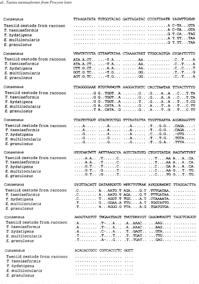
According to the alignment of the nucleotide sequences of the partial mitochondrial 12S rRNA gene of the present specimen and four species of taeniid cestodes (Fig. 2), the present specimen was identical to the sequence of T. taeniaeformis isolated from a Norway rat captured in Sapporo, Hokkaido (Yagi et al. 1999). DNA analysis for mitochondrial 12S rRNA gene of taeniid cestodes has shown that different species have different nucleotide sequences and identification of
these species is feasible by detection of these differences (Yagi et al. 1999). The sequence data supports the identification based on the morphological observations described above.
So far, T. taeniaeformis has not been known as a serious zoonotic pathogen, however, the related species, E. multilocularis is a known zoonotic pathogen in Hokkaido, Japan (Uchino and Sato 1996). Both T. taeniaeformis and E. multilocularis belong to the same family Taeniidae, and have similar life cycles; the cestode infections of definitive hosts (in the order Carnivora) are derived from eating intermediate hosts (in the order Rodentia) already having metacestodes (=bladder worms) (Abuladze 1964), suggesting that raccoons may also serve as a definitive host of E. multilocularis. Yamada et al. (2000) orally inoculated a juvenile raccoon with protoscoleces of E. multilocula-ris, but they found no parasites in the small intestine at fifteen days post-infection and excretion of E. multilo-cularis coproantigen was not detected during the course of the infection. Nevertheless, this should not be seen as confirmation that raccoons can not serve as a definitive host of E. multilocularis since the study by Yamada et al. (2000) was a preliminary investigation using only one animal. Since a feral raccoon is known to have eaten a red-backed vole Clethrionomys rufocanus, which is an important intermediate host for E. multilo-cularis in Hokkaido (Asakawa et al. 2000), and because of the present detection of the taeniid species, more precise experimental infection studies using more raccoons should be done in the future. Simultaneously, a large scale epidemiological investigation of natural infection with E. multilocularis and the food habits of raccoons should be undertaken under strict conditions of biological safety as soon as possible because raccoons have now spread to occur throughout Hokkaido (Ikeda 2002).
Acknowledgments: We wish to thank the many trappers who helped us collect raccoons. We also thank the staff of Raccoon Research Society for their support during this study. We also extend our gratitude to two anonymous referees and Dr. Mark A. Brazil for their help in improving this manuscript. We are also grateful to local government offices, EnVision, Hokkaido Forest Management Corporation and Nopporo Natural Forest Park Office all of which provided us with raccoon samples. This study was carried out in association with the Management Plan for the Feral Raccoon Population
Fig. 1. Scolex of Taenia taeniaeformis obtained from a raccoon in Hokkaido, Japan (Bars = 0.2 mm), apical (a) and lateral (b) views.
in Hokkaido, and was partly funded by the Nature Preservation Division of the Hokkaido Government.
This study was partly supported by the 2002 Hokkaido Kankyo-zaidan-jyosei, the Grant-in-Aid (no. 14560271),
Fig. 2. Alignment of the partial mitochondrial 12S rRNA gene sequences of the present taeniid cestode from a raccoon and four species of taeniid cestodes, Taenia taeniaeformis, Taenia hydatigena, Echinococcus multilocularis, Echinococcus granulosus. The nucleotide sequences of taeniid species are quoted from Yagi et al. (1999).
160 Mammal Study 28 (2003)
and High Technological Research Center (Rakuno Gakuen Univ.) of the Ministry of Education, Science and Culture of Japan.
References
Abuladze, K. I. 1964. Taeniata of Animals and Man and Diseases Caused by Them. Izdatel’stvo “Nauka”, Moscow (in Russian). Asakawa, M., Kurachi, T. and Wildlife Ecological Society. 1999.
Parasitic helminths of raccoons in Hokkaido, Japan. Japanese Journal of Zoo and Wildlife Medicine 4: 101–103 (in Japanese with English summary).
Asakawa, M., Matoba, Y., Yamada, D. and Kamiyama, T. 2000. Review of the parasitological state of feral raccoons captured in Nopporo National Park and its proximity, Hokkaido. Journal of Rakuno Gakuen University 25: 1–8 (in Japanese with English summary).
Dinkel, A., Nickisch Rosenegk, M., Bilger, B., Merli, M., Lucius, R. and Romig, T. 1998. Detection of Echinococcus multilocularis in the definitive host: Coprodiagnosis by PCR as an alternative to necropsy. Journal of Clinical Microbiology 36: 1871–1876. Ikeda, T. 2002. Procyon lotor. In (The Ecological Society of Japan,
ed.) Handbook of Alien Species in Japan. Pp. 70. Chijin-shokan, Tokyo (in Japanese).
Matoba, Y., Asano, M., Masubuchi, H. and Asakawa, M. 2002. First records of the genera Eimeria and Isospora (Protozoa: Eimeri-idae) obtained from feral raccoons (Procyon lotor) alien species in Japan and prevalence of serum antibodies to Toxoplasma gondii among the raccoons. Japanese Journal of Zoo and Wild-life Medicine 7: 87–90 (in Japanese with English summary). Singh, B. B. and Rao, B. V. 1966. Some biological studies on Taenia
taeniaeformis. Indian Journal of Helminthology 18: 151–160. Uchino, J. and Sato, N. (eds.) 1996. Alveolar Echinococcosis:
Strategy for Eradication of Alveolar Echinococcosis of the Liver. Fujishoin, Sapporo.
Yagi, K., Ohyama, T., Okamoto, M., Oku, Y., Kamiya, M. and Kimura, H. 1999. PCR-RFLP for identification of Echinococcus multilocularis and related taeniid cestodes based on the deter-mination of partial mitochondrial 12S rRNA gene. Report of Hokkaido Institute of Public Health 49: 163–166 (in Japanese). Yamada, D., Oku, Y., Nonaka, N., Asakawa, M., Ikeda, T., Asano, M.,
Akamatsu, R., Matoba, Y. and Kamiya, M. 2000. Studies on the parasite fauna of raccoon (Procyon lotor) naturalized in Hokkaido, Japan. Parasitology International 49 (Suppl.): 91.