56:37
はじめに
硬膜動静脈瘻(dural arteriovenous fistula; DAVF)は硬膜に
発生する異常な動静脈シャントを病態とする疾患である1)~3).
頭蓋内 DAVF の好発部位は海綿静脈洞部,横 -S 状静脈洞部で
あり頭蓋頸椎移行部は比較的稀である1)4).今回我々は右椎骨
動脈と右上行咽頭動脈を流入動脈とし脊髄静脈に流出する頭 蓋頸椎移行部硬膜動静脈瘻(craniocervical junction DAVF; CCJDAVF)の症例を経験した.本症例は対麻痺,下肢異常感 覚,膀胱直腸障害を呈しており,脊髄 MRI において胸髄以下 のレベルで異常信号を示す CCJDAVF は稀である. 症 例 患者:60 歳,女性 主訴:両下肢脱力,異常感覚 既往歴:糖尿病. 生活歴:喫煙歴なし. 家族歴:特記事項なし. 現病歴:2013 年 8 月中旬以降,数時間から半日程度で自然 に回復する両下肢の脱力を数回自覚した.脱力は軽度であり 歩行は可能であった.また両下肢の脱力に伴って残尿感も自 覚していたが脱力の回復とともに残尿感もなくなっていた. 10月上旬,仕事に出かけ座っている時に特に誘因なく下肢の 脱力が出現し,起立困難となったため受診し精査目的に入院 となった. 入院時身体所見:身長 160 cm,体重 58 kg.一般身体所見 に異常を認めず,四肢末梢動脈触知は良好であった.神経学 的所見では意識清明,脳神経,両上肢に異常所見を認めなかっ た.受診時には起立,立位保持は可能となっており,両側腸 腰筋,大腿四頭筋,前脛骨筋,ハムストリングの筋力は徒手 筋力テストで 4/5,腓腹筋筋力は正常であった.膝蓋腱反射, アキレス腱反射は両側とも亢進していた.バビンスキー徴候 は両側陰性で,ビーヴァー徴候は臍の移動はなかった.ロン ベルグ徴候は陰性であった.両側下肢は L5~S1 領域にビリ ビリとした異常感覚と温痛覚の低下を認め,振動覚は両側の 腸骨稜,膝蓋,第一趾で低下していた.触覚は保たれていた. また排尿はあるが残尿が多かった. 検査所見:第 1 病日に施行した全血算は異常なく,血液生 化学検査で随時血糖値 180 mg/dl,HbA1c 7.0%であった.ビ タミン B1 29 ng/ml,ビタミン B12 573 pg/ml,葉酸 12.3 ng/ml, 血清銅 112
μ
g/dl,血清亜鉛 76μ
g/dlであり,抗核抗体,抗 SS-A抗体,抗 SS-B 抗体,MPO-ANCA,PR3-ANCA,抗アク アポリン 4 抗体は陰性であった.髄液所見は無色透明,有核 細胞 14/3/μ
(N:L=1:9),蛋白 88 mg/dl,糖 76 mg/dl,IgG index l 0.57,ミエリン塩基性蛋白 < 31.3 pg/ml,オリゴクローナルバ症例報告
脊髄 MRI で胸髄から円錐部に異常信号を認めた
頭蓋頸椎移行部硬膜動静脈瘻の 1 例
上田 雅道
1)上田 美紀
1)3)竹内 有子
1)落合 淳
1)*
馬渕 千之
1)服部新之助
2) 要旨: 症例は 60 歳の女性である.6 週間にわたり間欠的に出現する両下肢脱力と残尿感があったが歩行障害が 増悪したため当院を受診した.対麻痺と下肢にビリビリとした異常感覚,排尿障害を呈していた.MRI では T2強 調像で胸腰髄の腫大,Th3 のレベルから脊髄円錐に高信号域,造影 T1強調像で脊髄前方に異常血管を認めた.血 管造影検査で右椎骨動脈と右上行咽頭動脈からの流入動脈と脊髄前面を下降する流出静脈を認め,右頭蓋頸椎移 行部硬膜動静脈瘻と診断した.動静脈瘻遮断術により症候と画像所見の改善を認めた.胸髄以下のレベルに病変を 呈する頭蓋頸椎移行部硬膜動静脈瘻は稀であり報告する. (臨床神経 2016;56:37-42) Key words: 頭蓋頸椎移行部硬膜動静脈瘻,脊髄症,動静脈瘻遮断術,椎骨動脈,上行咽頭動脈 *Corresponding author: 名古屋掖済会病院神経内科〔〒 454-8502 名古屋市中川区松年町 4-66〕 1)名古屋掖済会病院神経内科 2)名古屋掖済会病院脳神経外科 3)現:名古屋大学医学部附属病院神経内科(Received August 5, 2015; Accepted September 8, 2015; Published online in J-STAGE on November 30, 2015) doi: 10.5692/clinicalneurol.cn-000789
ンドは陰性であった.第 1 病日,胸椎単純 MRI では T2強調
像(以下 T2WI)で Th3 レベルから脊髄円錐部にかけて腫大
と髄内に高信号域を認めた(Fig. 1A).頭部 MRI では異常所 見を認めなかった.第 3 病日,頸椎単純 MRI では脊髄実質に 異常信号は明らかでなかったが脊髄前方にflow voidを認めた (Fig. 2B).第 8 病日,胸椎,頸椎造影 T1強調像で腰髄実質に 軽度の造影効果を認め,上位頸椎から胸腰椎移行部までの脊 髄前面に近接し蛇行した線状の造影効果を認めた(Fig. 1C, D). なお第 3 病日から第 4 病日にかけて施行した神経伝導検査は 脛骨神経,腓腹神経で正常,体性感覚誘発電位は脛骨神経で 正常,聴性脳幹反応,視覚誘発電位は正常であった.第 10 病 日,脊髄 CT angiography で脊髄前方に蛇行する線状の構造物 を認めた(Fig. 2).MRI と同様に蛇行する線状構造物は異常 血管と考えられた.第 24 病日,頭蓋内血管造影で右椎骨動脈 と右上行咽頭動脈からの数本の流入動脈と脊髄前面を下降す る流出静脈を認めた.脊髄血管造影では異常血管は認めず, 右 CCJDAVF と診断した(Fig. 3). 入院後経過:入院後に 2 回,両下肢筋力低下と異常感覚, 残尿感が出現した.病棟内での軽度の運動が誘因とも考えら れたが明確な誘因はなく,いずれも数時間から半日の安静に より脱力と残尿感は消失したが異常感覚は残存していた.動 静脈瘻遮断術の予定とし第 38 病日に一旦退院となったが,第 65病日,特に誘因なく対麻痺により歩行不能となり,下肢異 常感覚の悪化,尿閉のため再入院となった. 2回目入院後経過:翌日には対麻痺と尿閉はやや改善して いたが歩行はできず,その後も症候の増悪と改善を繰り返し, 便秘もあった.第 79 病日に後頭下開頭動静脈瘻遮断術を施行 し,下肢しびれは改善に乏しかったが対麻痺と残尿は改善し Fig. 1 MRI images of spinal cord.
(A): Thoracic sagittal T2-weightd images (1.5 T; TR 3,800 ms, TE 120 ms) on admission shows enlargement of the spinal cord and high intensity signals from Th3 to the conus medullaris (arrows). (B): Cervical sagittal T2-weightd images (1.5 T; TR 3,800 ms, TE 120 ms) in two days shows flow void in front of spinal cord, but shows no abnormal signals. (C) (D): Sagittal contrast-enhanced T1-weighted images (1.5 T; TR 484.30 ms, TE 10 ms) in eight days shows slight parenchymal enhancement in lumber spinal cord and abnormal vessels anterior cervical to lumbar spinal cord (arrows). ant: anterior.
脊髄 MRI で胸髄から円錐部に異常信号を認めた頭蓋頸椎移行部硬膜動静脈瘻の 1 例 56:39 歩行器歩行は可能となった.第 87 病日に施行した脊髄単純 MRIは T2WIで胸腰髄の高信号域,腫大の消失と造影 T1WIで 異常血管の消失を認め(Fig. 4A, B),第 118 病日に回復期リ ハビリテーション病院に転院した. 5カ月後に回復期リハビリテーション病院を退院し,平地 であれば歩行は可能であり,手すりを使って階段昇降もでき るようになり,膀胱直腸障害も改善していたが,下肢の異常 感覚,温痛覚障害,振動覚低下は改善していなかった.術後 5カ月で施行した脊髄 MRI では脊髄の腫大はなくなり,異常 血管も消失していた(Fig. 4C, D).その後は症候の再発する ことなく外来通院中である.
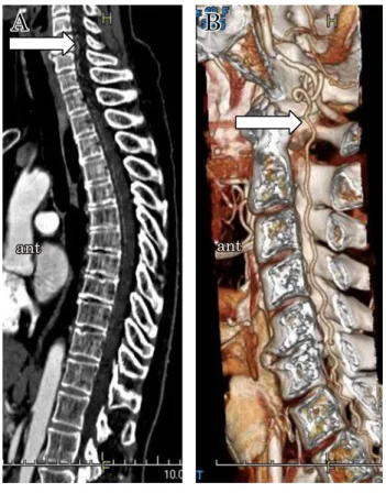
Fig. 2 CT angiography of spinal cord.
(A) (B): Saggital CT angiography of spinal cord imagies reveal the descending vessel (arrows). ant: anterior.
Fig. 3 Angiography of right vertebral artery and common carotid artery.
(A): Anterioposterior view of the right vertebral artery shows a dural arteriovenous fistula (DAVF) at the craniocervical junction fed by vertebral artery (arrow). (B): Lateral view of the right common carotid artery shows a DAVF at the craniocervical junction fed by ascending pharyngeal arteries (arrow). (C): Lateral view of the right common carotid artery, in venous phase, demonstrates the drainage into perimedullary veins (arrow). rt: right, ant: anterior.
考 察 CCJDAVFを含め頭蓋内 DAVF はくも膜下出血など頭蓋内 病変を呈することが多く,脊髄症は稀である1)3)5)~8).頭蓋内 DAVFで脊髄症を呈する症例の多くは MRI T2WIで延髄から頸 髄にかけて高信号域を認めており,胸髄以下に高信号を呈す ることは極めて稀である6)7)9).Asakawa ら7)によると頭蓋内 DAVFで脊髄症を呈した 31 例では下肢から始まる緩徐進行性 上行性脊髄症が特徴的である.このうち MRI を施行した 23 例のうち 21 例は延髄から頸髄にかけての病変を示し,胸髄以 下に病変を示したのは 2 例のみであった.また Vasdev ら10)は 頭蓋内 DAVF により MRI T2WIで胸腰髄のみに高信号を呈し た 2 例を報告している(Table 1).Mascalchi ら8)は後頭蓋窩 DAVFで胸髄病変と頸髄病変を呈した症例をそれぞれ報告 し,脊髄の障害部位の違いは頸胸椎移行部での perimedullary veinの流出路の流出パターンの違いと関連しているとしてい
る.頸胸椎移行部での perimedullary vein の流出路は convergent patternと divergent pattern があり,前者では頸髄の静脈血は 胸腰髄の perimedullary venous system と長軸方向につながり
尾側へと流出する8).一方後者では頸髄の静脈血は神経根静 脈を介して外側椎骨静脈叢と短軸方向につながる8).それぞ れの流出路の発達の程度には個人差があると考えられが,流 出静脈により静脈血の逆流が起こりconvergent patternの静脈 還流が障害されると重力の影響で脊髄下位レベルに静脈うっ 血が起こり,脊髄下位に障害が発生する8).また divergent patternの静脈還流が障害されても胸髄レベル以下には静脈 圧の上昇は伝播せず,頸髄レベルに静脈うっ血が起こり頸髄 の障害が発生する8).本症例は胸髄以下に病変を呈しており, convergent patternでの脊髄静脈還流障害が発生したと考えら れた.胸髄以下の脊髄症で発症する CCJDAVF は稀な疾患で あるが,MRI や血管造影検査,小別所ら12)の報告のように CT angiograpgyで診断可能であり,外科的治療により症候が Fig. 4
(A) (B): 8 days after surgery, saggital T2-weight images (1.5 T; 3,700 ms, TE 108 ms) show a decrease of the enlargement on the spinal cord and the abonormal signal. (C) (D): 5 months after surgery, sagittal contrast-enhanced T1-weighted images (1.5 T; 500 ms, TE10 ms) show that enhancement and abnormal vessels disappeared. ant: anterior.
脊髄 MRI で胸髄から円錐部に異常信号を認めた頭蓋頸椎移行部硬膜動静脈瘻の 1 例 56:41 改善する可能性がある.本症例も MRI,CT angiography,血 管造影検査が診断に有用であった. なお DAVF の治療目的は流入動脈とシャントの閉塞を行い 病変の永続的な消失を得ることである.治療法は血管内治療, 外科的治療,放射線治療があり,血管内治療が選択されるこ とが多い13)14).本症例のように頭蓋頸椎移行部の病変では頭 蓋内出血,静脈性梗塞,慢性頭蓋内圧亢進症候,脊髄症候を 呈することが多く積極的な治療適応となる.流入動脈は同側 の脊椎動脈や外頸動脈の硬膜枝,上行咽頭動脈であり,流入 動脈とシャント間の距離が短いことから血管内治療が困難で あり,外科的治療が標準とされている13)14).本症例は直達手 術による動静脈瘻遮断術によりシャントが消失し,両下肢異 常感覚の残存はあるが歩行障害と膀胱直腸障害,画像所見の 改善を認めた.ただ異常感覚,温痛覚障害,振動覚障害の改 善が乏しい明確な理由は不明である.小別所ら15)は CCJDAVF による脊髄症を呈した症例で治療後に歩行障害,MRI 所見が 改善後も感覚障害が残存した症例を報告し,治療前の神経線 維の障害が運動神経よりも感覚神経に強かったためと考察し ているが,本症例では治療前の症候から感覚神経の障害が強 かったどうか定かではない.感覚神経は運動神経より障害に 対する抵抗性が弱いまたは回復しにくいと考えられた. 本疾患で胸髄以下のレベルに病変を呈することは極めて稀 であり報告した. 本報告の要旨は,第 138 回日本神経学会東海・北陸地方会で発表 し,会長推薦演題に選ばれた. ※本論文に関連し,開示すべき COI 状態にある企業,組織,団体 はいずれも有りません. 文 献 1) 里見淳一郎,佐藤浩一.脳硬膜動静脈瘻の疫学・成因.Brain Nerve 2008;60:883-886.
2) Criscuolo GR, Long DM. Vascular anomalies of the spinal cord. In: Frymoyer JW, editor. The adult spine: principles and practice. 1st ed. New York: Raven Press; 1991. p. 679-695. 3) Mohr JP, Stein BM, Hilal SK. Arteriovenous malformations. In:
Toole JF, editor. Handbook of clinical neurology, Vol. 54. Revised
series. New York: Elsevier; 1989. p. 361-393.
4) Hiramatsu M, Sugiu K, Hishikawa T, et al. Epidemiology of dural arteriovenous fistula in Japan: analysis of Japanese registry of neuroendovascular therapy (JR-NET2). Neurol Med Chir (Tokyo) 2014;54:63-71.
5) Kinouchi H, Mizoi K, Takahashi A, et al. Dural arteriovenous shunts at the craniocervical junction. J Neurosurg 1998;89:755-761.
6) Brunereau L, Gobin YP, Meder JF, et al. Intracranial dural arteriovenous fistulas with spinal venous drainage: relation between clinical presentation and angiographic findings. AJNR Am J Neuroradiol 1996;17:1549-1554.
7) Asakawa H, Yanaka K, Fujita K, et al. Intracranial dural arteriovenous fistula showing diffuse MR enhancement of the spinal cord: case report and review of the literature. Surg Neurol 2002;58:251-257.
8) Mascalchi M, Scazzeri F, Prostetti D, et al. Dural arteriovenous fistula at the craniocervical junction with perimedullary venous drainage. AJNR Am J Neuroradiol 1996;17:1137-1141.
9) Trop I, Roy D, Raymond J, et al. Craniocervical dural fistula associated with cervical myelopathy: angiographic demonstration of normal venous drainage of the thoracolumbar code does not rule out diagnosis. AJNR Am J Neuroradiol 1998;19:583-586. 10) Vasdev A, Lefournier V, Bessou P, et al. Intracranial dural fistula
with spinal cord venous drainage. Apropos of 2 cases. J Neuro-radiol 1994;21:134-154.
11) Oishi H, Okuda O, Arai H, et al. Successful surgical treatment of a dural arteriovenous fistula at the craniocervical junction with reference to pre- and postoperative MRI. Neuroradiology 1999;41:463-467.
12) 小別所博,三井 茂,石原広之ら.Multidetector row helical CT(MDCT)をもちいた CT angiography が流入血管の同定に 有用であった脊髄硬膜動静脈瘻.臨床神経 2008;48:205-207. 13) 宮地 茂.脳硬膜動静脈瘻の血管内治療.Brain Nerve 2008; 60:907-914. 14) 桑山直也,久保道也,遠藤俊郎ら.わが国における硬膜動静 脈瘻の治療の現状.脳外誌 2011;20:12-19. 15) 小別所博,上坂義和,西宮理気ら.両下肢先端より上行する 異常感覚にて発症した頭蓋頸椎移行部の硬膜動静脈瘻の 68 歳男性例.臨床神経 2004;44:171-175.
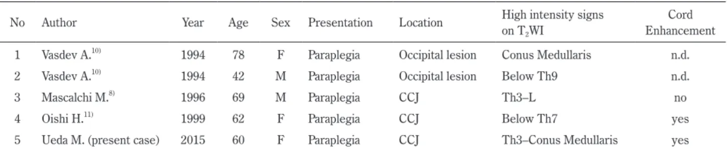
Table 1 Summary of cases of intracranial DAVF with thoracolumbar myelopathy. No Author Year Age Sex Presentation Location High intensity signs
on T2WI
Cord Enhancement 1 Vasdev A.10) 1994 78 F Paraplegia Occipital lesion Conus Medullaris n.d.
2 Vasdev A.10) 1994 42 M Paraplegia Occipital lesion Below Th9 n.d.
3 Mascalchi M.8) 1996 69 M Paraplegia CCJ Th3–L no
4 Oishi H.11) 1999 62 F Paraplegia CCJ Below Th7 yes
5 Ueda M. (present case) 2015 60 F Paraplegia CCJ Th3–Conus Medullaris yes M: male, F: femal, CCJ: craniocervical junction, n.d.: not described.
Masamichi Ueda, M.D.
1), Miki Ueda, M.D.
1)3), Yuko Takeuchi, M.D., Ph.D.
1),
Jun Ochiai, M.D., Ph.D.
1), Chiyuki Mabuchi, M.D., Ph.D.
1)and Shinnosuke Hattori, M.D., Ph.D.
2) 1)Department of Neurology, Nagoya Ekisaikai Hospital2)Department of Nuerosurgery, Nagoya Ekisaikai Hospital 3)Present Address: Department of Neurology, Nagoya University Hospital
A 60-year-old woman had transient weakness of the legs and urinary retention for six weeks. She presented with a
gait disorder and was admitted to the hospital. She showed symptoms of paraplegia, tingling in the lower extremities,
dysuria. She underwent an MRI, and T
2-weighted images showed an enlargement of the thoracolumbar spinal cord and
high intensity signal from Th3 to the medullary cone, and a contrast-enhanced T
1-weighted image showed abnormal
vessels anterior to the spine cord. Cervical and spinal angiography documented a dural arteriovenous fistula at the
craniocervical junction that was fed by the right vertebral artery and the right ascending pharyngeal arteries and drained
into the perimedullary veins. Surgical therapy improved her symptoms and MRI images. Craniocervical junction DAVF
with thoracic-medullary cones lesion is rare.
(Rinsho Shinkeigaku (Clin Neurol) 2016;56:37-42)
Key words: dural arteriovenous fistula at the craniocervical junction, myelopathy, arteriovenous fistula ligation, vertebral