• 検索結果がありません。

Influences of these procedures on the upper respiratory tract have also been widely studied.

N/A
N/A
Protected

Academic year: 2021

シェア "Influences of these procedures on the upper respiratory tract have also been widely studied."

Copied!
6
0
0

読み込み中.... (全文を見る)

全文

(1)

INTRODUCTION

Mandibular prognathism is among the most common types of jaw deformity and the degree of deformity and the severity level varies greatly from case to case.

Surgical procedures to correct mandibular prog- nathism are generally determined based on the de- gree of mandibular protrusion and maxillary retru- sion. Treatment options include the one jaw approach by mandibular sagittal split ramus osteotomy alone or the two jaw approach by a combination of mandibular setback and maxillary advancement with Le Fort I os- teotomy. Pre- and postoperative changes in facial es- thetics and occlusion, and postoperative skeletal sta-

bility have been investigated extensively.

1, 2

Influences of these procedures on the upper respiratory tract have also been widely studied.

3−6

However, few re- ports have addressed long-term changes in the pha- ryngeal airway space of patients treated by these pro- cedures. The purpose of this study was to investigate how different surgical procedures affect pharyngeal airway changes through a comparative analysis of skeletal mandibular prognathism patients who were treated by the one jaw and two jaw approaches. Pre- and postoperative measurements of the dentofacial skeleton, pharyngeal airway space and areas that af- fect pharyngeal airway space were used as the pa- rameters for measurements and comparison.

Effects of different surgical procedures on the pharyngeal space with mandibular prognathism

Yutaka Yamada and Naoyuki Matsumoto

Department of Orthodontics, Osaka Dental University, 8-1 Kuzuhahanazono-cho, Hirakata-shi, Osaka 573-1121, Japan

We investigated how different surgical procedures affect pharyngeal airway changes through a comparative analysis of skeletal mandibular prognathism patients treated by sag- ittal split ramus osteotomy surgery (SSRO) with and without Le Fort I osteotomy surgery.

Twenty patients who were diagnosed as having skeletal mandibular prognathism were di- vided into two groups : 10 patients who underwent mandibular setback surgery using SSRO (SSRO group) and 10 who underwent two-jaw surgery using Le Fort I osteotomy and SSRO surgery (Two-jaw group). To compare the jaw relationship, the position of the hyoid bone and pharyngeal airway morphology were examined in each group using lateral cephalometric ra- diographs that were taken before surgery (T0) and after more than one year and six months post-surgery (T1). The surgical changes between T0 to T1 were calculated and statistically compared for the two groups. The result, tongue position did not appear to change signifi- cantly between the two groups. The palatal pharyngeal space increased significantly in the Two-jaw group, while there was no significant difference in the SSRO group. The superior posterior and middle pharyngeal space decreased significantly in the SSRO group, while there was no significant difference in the Two-jaw group. All changes showed significant dif- ference between the two groups. In contrast, the epiglottic pharyngeal space decreased in both groups. The ratio was bigger in the SSRO group than in the Two-jaw group. We con- cluded that the surgical method influences the pharyngeal airway space for patients treated only with SSRO and for those who recieved two-jaw surgery. (J Osaka Dent Univ 2015 ; 49 : 143−148)

Key words : Pharyngeal space ; Orthodontic treatment ; Skeletal mandibular prognathism

(2)

MATERIALS AND METHODS Subjects

We selected 20 female patients who had been diag- nosed with skeletal mandibular prognathism and who had received orthognathic surgery at Osaka Dental University. Ten had achieved a mandibular setback of over 10 mm with sagittal split ramus osteotomy (SSRO group). Ten other patients had achieved a mandibular setback and maxillary advancement of over 10 mm in total by surgery using Le Fort I osteot- omy and SSRO surgery (Two-jaw group). Both groups were followed up for at least one year and six months after the surgery. The data for this study was acquired from the cephalograms taken immediately before surgery (T0) and after more than one year and six months post-surgery (T1). The mean age of the patients at T0 was 21 years and 3 months (SSRO group) and 21 years and 8 months (Two-jaw group).

The mean mandibular setback was 11.2 mm for the SSRO group. For the Two-jaw group the mean mandi- bular setback was 7.1 mm and the mean maxillary ad- vancement was 4.5 mm. The Committee of Medical Ethics, Osaka Dental University approved the proto- col of this study (No. 110801).

Methods

We analyzed the cephalograms at T0 and T1 in the two groups. The reference points and planes used in assessing the craniofacial skeleton, pharyngeal air- way morphology and hyoid position were defined based on the reports by Kouno et al.

6

and Mochida et al.,

7

and the following cephalometric measurements of the craniofacial skeleton were performed (Fig. 1) :

Table 1 Comparison of measurements at T0 and T1 for each group

SSRO group Two-jaw group

T0 T1 t-test T0 T1 t-test

∠SNA (° )

∠SNB (° )

∠ANB (° )

∠MP (° )

∠OPT-SN (° )

∠HSN (° )

∠SP (° ) S-H (mm) C3-H (mm) D1 (mm) PPS (mm) SPPS (mm) MPS (mm) IPS (mm) EPS (mm)

78.2±3.0 85.1±3.1

−6.2±2.2 31.2±4.5 98.8±7.5 88.2±2.8 56.8±6.0 117.1±8.7 39.1±5.5 16.8±3.5 25.5±4.3 14.8±2.8 13.3±2.1 17.2±4.9 17.1±3.8

78.3±2.8 77.6±3.8 0.5±1.8 33.5±4.7 99.0±7.2 91.0±4.2 53.2±7.3 119.2±7.8 38.8±7.2 11.8±3.8 25.7±3.8 12.1±1.8 10.8±3.7 13.1±4.2 13.2±4.5

NS

*

*

* NS

*

* NS NS

* NS

*

*

*

*

76.4±2.8 83.1±5.1

−6.8±3.2 30.8±8.5 98.5±8.5 89.1±6.8 57.1±6.0 118.0±7.7 39.4±3.5 18.8±4.5 24.2±3.5 13.1±2.2 13.2±2.5 16.6±3.8 17.7±2.8

79.3±3.8 76.8±4.8 2.2±2.3 31.7±7.7 99.0±8.2 90.4±6.2 52.3±7.1 119.1±7.4 38.9±3.2 14.8±2.8 28.4±2.1 13.3±2.9 13.3±2.7 14.2±3.2 16.1±4.0

*

*

*

* NS NS

*

* NS

*

* NS NS

*

* Mean±SD, NS : Not significant, *p<0.01, n=10 for each group.

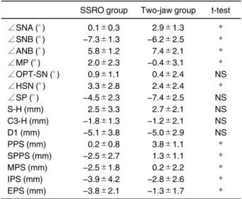
Table 2 Comparison of the differences from T0 to T1 between two groups

SSRO group Two-jaw group t-test

∠SNA (° )

∠SNB (° )

∠ANB (° )

∠MP (° )

∠OPT-SN (° )

∠HSN (° )

∠SP (° ) S-H (mm) C3-H (mm) D1 (mm) PPS (mm) SPPS (mm) MPS (mm) IPS (mm) EPS (mm)

0.1±0.3

−7.3±1.3 5.8±1.2 2.0±2.3 0.9±1.1 3.3±2.8

−4.5±2.3 2.5±3.3

−1.8±1.3

−5.1±3.8 0.2±0.8

−2.5±2.7

−2.5±1.8

−3.9±4.2

−3.8±2.1

2.9±1.3

−6.2±2.5 7.4±2.1

−0.4±3.1 0.4±2.4 2.4±2.4

−7.4±2.5 2.7±2.1

−1.2±2.1

−5.0±2.9 3.8±1.1 1.3±1.1 0.2±2.2

−2.8±2.6

−1.3±1.7

*

*

*

* NS

* NS NS NS NS

*

*

*

*

*

*p<0.01, n=10 for each group.

(3)

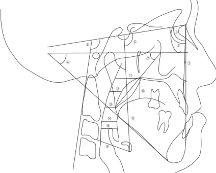
∠SNA, ∠SNB, ∠ANB, and mandibular plane angle (∠MP). The following measurements of angles and distances that may affect the pharyngeal airway were performed, ∠OPT-SN : Angle formed by the SN plane and the line through the most superior point and the most inferior point of the posterior part of the cervi- cal vertebra, ∠HSN : Angle formed by the SN plane and the line through S and the most inferior point (H) of the hyoid bone, ∠SP : Angle formed by the FH plane and the line through PNS and the most inferior point of the velum (PSP), S-H : Shortest distance from S to the He, C3-H : Distance from the most infe- rior point of the anterior part of the 3

rd

cervical vertebra to H, D1 : Distance from PNS projected on the line perpendicular to the FH plane to the dorsum of the tongue, Palatal pharyngeal space (PPS) : Distance between the posterior wall of the pharynx and PNS on the line parallel to the FH plane through PNS, Supe- rior posterior pharyngeal space (SPPS) : Distance between the posterior wall of the pharynx and the ve- lum on a line parallel to the FH plane through PNS and the midpoint between PNS and PSP, Middle pharyn- geal space (MPS) : Distance between the posterior

wall of the pharynx and PSP on a line parallel to the FH plane through the PSP, Inferior pharyngeal space (IPS) : Distance between the posterior wall of the pharynx and the tongue on a line parallel to the FH plane through the most inferior point of the anterior part of the second cervical vertebra, Epiglottic pharyn- geal space (EPS) : Distance between the posterior wall of the pharynx and the tongue on the line parallel to the FH plane through the epiglottis. Measurements for both groups at T0 and T1 were analyzed and com- pared, and the statistical significance of the differ- ences was determined using the t-test. In addition, the changes between T0 and T1 for the two groups were analyzed and compared, and the statistical signifi- cance was determined using the unpaired t-test.

RESULTS

Measurements of the dentofacial skeleton

Between T0 and T1, the SSRO group showed a sig- nificant decrease in ∠SNB and a significant increase in ∠ANB and ∠MP. In the Two-jaw group, ∠SNA,

∠ANB and ∠MP showed a significant increase, whereas ∠SNB showed a significant decrease.

Fig. 1 Measurements on the lateral cephalogram.

①∠SNA (° ), ②∠SNB (° ), ③∠ANB (° ), ④∠MP (° ), ⑤∠OPT-SN (° ),

⑥∠HSN (° ), ⑦∠SP (° ), ⑧S-H (mm), ⑨C3-H (mm), ⑩D1 (mm),

⑪PPS (mm), ⑫SPPS (mm), ⑬MPS (mm), ⑭IPS (mm), ⑮EPS (mm).

(4)

When comparing the amount of changes between T0 and T1 for the two groups, the changes in ∠SNA and

∠ANB for the Two-jaw group were significantly greater than for the SSRO group and the changes in

∠SNB and ∠MP were significantly smaller for the Two-jaw group.

Measurement of the areas that affect the pharyn- geal airway

Between T0 and T1, the SSRO group showed a sig- nificant increase in ∠HSN and a significant decrease in ∠SP and D1. In the Two-jaw group, the value S-H showed a significant increase, whereas ∠SP and D1 showed a significant decrease. When comparing the amount of changes between T0 and T1 for the two groups, the change in ∠HSN of the Two-jaw group was significantly smaller than that in the SSRO group.

Measurement of pharyngeal airway space

In the SSRO group, SPPS, MPS, IPS and EPS de- creased significantly from T0 and T1. In the Two-jaw group, PPS increased significantly, whereas IPS and EPS showed a significant decrease. When comparing the amount of changes between T0 and T1 for the two groups, significant differences were observed in every parameter.

DISSCUSSION Study method

When treating patients with skeletal mandibular prog- nathism, it is important to achieve occlusal and es- thetic improvements while minimizing the impacts of mandibular setback on the morphology and function of the pharyngeal airway. We investigated how and where the morphology and function of the pharyngeal airway are affected by SSRO with mandibular setback alone, and by two-jaw surgery with a combination of mandibular setback and maxillary advancement. In- cluded in this study were patients with severe skeletal mandibular prognathism requiring a relative positional bony change greater than 10 mm. A significant in- crease in ∠ANB was observed in both groups, indi- cating that surgery had produced a considerable im- provement in the relative position of maxilla and man- dible. It has been reported that the head position on

the lateral cephalograms influences the pharyngeal airway.

8

After mandibular setback surgery, the cervi- cal spine undergoes compensatory changes to se- cure pharyngeal airway space that has been nar- rowed. Kouno

6

investigated changes in the pharyn- geal airway space in relation to the changes in the cer- vical spine at a natural head position. On the other hand, a report has suggested the need of evaluating pharyngeal airway measurements by excluding the pharyngeal airway changes caused by cervical spine movements.

4

In this study, conventional imaging was performed using the FH plane as reference, and the measurements at T0 and T1 were compared. Neither group showed any significant difference in ∠OPT- SN, indicating that the head position is unlikely to have a significant impact on the pharyngeal airway space.

Changes in pharyngeal airway morphology Kawakami et al.

9

investigated the pharyngeal airway space after mandibular setback surgery and reported a decrease of 2.2 mm in IPS, which is a significant fac- tor in this study, one year after the surgery. Achilleos et al.

4

reported a decrease in PPS and no change in EPS 3 years after the surgery. Both PPS and EPS are the significance factors in this study. Although slight differences are observed in the results due to different study protocols such as the postoperative follow-up period, the method of cephalograms and the amount of mandibular setback, there seems to be a basic con- sensus that pharyngeal airway space decreases with mandibular setback surgery.

In this study, a significant decrease in SPPS and

MPS, which are in the posterior area of the soft pal-

ate, and IPS and EPS, which are in the lower area of

the oropharyngeal airway, was confirmed in the

SSRO group. For Le Fort I osteotomy, on the other

hand, Kouno et al.

6

reported a significant increase in

PPS and a significant decrease in IPS and EPS. Sakai

et al.

10

also reported a decrease of EPS, a significant

factor in this study, at follow-ups of over 2 years after

surgery. Although the Two-jaw group in this study

showed no significant increase in SPPS and MPS,

there was a decrease in IPS and EPS and an increase

of PPS. Based on the significant difference observed

(5)

in the amount of change in every parameter, the post- operative pharyngeal airway space narrowing in the Two-jaw group seems less than that in the SSRO group, which is consistent with the findings of Kouno et al.

Changes of hyoid bone position

It has long been thought that it is extremely difficult to accurately capture the hyoid bone position because it easily changes with changes in the head/neck pos- ture when cephalograms are expressed.

11, 12

It is be- lieved that a substantial margin of error should be ac- cepted when determining the position of hyoid bone or tongue on lateral cephalograms.

13, 14

Analyzing the hyoid bone position on lateral cephalograms by defin- ing various measurement parameters is believed diffi- cult due to the considerable variation and poor repro- ducibility of the measured values. With this in mind, Nagai et al.

15

investigated how the head position influ- ences hyoid bone position and suggested parameters that can be used to determine hyoid bone position that are not affected by head position.

According to the report by Nagai et al.,

15

the hyoid bone position in this study was evaluated using S-H for vertical and ∠HSN and C3-H for horizontal meas- urements. Tongue movements resulting from posi- tional changes in the hyoid bone have been reported as a potential cause of pharyngeal airway narrow-

ing.

9, 10

Kawakami et al.

11

indicated that the hyoid bone

is pressed inferiorly and the tongue is pushed posteri- orly after SSRO, resulting in narrowing of the pharyn- geal airway space. Kouno et al.

6

reported that al- though the hyoid bone after surgery in the SSRO group was posterior to its position in the Two-jaw group, there was no changes in its vertical relation- ship. They concluded that the pharyngeal airway space narrowing was caused by the tongue being compressed towards the pharyngeal airway. Sakai et al.

10

reported that the hyoid bone after SSRO was slightly posterior to its preoperative position. For the Le Fort I osteotomy, on the other hand, they reported that the postoperative hyoid bone was located close to the preoperative position. They suggested that ad- aptation of the upper respiratory tract to the new post- operative environment created by the soft palate and

nasal cavity changes resulted from the maxillary ad- vancement. In our study, although slight shifts in the hyoid bone toward the inferior-posterior direction were observed in both groups, the changes were not statistically significant. There was no significant differ- ence of the changes between the SSRO group and the Two-jaw group.

From these observations, the difference in the amount of mandibular setback is thought to be a factor contributing to the positional changes in the hyoid bone. The parameters used in this study to measure the hyoid bone position and the soft palate inclination were D1 and ∠SP. Reports have indicated that changes in the soft palate morphology after mandibu- lar setback surgery lead to a decrease in oral cavity volume and compression of the soft palate by the tongue being pressed in a posterior direction. This pushes up the posterior region of the soft palate, lead- ing to a narrowing of the pharyngeal airway, which is located in the posterior of the soft palate.

16−18

In contrast, it has been reported that maxillary ad- vancement leads to an increase in oropharyngeal air- way space as a result of the anterior shift of the soft plate. Based on the findings of this study such as the significant decreases in D1, ∠SP, SPPS and MPS in the SSRO group, we think that the tongue, which was upthrusted by the decrease in the oral cavity volume, pushed up the posterior area of the soft palate and re- sulted in a decrease in the oropharyngeal airway space. In the Two-jaw group there was a decrease in the D1 and ∠SP, no significant change in SPPS or MPS, and a significant increase in PPS. Therefore, an anterior shift of the soft palate caused by the maxillary advancement partially contributed to the prevention of oropharyngeal airway narrowing.

Skeletal relapse after correction of mandibular prognathism

Reports have investigated how the amount of mandi-

bular setback causes skeletal relapse after surgical

correction of mandibular prognathisum.

2, 19−23

No in-

vestigation of skeletal relapse was performed in this

study. However, the subjects in this study all had se-

vere mandibular prognathism requiring mandibular

setback of over 10 mm, and the amount of change in

(6)

the pharyngeal airway space was greater in the SSRO group than in the Two-jaw group. Therefore, reduced pharyngeal airway space may impact post- operative skeletal stability. To maintain long-term sta- bility, it is necessary to train the perioral muscles and tongue to facilitate their adaptation to the new envi- ronment.

REFERENCES

1. Yamada A, Tamaoki S, Hisanaga Y, Ishikawa H. Relationship between hard tissue and soft tissue changes in surgical ortho- dontic treatment. Orthod Waves Jpn Ed 2004 ; 63 : 85−94.

(Japanese)

2. Kuroda T, Shimizu T, Higuchi K, Mita K, Toneki T, Suzuki K, Tsuruki T, Ichinokawa Y, Nomura M, Yamaguchi H. Longitudi- nal observation of postoperative stability in correction of man- dibular prognathism–Comparison of sagittal split ramus os- teotomy and two-jaw surgery–. J Tokyo Dent Coll Soc 2002 ; 102 : 583−596. (Japanese)

3. Liukkonen M, Vahatalo K, Peltomaki T, Tiekso J, Happonen R. Effect of mandibular setback surgery on the posterior airway size. Int J Adult Orthodon Orthognath Sug 2002 ; 17 : 41−

46.

4. Achilleous S, Krogstad O, Lyberg T. Surgical mandibular set- back and changes in uvuloglossopharyngeal and head pos- ture : a short- and long- term cephalometric study in males.

Eur J Orthod 2000 ; 22 : 383−394.

5. Gu GM, Nagata J, Sato M, Anraku Y, Nakamaura K, Kuroe K, Ito G. Hyoid position, pharyngeal air way and head position in relation to relapse after the mandibular setback in skeletal Class III. Clin Orthod Res 2000 ; 3 : 67−76.

6. Kouno K, Igawa K, Takamori K, Kurokawa H, Sakoda S. Mor- phological changes in pharyngeal airway space after mandibu- lar setback surgery with and without Le Fort I osteotomy in skeletal Class III patients. Jpn J Jaw Deform 2006 ; 16 : 184

−189. (Japanese)

7. Mochida M. Effects of maxillary distraction osteogenesis on the upper-airway size and nasal resistance in subjects with cleft and palate. Orthod Craniofacial Res 2004 ; 7 : 189−

197.

8. Muto T, Takeda S, Kanazawa M, Yamazaki A, Fujiwara Y, Mi- zoguchi I. The effect of head posture on the pharyngeal airway space (PAS). Int J Oral Maxillofac Surg 2002 ; 31 : 579−583.

9. Kawakami M, Yamamoto Y, Fujimoto M, Ohgi K, Inoue M, Kirita T. Changes in tongue and hyoid positions, and posterior airway space following mandibular setback surgery. J Cra- niomaxillofac Surg 2005 ; 33 : 107−110.

10. Sakai K, Shimazaki K, Kokai S, Fukuyama E, Ono T. Effects

of different surgical procedures on the upper-airway dimension in subjects with mandibular prognathism. Jpn J Jaw Deform 2012 ; 22 : 239−243. (Japanese)

11. King EW. A roentgenographic study of pharyngeal growth. An- gle Orthod 1952 ; 22 : 23−37.

12. Stepovich ML. A cephalometric positional study of the hyoid bone. Am J Orthod 1965 ; 51 : 882−900.

13. Matsumoto H. X-ray TV cineramatographical observation of positioned changes in the hyoid bone and the base of epiglottis in swallowing, maximum opening and closing of the mouth, and tongue movement. J Tokyo Dent Coll Soc 1976 ; 75 : 1713−1770. (Japanese)

14. Torikai Y, Morita S, Hoshi T, Hiruma Y, Hanada K, Hayashi T.

The positional relationship between the mandibule and hyoid bone in patients with mandibular protrusion. Jpn J Jaw Deform 2001 ; 11 : 1−10. (Japanese)

15. Nagai M, Kudo A, Matsuno T, Yokoyama M, Manabe J, Hase- gawa S, Nakamura S. A study of hyoid bone position and air- way accompanied with influence of head posture. J Jpn Orthod Soc 1989 ; 48 : 214−225. (Japanese)

16. Saitoh K. Long-term changes in pharyngeal airway morphol- ogy after mandibular setback surgery. Am J Orthod Dentfac Orthop 2004 ; 125 : 556−561.

17. Turnbull N, Battagel J. The effects of orthognathic surgery on pharyngeal airway dimensions and quality of sleep. J Orthod 2000 ; 27 : 235−247.

18. Hochban W, Schurmann R, Brandenburg U, Conradt B. Man- dibular setback for surgical correction of mandibular hyperpla- sia–does it provoke sleep-related breathing disorders? Int J Oral Maxillofac Surg 1996 ; 25 : 333−338.

19. Kobayashi T, Kanoh H, Honda K, Shingaki S, Yamada K, Saito I, Hayashi T, Saito C. Temporomandibular joint symptoms and mandibular stability after orthognathic surgery in patients with mandibular retrognathism. Jpn J Jaw Deform 2001 ; 12 : 9−

14. (Japanese)

20. Soya T, Kanda N, Sugawara J, Kawamura H, Takahashi Y, Hayashi S. Evaluation of postoperative stability in skeletal class Ⅲ patients corrected by sagittal splitting osteotomy. J Jpn Orthod Soc 1983 ; 42 : 288−296. (Japanese)

21. Iwami-Morimoto Y, Yamada T, Tanne K. Long-term changes in craniofacial morphology and occlusal stability in mandibular prognathism patients treated with sagittal splitting ramus os- teotomy. Jpn J Jaw Deform 1996 ; 6 : 21−31. (Japanese) 22. Tamura H, Noma H, Takaki T. Cephalometric evaluation of

stability of mandibular position after treatment of mandibular prognathism by sagittal splitting ramus osteotomy. Jpn J Jaw Deform 1998 ; 8 : 1−17. (Japanese)

23. Mobarak KA, Krogstad O, Espeland L, Lyberg T. Long-term

stability of mandibular setback surgery : A follow-up of 80 bi-

lateral sagittal split osteotomy patients. Int J Adult Orthodon

Orthognath Surg 2000 ; 15 : 83−95.

Table 2 Comparison of the differences from T0 to T1 between two groups
Fig. 1 Measurements on the lateral cephalogram.

参照

関連したドキュメント

Provided that the reduction of the time interval leads to incomparableness of normalized bubble-size distributions and does not change the comparable distributions in terms of

Asymptotic properties of solutions of homogeneous equations with constant coef- ficients and unbounded delays have been investigated also in [3]... The statement can be proved by

Eskandani, “Stability of a mixed additive and cubic functional equation in quasi- Banach spaces,” Journal of Mathematical Analysis and Applications, vol.. Eshaghi Gordji, “Stability

The purpose of this review article is to present some of the recent methods for providing such series in closed form with applications to: i the summation of Kapteyn series

This approach is not limited to classical solutions of the characteristic system of ordinary differential equations, but can be extended to more general solution concepts in ODE

As an application, we present in section 4 a new result of existence of periodic solutions to such FDI that is a continuation of our recent work on periodic solutions for

As a result, we are able to obtain the existence of nontrival solutions of the elliptic problem with the critical nonlinear term on an unbounded domain by getting rid of

Using a method developed by Ambrosetti et al [1, 2] we prove the existence of weak non trivial solutions to fourth-order elliptic equations with singularities and with critical