Acta Med. Nagasaki 36: 176-184
Lectin Immunohistochemistry in Human Non-Malignant
and Malignant Gallbladder Tissuses
Faisal MUAZZAM 2, Takayoshi TODA 1, Shuji TOMITA 2 Shigeru DEGUCHI 2, Yoshihiro MUTO 2
and Iwao ONO 3
1. Department of Clinical Laboratory Medicine, School of Medicine, University of the Ryukyus, Okinawa
2. Department of Surgery, School of Medicine, University of the Ryukyus, Okinawa 3. Pathology Section, Nakadori Hospital, Akita
Received for publication, May 11, 1991
SUMMARY : Changes in the lectin binding pattern in non-malignant and malignant gallbladder tissues were examined using the following eight types of carbohydrate binding lectins : Ulex europaeus-1 (UEA-1), Arachis hypogaea (PNA), Griffonia simplicifolia (GS-1), Glycine maximum (SBA), Bauhinia purpurea (BPA), Dolichos biflorus (DBA), Canavalia ensiformis (Con-A), and Triticum vulgare (WGA). We used a total of 109 tissues including 31 normal tissues, 25 metaplasias, and 53 carcinomas. Lectin staining pattern was evaluated using the Hamada's crieria of the following four types : apical type, cytoplasmic type with polarity, cytoplasmic type without polarity, and stromal type. Normal cases showed apical type and cytoplasmic type with polarity, while carcinoma cases revealed cytoplasmic type with or without polarity. In carcinoma cases, GS-I and DBA lectins showed higher immunohistochemical positive rate and more frequent cytoplasmic type with polarity pattern of immunohistochemical localization than the other types of lectins.
These results suggest that the GS-I and DBA are the most reliable lectin marker for malignant transformation of the gallbladder tissues.
Key words : Lectin, immunohistochemistry, gallbladder carcinoma.
INTRODUCTION
Lectins sre sugar-binding and membrane bound proteins that are of bacterial, animal and plant origin, and glycosilation of protein in- volves sequential action of numerous enzyme to regulate structural and functional proper- ties of various cytoplasmic organelles" 2'. The usefulness of lectins as hitochemical tools is
based on their ability to bind specific mono- saccharides or oligosaccharides in complex car- bohydrates'). Many studies have been carried out regarding the changes of glycoprotein
structure in normal and neoplastic tissues of various organs such as the colon" 5. 6, 7), stom- ach", kidney- 10', prostate'and lung"). This study was conducted to evaluate the significance of lectin expression in non-neoplastic and neoplastic lesions of the gallbladder examining their qualitative and quantitative differences in lectin expressions.
MATERIALS AND METHODS
Tissues specimens were obtained from a total
of 109 cholecystectomized and autopsied cases
including 31 normal control (N), 25 metaplasia
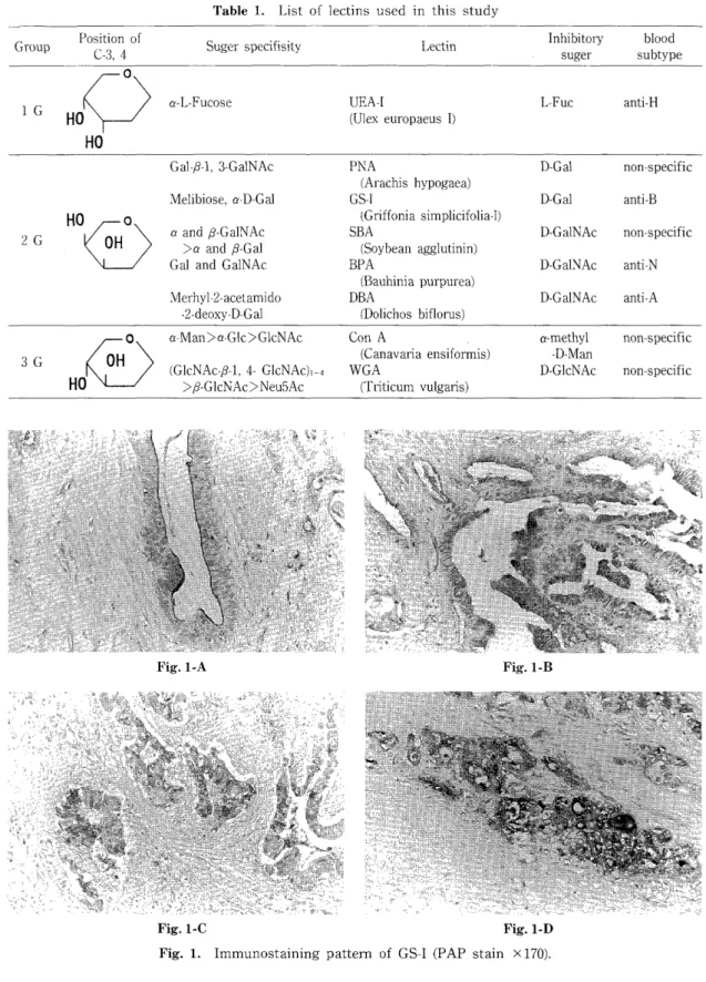
Table 1. List of lectins used in this study
Group Position of C-3 , 4 Suger specifisity Lectin Inhibitory suger subtype blood 0
1 G HO a-L-Fucose UEA-I (Ulex europaeus 1) L-Fuc anti-H
HO
Gal-/3-1, 3-GaINAc PNA D-Gal non-specific
(Arachis hypogaea)
Melibiose, a-D-Gal GS-1 D-Gal anti-B
HO O (Griffonia simplicifolia-1)
2 G OH a and 13-Ga1NAc > a and P-Gal SBA (Soybean agglutinin) D-Ga1NAc non-specific
Gal and Ga1NAc BPA D-Ga1NAc anti-N
(Bauhinia purpurea)
Merhyl-2-acetamido DBA D-Ga1NAc anti-A
-2-deoxy-D-Gal (Dolichos biflorus)
O a-Man>a-Glc>GlcNAc Con A a-methyl non-specific
3 G OH (G1 (Canavaria ensiformis) -D-Man
cNAc-(3-1, 4- GIcNAc),-9 WGA D-G1cNAc non-specific
HO >#-GIcNAc>Neu5Ac (Triticum vulgaris)
Fig. 1-A Fig. 1-B
Fig. 1-C Fig. 1-D
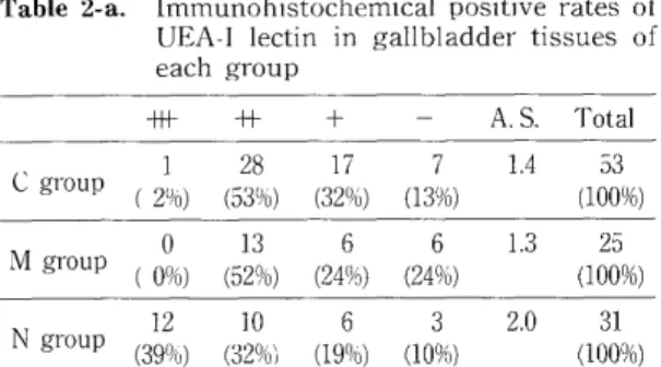
Fig. 1. Immunostaining pattern of GS-I (PAP stain x 170).
(M), and 53 carcinoma (C). All surgically re- moved tissues were fixed in 3% formaldehyde solutoin, washed, dehydrated and paraffin em- bedded. Nine 4 micro meter sections were cut from each blocks (Eight for lectin binding and one for routine hematoxylin and eosin stain- ing), placed on acetone cleaned slides, deparaf- finated in xylene and passed from absolute ethanol into phosphate buffered saline (PBS) pH 7.2. Lectin binding was performed using the method of Hsu et a113'. with a slight modifica- tion. Endogenous peroxidase were inhibited by treatment with 1% hydrogen peroxide (H202) in PBS for 5 Minutes. Sections were then repeatedly washed in PBS for three times, 5 minutes each. Then the sections were incubat- ed in biotinated lectin at the concentration of 0.004mg/ml, dissolved in 1%BSA-NaCI (To min- imize background stains), in a moist chamber for overnight (around 12 hours) at 4°C. Table 1 shows the sugar specificity and sugar recognizing eight biotinated lectins including UEA-I, PNA, GS-I, SBA, BPA, DBA, Con-A, and WGA (E. Y. Labs. INC. San Mateo Cal.). After the overnight incubation the sections were washed three times in PBS (Five minutes each wash) and then were incubated in avidin peroxidase in a moist chamber for 30 minutes in room temperature. Peroxidase activity of the bound lectin conjugates were shown by treating with 3,3 diaminobenzidine (DAB) for a maxi- mum period of two minutes. Sections were then washed, counterstained lightly with hema- toxylin, dehydrated and mounted. The inten- sity of immunoreaction of lectins was evaluated by light microscope at the magnification of 100 times. Irrespective of staining patterns, the following 4 gradings of intensity of im- munoreaction were adopted : More than 50% of positive tumor cells, (+++) ; 25% to 50% of positive tumor cells, (++) ; less than 25% of positive tumor cells, (+) ; no positive cells (-).
According to these criteria, average scores of immunohistochemical positive rates were quantified as + =1, + + =2, and +++ =3.
For the evaluation of immunohistochemical staining pattern of CA19-9, Hamada's criteria 14) was applied as follows : 1) Only luminal cyto- plasmic surface is positively stained (Apical type), 2) Supranuclear portion of the cytoplasm
is intensely stained (Cytoplasmic type, with polarity), 3) Total area of the cytoplasm is positively stained (Cytoplasmic type, without polarity), 4) Surrounding stromal cells are positively stained (Stromal type) (Figure 1).
RESULTS
Ulex europaeus-1 (UEA-1) : In normal mucosa, UEA-1 reacted mostly in the apical portion of the cytoplasm and occasionally in the whole
cytoplasm with polarity. However, in carci- noma, UEA-1 was seen in the whole cytoplasm without polarity. Table 2 repesents immuno- histochemical positive rates (-a) and localiza- tion (-b) of UEA-1 lectin in gallbladder tissues of each group. The immunohistochemical negative rate of UEA-I was rather higher in N group than in M and C groups. The sup-
ranuclear area of the cytoplasm of normal epithelium was constantly stained by the UEA-I (53%). Apical pattern was predominantly seen in both C (50%) and M (47%) groups.
Arachis hypogaea (PNA) : Immunohistochem- ical positive rates of PNA was similar in all groups (Table 3-a). In N group, PNA ataining was limited to the supranuclear region of the
cytoplasm. In M group, PNA staining was noted in the apical (30%) and supranuclear area (65%).
Entire cytoplasm was stained in 20 cases in C group (41%) (Table 3-b).
Griffonia simplicifolia (GS-1): Negative rates of GS-I lectin was 41%, 20%, 74% in C, M, and N groups, respectively (Table 4-a). The average score of positive immunostaining was the lowest in N group. Table 4-b presents staining pattern in each group. More than half of the carcinoma cases showed cytoplasmic pattern without po- larity.
Glycine max (SBA) : SBA showed more in- creased affinity for mucin of N and M groups than C group. SBA was negatively stained in 10 out of 53 cases (19%) in C group (Table 5-a).
The apical luminal border of columnar cells and glycocalyx were extensively stained in N and C groups. SBA stained the cytoplasm of adeno- carcinoma in 18 out of 43 cases of C group (34%) (Table 5-b).
Bauhinia purpurea (BPA) : The positive rate
was generally lower in all three groups com-
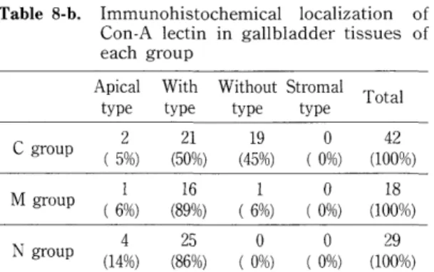
Table 2-a. Immunohistochemical positive rates of UEA-I lectin in gallbladder tissues of
each group
-+- -H- + - A. S. Total
1 28 17 7 1.4 53 C gro
up (21/6) (531/6) (32%) (13%) (100%) M group (0%) 0 (52%) (24%) (24%) 13 6 6 1.3 (100%) 25 N group (390 12 10 6 3 2.0 31
0) (32%) (19°ro) (10%) (100%) A. S. ; Average score of positive immunostaining Table 2-b. Immunohistochemical localization of
UEA-I lectin in gallbladder tissues of
each group
Apical With Without Stromal Total type type type type
23 21 2 0 46
C
group (50%) (46%) (4%) (0%) (100%) M group (47%) 9 (530 10 0 0 19
0) (0%) (0°o) (100%) N group (21%) 6 (79°l 22 0 0 28
o) (0%) (0%) (100%) With type ; Cytoplasmic type, with polarity
Without type ; Cytoplasmic type, without polarity Table 3-a. Immunohistochemical positive rates of
PNA lectin in gallbladder tissues of
each group
-H+ -H- + - A. S. Total
C group P (19%) (43%) (30%) (8%) 10 23 16 4 1.7 (100%) 53 M group ( 8%) (48%) (32%) (12%) 2 12 8 3 1.5 (100%) 25 N group (26%) (45%) (13%) (16%) 8 14 4 5 1.8 (100%) 31
A. S. ; Average score of positive immunostaining Table 3-b. Immunohistochemical localization of
PNA lectin in gallbladder tissues of
each group
Apical With Without Stromal T otal t
ype type type type
C group ( 6%) 3 ( 53%) 26 (39%) 19 (2%) 1 (100%) 49 M group (30%) 7 ( 65%) 15 (5%) 1 (0%) 0 (100%) 23 N group (0%) 0 (100%) 26 (0%) 0 (0%) 0 (100%) 26
With type ; Cytoplasmic type, with polarity Without type ; Cytoplasmic type, without polarity
Table 4-a. Immunohistochemical positive rates of GS-I lectin in gallbladder tissues of each
group
+H- +- + - A. S. Total
C group (25%) (9%) 13 5 (25%) (41%) 13 22 1.2 (100%) 53 M group (32%) (24%) (24%) (20%) 8 6 6 5 1.7 (100%) 25 N group ( 0%) ( 6%) (20%) (74°l 0 2 6 23 0.3 31
o) (100%)
A. S. ; Average score of positive immunostaining Table 4-b. Immunohistochemical localization of
GS-I lectin in gallbladder tissues of each
group
Apical With Without Stromal T otal t ype type type type
C group (0%) 0 (29%) 9 (58%) 18 (13%) 4 (100%) 31 M group (15%) 3 (80%) 16 (5%) 1 (0%) 0 (100%) 20 N group (75%) 6 (25%) 2 (0°/ 0 0 8 o) (0%) (100%) With type ; Cytoplasmic type, with polarity
Without type ; Cytoplasmic type, without polarity Table 5-a. Immunohistochemical positive rates of
SBA lectin in gallbladder tissues of each
group
-H+ -I+ + - A. S. Total
C group (13%) (45%) (23%) (19%) 7 24 12 10 1.5 (100%) 53 M group (68%) (24%) (0%) 17 6 0 (8%) 2 2.5 (100%) 25
N group (61%) (19%) (19%) (0%) 19 6 6 0 2.4 (100%) 31 A. S. ; Average score of positive immunostaining Table 5-b. Immunohistochemical localization of
SBA lectin in gallbladder tissues of each
group
Apical With Without Stromal T otal type type type type
C group (21%) 9 ( 42%) 18 (35%) 15 (2%) 1 (100%) 43 M group (0%) 0 (100%) 23 (0%) 0 (0%) 0 (100%) 23 N group (19%) 6 (81%) 25 (0%) 0 (0%) 0 (100%) 31
With type ; Cytoplasmic type, with polarity
Without tvne : Cvtoulasmic tvne. without polarity
Table 6-a. Immunohistochemical positive rates of BPA lectin in gallbladder tissues of
each group
+H- -H- + - A. S. Total
C group ( 0%) (10%) (28%) (62%) 0 5 15 33 0.5 (100%) 53 M group ( 8%) (20%) (20%) (52%) 2 5 5 13 0.8 (100%) 25 N group (0%) 0 (6%) 2 (23%) (71%) 7 22 0.4 (100%) 31
A. S. ; Average score of positive immunostaining Table 6-b. Immunohistochemical localization of
BPA lectin in gallbladder tissues of
each group
Apical With Without Stromal Total type type type type
C group (40%) 8 (35%) 7 (25%) 5 (0%) 0 (100%) 20 M group (58%) 7 (42%) 5 (0%) 0 (0%) 0 (100%) 12
N group (22%) (78%) (0%) (0%) (100%) With type ; Cytoplasmic type, with polarity
Without type ; Cytoplasmic type, without polarity Table 7-a. Immunohistochemical positive rates of
DBA lectin in gallbladder tissues of
each group
+H- ++ + - A. S. Total
C group (15%) (19%) (40%) (26%) 8 10 21 14 1.2 (100%) 53 M group (28%) (28%) (24%) (20%) 7 7 6 5 1.6 (100%) 25 N group (6%) 2 (19%) (26%) (48%) 6 8 15 0.8 (100%) 31
A. S. ; Average score of positive immunostaining Table 7-b. Immunohistochemical localization of
DBA lectin in gallbladder tissues of
each group
Apical With Without Stromal T otal t ype type type type
C group (15%) 6 (54%) 21 (21%) 8 (10%) 4 (100%) 39 M group (0%) 0 (90%) 18 (10%) 2 (0%) 0 (100%) 20
8 8 0 0 16
N group
(50%) (50%) (0%) (0%) (100%) With type ; Cytoplasmic type, with polarity
Without type ; Cytoplasmic type, without polarity
Table 8-a. Immunohistochemical positive rates of Con-A lectin in gallbladder tissues of
each group
+I+ +I + - A. S. Total
C group P (26%) (32%) (21%) (21%) 14 17 11 11 1.6 (100%) 53 M group (4%) 1 (32%) (36%) (28%) 8 9 7 1.1 (100%) 25 N group (6%) 2 (68%) (19%) (6%) 21 6 2 1.7 (100%) 31
A. S.; Average score of positive immunostaining Table 8-b. Immunohistochemical localization of
Con-A lectin in gallbladder tissues of
each group
Apical With Without Stromal T otal type t
ype type type
C group 2 21 19 0 42
( 5%) (50%) (45%) (0%) (100%) M group (6%) 1 (89%) 16 (6%) 1 (0%) 0 (100%) 18 N group (14%) 4 (86%) 25 ( 0%) 0 ( 0%) 0 (100%) 29
With type ; Cytoplasmic type, with polarity Without type : Cvtoplasmic type. without polarity
Table 9-a. Immunohistochemical positive rates of WGA lectin ii: gallbladder tissues of
each group
+F- * + - A. S. Total
C group (23%) (40%) (17%) (21%) 12 21 9 11 1.6 (100%) 53 M group (44%) (28%) (12%) (16%) 11 7 3 4 2.0 (100%) 25 N group (61%) (19%) (19%) (0%) 19 6 6 0 2.4 (100%) 31
A. S. ; Average score of positive immunostaining Table 9-b. Immunohistochemical localization of
WGA lectin in gallbladder tissues of
each group
Apical With Without Stromal T otal t ype type type type
C group (21%) 9 (36%) 15 (43%) 18 (0%) 0 (100%) 42 M group (19%) 4 (81%) 17 (0%) 0 (0%) 0 (100%) 21
N group (32%) 10 (68%) 21 ( 0%) 0 ( 0%) 0 (100%) 31 With type ; Cytoplasmic type, with polarity
Without type ; Cytoplasmic type, without polarity
pared to the other types of lectins (Table 6-a).
However, C group tended to show staining pattern toward bidirectional localization includ- ing apical and cytoplasmic without polarity (Table 6-b).
Dolichos biflorus (DBA) : Although DBA lectin showed more frequent reaction in M group than C group, the strong positive rate of this type of lectin in C and M groups was almost twice as frequent as that in N group. In normal group, positivity rate was 52%, but their reaction was very weak (Table 7-a). Immunohistochemical localization pattern of DBA lectin was similar to that of BPA lectin.
Canavalia ensiformis (Con-A) : Positive rates were higher in C and N groups than M group.
C group tended to show more frequent positive rates than M group (Table 8-a). Table 8-b shows immunohistochemical localization of Con A lectin. Cytoplasmic with polarity was very high in both M and N groups. Cytoplasmic without polarity type was frequently seen in C group.
Triticum vulgare (WGA) : Immunohistochem- ical positive rates of WGA lectin are listed in Table 9. The average positive score was highest in N group. In M and N groups, WGA staining was characterized by staining of glycocalyx and
Golgi cisternae and there was no staining of cytoplasm without polarity and stromal type.
In C group, WGA stained the entire cytoplasm as well as the apical portion and supranuclear area.
DISCUSSION
UEA-I binds specifically to L-alpha-fucose residues"'. Fucose is an important component
of blood group substances16'. It is situated terminally attached via its reducing end to either galactose or N-acetylglucosamine residues in
both ABH and Lewis antigens. Increased af- finity of adenocarcinoma for UEA-I has been demonstrated indicating that the increased expression of UEA-I binding sites may, in part, reflect an increased appearance of blood group substances in carcinoma of the colon"). In contrast, the present result shows no significant difference in the affinity of UEA-I for the normal
and carcinoma of the gall bladder.
PNA has a high specificity for the disac-
charide B-D-Gal (1-3)-D-Ga1NAc, although it also reacts with terminal nonreducing D-Gal.
Immunohistochemical positive rates of PNA lectin were not different in C, M, and N groups, while immunohistological localization pattern
was different in these three groups. Ultrastruc- tural studies have demonstrated the presence of Gal and galactosyl-transferases in the Golgi cistern""". Thus, the PNA reaction in the supranuclear region shown in this syudy probably indicates the presence of Gal terminal residues in the nascent chain of oligosac- charides in the Golgi complex. The present study demonstrates a cellular redistribution of the receptors for the PNA from the supra- nuclesr zone in normal mucosa to the entire cytoplasm in carcinomas. The modification in the distribution of PNA receptors in carcinomas
probably indicates a synthesis of incomplete glycoproteins. It has been reported that in an experimental and a human tumor system, most tumor cells which metastasize showed prefer- ential binding of PNA20'. However, the expres- sion of the PNA-labeled carbohydrate or its intracellular reorganization seems to be a rather common change associated with the neoplastic transformation of several tissues.
FITC-coupled GS-I B4 isolectin has been reported to be a reliable histochemical probe for alpha-D-Gal groups which are found in base- ment membrane and certain epithelial cells of mouse, rat, rabbit, and TA3 murine mammary carcinoma"'. Kizaki et a122'. also reported that GS-I binding glycocompounds appear in the mesangium in the diseased glomeruli in MRL/l mice. They speculated that mesangial cells, macrophages, and endothelial cells are involved in the appearance of GS-I-binding glycocom- pounds in the diseased mesangium. In the present study, immunohistochemical positive rates of GS-I was higher in C and M groups than N group. Immunohistochemical localiza- tion of GS-I was different in N and C groups.
SBA which recognize alpha- and beta- forms of Ga1NAc reacts with suprabasal cell lay- ers23' 24) This type of lectin demonstrated no significant difference of immunohistochemical positive rates and localization in C, M, and N groups.
Nonpregnant uteri bind the Fuc specific lectin
UEA- I. On the other hand, pregnant uteri is bound to BPA, suggesting that pregnancy hormones cause appearance of Gal/GalNAc-rich glycoconjugates and concomitant loss of Fuc residues25' 2"'. Although immunohistochemical localization pattern of C group showed slight shift toward cytoplasmic type without polarity, immunohistochemical positive rates of BPA were very low in C, M, and N groups
Several workers", 28) reported that the seeds of Dolichos biflorus contain lectin (DBA) that agglutinates type All red blood cells and specifically precipitates with blood group A substance. Etzler et al."" have reported that the reactivity of lectin purified from Dolichos biflorus seeds with blood group A ascribed to terminal nonreducing alphalinked D-GalNAc residues. Positive rates of DBA lectin were higher in C and M groups than N group.
Immunohistochemistry of DBA tended to localize in the apical area of N group and it showed staining of the entire cytoplasm in C group. Thus, this type of lectin seemed to be related to neoplastic transformation of gall bladder tissues.
Concanavalian A (Con A), the lectin obtained from Jack beans (Canavalia ensiformis), reacts with D-mannose, D-glucose, and N-acetyl-D- glucosamine30'. Caccamo, et a1.31 reported that mannose is the specific sugar that interacts with the lectin in the paradoxic Con A stain. They further stated that transitional and malignant colonic mucosa produce Con A-positive ab- normal mucins whose histochemical patterns represent a re-emergence of the fetal type found during develoment. Negative rates of Con-A were higher in M and C groups than in N group.
The reactivity was seen in the supraunclear zone in N and M groups, while the positive reaction was seen in the entire eytoplasm without polarity in C group. Thus, phenomena observed by Caccamo et al") were not clear in the present study.
WGA labels G1cNAc and sialic acid groups, two important components of the oligosac- charides of the mucous secretion. WGA reacts wite G1cNAc group and sialic acid, which are present in several oligosaccharidesl5, 32, 33, The extensive background stain observed by WGA lectin were probably due to its internal GIcNAc
residues which are typical components of hy- aluronic acid, accounting for the intensive bind- ing to connective tissue stroma''. Therefore, it is not possible to know whether the different patterns seen in normal mucosa and carcinomas are due to the cytologic redistribution of the same glycoprotein or the expression of different glycoproteins in both situations. Immunohisto- chemical negative rates of WGA lectin are higher in C and M groups than N group, while C group showed shift to the cytoplasmic type without polarity in immunohistochemical lo- calization of WGA lectin.
It seems that glycoperotein alterations as- sociated with cancer are non-specific. And they may be an epiphenomenon reflecting an under- lying disturbance in Glycoprotein metabolism suggesting alterations in cellular function or differentiation. This disturbance can occur in situations where cells are less differentiated owing to developmental immaturity, rapid cellular division or neoplastic dedifferentiation.
Further study warrants the potential usefulness of lectin expression as carcinogensis and a predictive indicator of biological behavior of adenocarcinoma of the gallbladder.