Acta Med. Nagasaki 39: 76-81
Regulation of Colonic Mucosal Blood Flow by Exogenous Ecosanoids in the Rat
Noboru YOSHIDA, Kazuya MAKIYAMA, Tatsuhide HOSHIKO
The Second Department of Internal Medicine, Nagasaki University School of Medicine, Nagasaki 852, Japan
We measured colonic mucosal blood flow using a reflec- tance spectrophotometry in Wistar rats during and after continuous injection of prostaglandins (PGs: PGE1, PGE2 and PGI2), as vasodilators, and thromboxane A2 (TXA2) as a vasoconstrictor. Administration of PGs increased colonic mucosal blood volume and oxyhemoglobin saturation of the colonic mucosal tissue, representing a parameter of mucosal oxygenation. The dose used did not change arterial blood pressure. These results suggest that PGs regulate colonic mucosal hemodynamics and oxygenation, and may thus act as cytoprotective substances. On the other hand, TXA2 injection diminished colonic mucosal blood flow compared with the same dose of PGI2 injection and acted against PGI2.
Our results suggest that TXA2 and PGI2 regulate colonic mucosal hemodynamics.
Key words : Prostaglandins, inflammatory bowel disease, colonic vascularization, thromboxane A2 ,
cyclic adenosine monophosphate.
INTRODUCTION
Prostaglandins (PGs) are important substances influ- encing vascular reactivity, and may provide cyto- protection to the microcirculation. The cytoprotective role of PGs was initially reported in the gastric mucosa (1).
Subsequent studies indicated that PGE2, PGI2 and dmPGE2 increase gastric mucosal blood flow when administered at non-secretory doses (2-4). Furthermore, the cytoprotective activity of PGs has also been demonstrated in an animal model of inflammatory bowel diseases 0131)) (5, 6). It has been recognized that the vasodilatory action of PGs enhances colonic blood flow in rabbit colitis (7), and that the administration of PGE, in the dog increases mesenteric blood flow (8). These studies suggest that PGs play a significant cytoprotective role in both gastric and colonic mucosa. However, the relationship between colonic mucosal microcirculation and exogenous PGs is still unclear.
On the other hand, the combination of thromboxane A2 (TXA2) and PGI2 has been demonstrated to regulate the cardiovascular system (9), while a reduction of 6-keto-
PGF,Q (PGI2 metabolite) thromboxane B2 (TXA2 metabo- lite) ratio may alter the cytoprotective capacity of the mucosa of active inflammatory bowel disease (10, 11). We examined the effect of several PGs (PGE,, PGE2, PGI2) and TXA2 on the pattern of colonic mucosal blood flow in normal rats.
METHODS
Ecosanoids (PGE combined cyclodextrin : PGE,- a-cyclo- dextrin and PGE2-/3-cyclodextrin), PGE metabolites (15- keto-PGE, and 15-keto-PGE2 ), PGI2 sodium salt and TXA2 (ONO-3708, STA2) were supplied by ONO Phar- maceutical Co.,(Osaka, Japan). Male Wistar rats of approximately 300g body weight were anesthetized with i. p. injection of pentobarbitar (50-mg/kg). SHR (Sponta- neous hypertensive rat) and WKY (Wistar Kyoto rat) rats were also used as control groups to examine sympathetic nerve activity. PGs or TXA2 were administered, in normal saline, at an infusion rate of 0.1 ml/min, through a cannula inserted into the femoral vein. Control rats received the vehicle only (normal saline). We also moni- tored arterial blood pressure through a cannula inserted into the common cervical artery. Infusion of the drug continued for 30 min, and the experiment was followed by another period of 60 min of observation.
The tip of an opticfiber was attached to the caecal mucosa before the injection of ecosanoids. The colonic mucosal hemoglobin concentration index (IHb), a parame- ter of microcirculatory blood volume, and mucosal oxyhemoglobin saturation index (ISO2), a parameter of mucosal oxygenation, were measured by a tissue spectrum analyzer TS-200 (Sumitomo Electric Industries, Ltd.
Osaka, Japan ). IHb and ISO2 were calculated according to the equation provided by Sato et al.(12). These experiment were kinetically performed for 90 min commencing with the start of ecosanoids injection. IHb and ISO2 were expressed as a percent change to the basal value before the administration of ecosanoids. The changes of IHb and ISO2 caused the ecosanoids injection were compared with the changes of those caused the saline injection as the
control. Furthemore, the serum PGE2 and cAMP (cyclic adenosine monophosphate) were measured using radio immune assay.
Data were expressed as mean ± SEM. Statistical analysis was performed using Student's t-test, and a p value of < 0.05 was considered to indicate the presence of a significant defference.
RESULTS
Effect of PG administration on colonic blood flow.
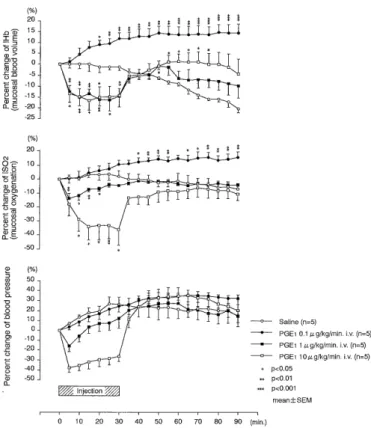
Injection of 0.1 #g/kg/min of PGE1 caused a progressive increase in IHb and ISO2. IHb increased maximally to 14.7
±8.6 % at 80 min after the start of injection while ISO, was 15.6±8.0 % at 75 min. On the other hand, a dose of 1.0 ,ug/kg/min or 10,ug/kg/min of the same substance caused a decrease in systemic blood pressure and the level of IHb and ISO,. However, under these conditions, IHb increased
slowly to the higher level than the control level and ISO2 returned to the control level following the recovery of blood pressure (Fig. 1).
Infusion of 0.1 ,ug/kg/min of PGE2 caused a gradual increase in IHb and ISO,. The maximal increase of IHb was 10.7±4.5 % at 70 min after the start of infusion, while ISO2 was 12.5 ± 8.1 % at 85 min after the start of PGE2 injection. When infused at 1.0ug/kg/min or 10,ug/kg/min, PGE2 reduced systemic blood pressure and the level of IHb and ISO,, but IHb and ISO, returned slowly to the control level following the recovery of blood pressure (Fig. 2). We also examined the effect of PGE metabolites, 15-keto- PGE1 and 15-keto-PGE2, at a dose of 5 ,ug/kg/min. Such dose failed to influence IHb. The injection of 1.O,ug/kg/min of PGI2 had an effect similar to that of 0.1 ,ug/kg/min of PGE, and did not change systemic blood pressure. The maximal increase of IHb was 11.5±8.2 % at 50 min while that of ISO2 was 10.9± 7.6 % at 85 min after the start of PGI2 injection in a dose of 1.0,ug/kg/min. The level of IHb and ISO2 did not change significantly during and after the administration of 0.1 ,ug/kg/min of PGI2. However, a severe hypotension and reduction in IHb and ISO, levels were observed during injection of 10 gg/kg/min of PGI2, but both IHb and ISO2 increased slowly to the higher level than the control level following the recovery of blood pressure (Fig. 3).
Figure 1. The effect of exogenous PGE, on colonic mucosal blood volume, mucosal oxygenation and blood pressure.
PGE1 was continuously injected for 30 min at a concentration of 0.1 ,ug/kg/min (closed circles), 1.0 ,ug/kg/min (closed squares) and -10jg/kg/min (open squares). Open circles : saline injection to control rats. Solutions were administered using the same flow rate in the femoral vein of anesthetized rats. Index of colonic mucosal hemoglobin concentration (upper panel) and colonic mucosal oxyhemoglobin satura- tion (middle panel) were measured by a tissue spectrum analyzer during monitoring of blood pressure (lower panel) during 90 min.
Figure 2. The effect of exogenous PGE2 on colonic mucosal blood volume, mucosal oxygenation and blood pressure. The conditions were similar to those in Fig. 1.
Figure 3. The effect of exogenous PGI2 on colonic mucosal blood volume, mucosal oxygenation and blood pressure. The conditions were similar to those of Fig. 1.
Effect of TXA2 administration on colonic blood flow.
Whereas the injection of 0.1,ug/kg/min of TXA2 failed to influence systemic blood pressure, the drug reduced IHb significantly during injection to -12.2+-4.7% at 30 min.
The level of IHb returned, however, to the control level at 60 min after the commencement of TXA2 injection. In contrast, injection of a larger dose of TXA2 (1.0 ,ug/kg/
min) caused a significant fall in systemic blood pressure and further reduced the level of IHb and ISO2 (IHb, -28.4
±11.8 % ; ISO2, -46.5 ± 24.6 %, at 30 min after the start of TXA2 injection). However, as the blood pressure started to gradually recover, IHb and ISO2 also progressively in- creased (Fig. 4). Injection of 0.01 #g/kg/min of TXA2 failed to have an effect on colonic blood flow. Injection of
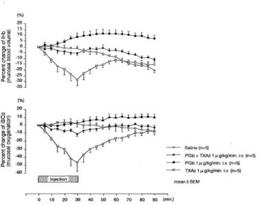
10 ,ug/kg/min of TXA2 caused a severe and rapid fall in systemic blood pressure and death. Furthermore, the simultaneous injection of 1.0 ,ug/kg/min of PGI2 and TXA2 was also examined. A combination of PGI2 and TXA2 showed compromised values with little or on change in blood pressure, IHb and ISO2 (Fig. 5).
Kinetics of residual PGE2 following the administration of PGE2. Administration of PGE2 (10,ug/kg/min) caused an initial decrease in IHb and ISO2, but the level of both
compounds increased slowly after the cessation of injec- tion. The serum concentration of PGE2 reached a peak level
Figure 4. The effect of exogenous TXA2 on colonic mucosal blood volume, mucosal oxygenation and blood pressure. The conditions were similar to those in Fig. 1.
Figure 5. Effects of combination of PGI2 and TXA2 on colonic mucosal blood volume and mucosal oxygenation.
The effect of 1.0 ag/kg/min of PGI2 and a similar dose of TXA2 administered together was compared with that of 1.0 ,ug/kg/min of PGI2 and 1.0 ,ug/kg/min of TXA2 injection, administered separately, on colonic mucosal hemoglogbin concentration (upper panel) and colonic mucosal oxyhemo- globin saturation (lower panel).
Figure 6. The concentration of serum PGE2 following the administration of exogenous PGE2 and the effect of such level on colonic mucosal blood volume, mucosal oxygenation and blood pressure. The concentration of serum PGE2 was measured after the injection of 10 ,ug/kg/min of PGE2 to confirm whether the residual PGE2 (upper panel) influences recovery of colonic mucosal hemoglobin concentration, colonin mucosal oxyhemoglobin saturation and blood pressure (lower panel).
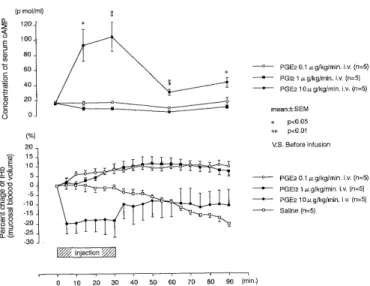
Figure 7. Serum cAMP level and effect on colonic mucosal blood volume after administration of exogenous PGE2 and PGI2. The concentration of serum cAMP (upper panel) was measured after the injection of 0.1,ug/kg/min and 10 ,ug/kg/
min of PGE2 and 1.0 ,ug/kg/min PGI2 to examine whether cAMP influences the increase in colonic mucosal hemoglobin concentration (lower panel).
Figure 8. A comparison of the effect of PGE2 on colonic blood volume and blood pressure between SHR and WKY rats. The effect of 10,ug/kg/min of PGE2 injection was com- pared with saline injection in SHR and WKY to confirm the correlation between IHb of colonic mucosa and sympathetic nerve activity. Upper panel : colonic mucosal hemoglobin concentration during and after saline injection in SHR and WKY rats. Middle panel: colonic mucosal hemoglobin concentration during and after injection of 10,ug/kg/min of PGE2 in SHR and WKY rats. Lower panel : blood pressure level during and after saline and 10 ,ug/kg/min of PGE2 injection in SHR and WKY rats.
at 30 min after the start of injection, then diminished to the initial value (Fig. 6).
Kinetics of serum cAMP following the administration of PGE2 or PGI2. Injection of PGE2 (0.1 ,ug/kg/min) or PGI2 (1.0 ,ug/kg/min) caused a gradual increase of IHb until 60 min after injection, but failed to influence the level of
serum cAMP. However, a higher dose of PGE2 (10 ,ug/kg/
min) further reduced IHb during injection but the level returned slowly following cessation of PGE2 injection.
Injection of such a high dose of PGE2 markedly elevated serum cAMP during infusion of PGE2, but the level rapidly decreased after the end of injection, although it did not return to baseline value (Fig. 7).
Effect of PGE2 administration on colonic blood flow in SHR and WKY rats. IHb was measured in hypertensive
(SHR) and nonhypertensive rats (WKY) in order to examine the correlation between IHb and sympathetic nerve activity. As shown in Fig. 8, under control condi-
tions, IHb diminished in SHR rats compared with WKY rats. Administration of 10,ug/kg/min of PGE2 reduced IHb acutely in "both groups of rats, but the level recovered gradually to the initial level following cessation of injec- tion. There were no statistical differences between SHR and WKY rats.
DISCUSSION
The major finding of our study is a gradual increase in colonic mucosal blood volume caused by the administra- tion of exogenous PGs (PGE,, PGE2 and PGI2). The in- crease was not due to changes in blood pressure.
Furthermore, we also observed an increase in colonic mucosal volume with the administration of a high dose of
PGs, without affecting the systemic blood pressure. Our results suggest that PGs may be one of the cytoprotective substances involved in increasing colonic mucosal blood flow.
Reflectance spectrophotometry can determine not only tissue blood volume but also the average oxy-and deoxyhemoglobin equilibrium in tissue by measuring the absorbance of hemoglobin and tissue oxygen saturation, respectively (13). The present study also demonstrated that exogenous PGs also increased ISO,, representing increased tissue oxygen saturation, without influencing blood pressure. Several investigators described a vasodilatory effect for PGs by demonstrating an increase in gastric mucosal blood flow (1, 2). In this regard, Dousa (14) reported that PG stimulates cAMP formation, which in turn accelerates the production of glycoprotein and glycosaminoglycan. It was also suggested that PG could provide mucosal cytoprotection by providing a sufficient level of tissue oxygen in mucosa. Thus, our results suggest a possible colonic mucosal cytoprotective role for exoge- nous PGs in addition to that already described in the gastric mucosa. Our conclusion is based on the finding that several PGs increased colonic mucosal blood flow and local tissue oxygen saturation.
TXA2 induces platelet aggregation and constriction of vascular smooth muscles, while PGI2 markedly suppresses platelet aggregation and dilates vascular smooth muscles.
Thus, TXA2 and PGI2 play an important role in regulating mucosal circulation by maintaining homeostasis in the cardiovascular system (15). Our results demonstrated that PGI2 increased colonic mucosal blood volume while maintaining a normal blood pressure, while TXA2 reduced it. These results suggest that the colonic microcirculation is regulated by a balance between PGI2 and TXA2.
Several studies have examined the effect of exogenous PGs on vascular smooth muscles (16-18). By using SHR and WKY rats in the present study, we were able to rule out any action of exogenous PG on the sympathetic nervous system, since no difference in colonic hemo-
dynamics was observed between the two groups of rats even when a high dose of PGE2 was administered.
Furthermore,, our findings suggest that PGE metabolites (15-keto-PGE) have no effect on colonic blood flow.
Residual PGE2 did not influence the recovery of colonic mucosal blood flow in our study, because the serum concentration of PGE2 diminished to the initial value following injection of a high dose of PGE2. Such recovery may be due to autoregulatory mechanisms acting after a reduction of blood pressure (19), and a change in the distribution of colonic blood flow. We failed to demon- strate increased levels of serum cAMP by PGE2 or 'PGI2 injection under normal level of systemic blood pressure, whereas serum cAMP increased during the administration of a high dose of PGE2. Although serum cAMP was too low to detect at normal blood pressure, it is possible that exogenous PGs may have increased cAMP, which may have dilated colonic mucosal capillaries.
IBD are characterized by ulceration or inflammation of the colonic mucosa and their etiology are still unknown.
Patients with active ulcerative colitis have higher produc- tion of PGI2 and TXA2 metabolites (6-keto-PGF,a and TXB2) in colonic mucosa than normal subjects (10).
Furthermore, abundant TXB2, and a low 6-keto-PGF,a/
TXB2 ratio are also discribed in Crohn's disease (11). These results suggest that deceleration of PGI2 metabolism and acceleration of TXA2 metabolism reduce colonic mucosal flow and diminish the cytoprotective capacity in patients with active IBD. Several investigators have discribed the protective effect of PGE in gastric mucosa (1, 20), and, in fact, PGE is used as a treatment for gastric ulceration. It is of interest that PGE2 was initially regarded as an inflammatory factor in the mucosa of IBD, but only
recently was re-considered as an anti-inflammatory substance inducing suppressor T cells (21). Sharon et
al. (22) reported that the level of PGE2 in rectal mucosa of patients with ulcerative colitis was higher than that found in a normal mucosa. In addition, Hulten et al. (23) re- ported that colonic mucosal blood flow increased during the active phase, whereas a little or no change in blood flow occurred during the remission phase in patients with ulcerative colitis and Crohn's disease. These results sug- gest that mucosal restoration in active IBD may possibly be due to the accumulation of endogenous PGE in colonic mucosa, increase in colonic blood flow and oxygenation, stimulation of cellular metabolism, and/or the transport of anti-inflammatory chemical mediators to tissues. The recent demonstration of an effective therapeutic role of PG in an experimental model of colitis (5,6), suggest that exogenous PGs may prove useful in the therapy of imflammatory bowel diseases.
ACKNOWLEDGEMENTS
The auther would like to thank Professor Kohei Hara for adequate advise and critical reading of the manuscript, and all of reseach assistants for their cooperation, the Second Department of Internal Medicine, and also mem- bers of the Laboratory Animal Center of Nagasaki University School of Medicine for their care of animals used in the experiment.
REFERENCE
1) Sewell RB : Prostaglandins-role in gastric mucosal cytoprotection. Med J Aust 142: 510-513, 1985.
2) Gerkens JF, Gerber JG, Shand DG and Branch RA : Effect of PG-I2, PG-E2 and blood flow and acid secretion. Prostaglandins 16: 815-823,
1978.
3) Gerber JG and Nies AS: Canine gastric mucosal vasodilation with prostaglandins and histamine analogs. Dig Dis Sci 27: 870-874, 1982.
4) Cheung LY : Topical effect of 16, 16-dimethyl prostaglandin E2 on gastric blood flow in dogs. Am J Physiol 238: G514-G519, 1980 5) Fedorak RN, Empey LR, MacArthur C and Jewell LD : Misoprostol
provides a colonic mucosal protective effect during acetic acid-induced
colitis in rats. Gastroenterology 98: 615-625, 1990.
6) Torsher KJ, Empey LR and Fedorak RN: Misoprostol therapy following trinitrobenzene sulfonic acid-induced colitis accelerates
healing. Prostaglandins leukot Essent Fatty Acids 45: 275-281, 1992 7) Brown JA and Zipser RD: Prostaglandin regulation of colonic blood
flow in rabbit colitis. Gastroeneterology 92: 54-59, 1987.
8) Shehades Z, Price WE and Jacobson ED: Effect of vasoactive agents on intestinal blood flow and motility in the dog. Am J Physiol 216:
386-392, 1969.
9) Moncada S and Vane JR : The role of prostacyclin in vascular tissue.
Fed Proc 38: 66-71, 1979.
10) Ligumsky M, Karmeli F, Sharon P, Zor U, Cohen F and Rachmilewitz D : Enhanced thromboxane A2 and prostacyclin production by cultured
rectal mucosa in ulcerative colitis and its inhibition by steroids and sulfasalazine. Gastroenterology 81 : 444-449, 1981.
11) Hawkey CJ, Karmeli F and Rachmilewitz D : Imbalance of pro- stacyclin and thromboxane synthesis in Crohn's disease. Gut 24:
881-885, 1983.
12) Sato N, Shichiri M, Hayashi N, Matsumura T, Kameda T, Abe I-I and Hagihara B : Non-destructive measurement of tissue oxidative
functions. In: Frontiers of Biological Energetics. Academic Press, New
York. p 1507-1514, 1978.
13) Sato N, Kameda T, Shichiri M, Kawano S, Abe H and I-Iagihara B : Measurement of hemoperfusion and oxygen sufficiency in gastric
mucosa in vivo. Gastroenterology 76: 814-819, 1979.
14) Dousa TP : Pathogenesis of ulcer disease : comments and perspectives.
In: Advances in ulcer disease. Excepta Medica, Amsterdam, Oxford, Princeton. p 128-136, 1980.
15) Moncada S and vane JR : Pharmacology and endogenous roles of prostaglandin endoperoxides, thromboxane A2 , and prostacyclin.
Pharmacol Rev 30: 293-331, 1979.
16) Kitamura K, Suzuki H and Kuriyama H : Prostaglandin action on the main pulmonary artery and portal vein of the rabbit. Jap J Physiol 26:
681-692, 1976.
17) Hedqvist P : Further evidence that prostaglandins inhibit the release of noradrenaline from adrenergic nerve terminals by restriction of
availability of calcium. Br J Pharmacol 58: 599-603, 1976.
18) Moncada S and Vane JR : Unstable metabolites of arachidonic acid and their role in haemostasis and thrombosis. Br Med Bull acid 34:
129-135, 1978.
19) Lundgren 0 : Autoregulation of intestinal blood flow : Physiology and pathophysiology. J Hypertens 7: s 79-s 84, 1989.
20) Wright JP, Young GO, Klaff LJ, Weers LA, Price SK and Marks IN:
Gastric mucosal prostaglandin E levels in patients with gastric ulcer
disease and carcinoma. Gastroenterology 82: 263-267, 1982.
21) Fischer A, Deist FL, Durandy A and Griscelli C : Separation of a population of human T lymphocytes that bind prostaglandin E2 and exert a suppressor activity. J Immunol 134: 815-819, 1985.
22) Sharon P, Ligumsky M, Rachmilewitz D and Zor U : Role of prostaglandins in ulcerative colitis : Enhanced production during active
disease and inhibition by sulfasalazine. Gastroenterology 75: 638-642,
1978.
23) Hulten L, Lindhagen J, Lundgren 0, Fasth S and Ahren C : Regional intestinal blood flow in ulcerative colitis and Crohn's disease.
Gastroenterology 72 : 388-396, 1977.