CD34-negative mesenchymal stem-like cells may act as the cellular origin of
human aortic valve calcification
Anan Nomuraa,b, Kazuhiko Seyaa, Zaiqiang Yua,b, Kazuyuki Daitokub, Shigeru Motomuraa, Manabu Murakamia, Ikuo Fukudab, and Ken-Ichi Furukawaa
aDepartment of Pharmacology, Hirosaki University Graduate School of Medicine, 5 Zaifu-cho, Hirosaki 036-8562, Japan
bDepartment of Thoracic and Cardiovascular Surgery, Hirosaki University Graduate School of Medicine, 5 Zaifu-cho, Hirosaki 036-8562, Japan
Corresponding author:
Ken-Ichi Furukawa
Department of Pharmacology, Hirosaki University Graduate School of Medicine, 5 Zaifu-cho, Hirosaki, 036-8562, Japan.
E-mail: [email protected]
TEL: +81-172-39-5023, FAX: +81-172-39-5022
Abstract
Although various osteogenic inducers contribute to the calcification of human aortic valve interstitial cells, the cellular origin of calcification remains unclear. We immunohistochemically investigated the cellular origin of valve calcification using enzymatically isolated cells from both calcified and non-calcified human aortic valve specimens. CD73-, 90-, and 105-positive and CD45-negative mesenchymal stem-like cells (MSLCs) were isolated from both types of valve specimens using fluorescence-activated cell sorting. MSLCs were further sorted into CD34-negative and -positive cells. Compared with CD34-positive cells, CD34-negative MSLCs were significantly more sensitive to high inorganic phosphate (3.2 mM), calcifying easily in response. Furthermore, immunohistochemical staining showed that significantly higher numbers (~7–9-fold) of CD34-negative compared with CD34-positive MSLCs were localized in calcified aortic valve specimens obtained from calcified aortic stenosis patients. These results suggest that CD34-negative MSLCs are responsible for calcification of the aortic valve.
Keywords: calcific aortic stenosis, aortic valve, mesenchymal stem-like cell, inorganic phosphate, CD34
1. Introduction
Calcific aortic stenosis (CAS) often develops in people over 65 years old, and its frequency increases with age [1]. CAS is characterized by valve thickening and stenosis of the orifice area triggered by an overactive inflammatory process involving expression of cytokines, including tumor necrosis factor-a (TNF-a) [2], transforming growth factor-beta 1 [3], and interleukin-1 beta (IL-1beta) [4]. These cytokines contribute to extracellular matrix formation, remodeling, and ectopic calcification, which are accelerated in the presence of high inorganic phosphate (2.2–3.2 mM) [5–7]. Although we recently demonstrated that TNF-a-induces aortic valve calcification in patients with CAS [8], the cellular origin of ectopic calcification remains unclear.
Osteogenic endothelial progenitor (EP) cells, such as vascular endothelial growth factor receptor-2/CD34-positive cells, contribute to aortic valve calcification in atherosclerosis patients [9–11]. Furthermore, two recent studies reported that aortic valve calcification is associated with ectopic ossification induced by various bone marrow-derived circulating osteogenic progenitor (COP) cells, such as type I collagen/CD45-positive and/or osteocalcin/CD45-positive cells [12,13]. However, whether these progenitor
cells that reside in the aortic valve trigger valve calcification has yet to be confirmed. In addition, the detailed mechanism of aortic valve calcification induced by these cell types remains unclear [14].
In this study, we investigated the cellular origin of human aortic valve calcification using fluorescence-activated cell sorting (FACS) of human aortic valve interstitial cells (HAVICs). We demonstrated that most HAVICs obtained from both calcific and non-calcific aortic valve specimens were characterized as CD73-, 90-, and 105-positive and CD45-negative mesenchymal stem-like cells (MSLCs). These MSLCs contained both CD34-positive and -negative cells, and the CD34-negative cells were rapidly calcified by a high concentration of inorganic phosphate (3.2 mM). Furthermore,
CD34-negative MSLCs were more abundant in calcified valve specimens than in non-calcified specimens.
2. Materials and methods
2.1. Isolation and culture of human aortic interstitial cells
Calcified aortic valves were obtained from eight patients with CAS, and non-calcified valves with no signs of calcification were obtained from eight patients with aortic regurgitation (AR) after informed consent. This study was approved by the institutional review board of Hirosaki University Hospital. There were no statistically significant differences in clinical factors associated with CAS between these two groups (data not shown).
Human aortic valve specimens were gently cut into pieces 2 ± 1 mm long. HAVICs were isolated by collagenase digestion of the aortic valve pieces as previously described [8] and cultured on a plastic dish in alpha minimal essential medium containing 10%
fetal bovine serum (FBS). The medium was changed every 3 days. All chemicals were of analytical grade, and were obtained from Wako Pure Chemicals (Osaka, Japan).
2.2. Isolation of CD34-negative and -positive mesenchymal stem-like cells
After proliferation, confluent passage 1 HAVICs were detached with 0.05% trypsin.
Approximately 3 _ 107 cells were washed with PBS containing 2% FBS. To confirm the presence of MSLCs among the isolated HAVICs, the cells were incubated with four fluorochrome-conjugated mouse anti-human monoclonal antibodies against CD markers: CD45-FITC, CD73-PE, CD90-APC, and CD105-PerCP-Cy5 (BD Biosciences, San Jose, CA, USA). According to the manufacturer’s recommendations, mouse monoclonal isotype antibodies were used to detect nonspecific fluorescence. After
incubation in the dark for 40 min at 4 _C, cell fluorescence was evaluated using a FACSAria™ cell sorter (BD Biosciences), and data were analyzed with CellQuest™
software (BD Biosciences).
To separate the CD34-negative and -positive MSLC populations, detached HAVICs
were incubated with four fluorochromeconjugated mouse anti-human monoclonal antibodies: CD34-FITC, CD73-PE, CD90-APC, and CD105-PerCP-Cy5 (BD Biosciences). Cell fluorescence was evaluated and analyzed as described above.
CD34-negative and -positive MSLCs were sorted using a highspeed sorter (FACSAria™, BD). The separated MSLC populations were centrifuged, transferred to culture medium, and incubated on plastic plates for the following calcification experiments.
2.3. Identification of calcification
CD34-positive and -negative MSLCs, sorted by FACS from the HAVICs enzymatically isolated from valve specimens as described above, were used in all experiments. After reaching confluence, cells were further cultured on 12-well plates in an osteogenic condition using inorganic phosphate for 7 days. The degree of calcification was measured by Alizarin Red S staining [15] at 0 and 7 days. The stained cells were imaged using a digital camera (Nikon, Tokyo, Japan). The amount of Alizarin Red S dye released from the extracellular matrix after incubation in 100 mM aqueous cetylpyridinium chloride solution was quantified by spectrophotometry at 550 nm [16].
2.4. Immunohistochemical staining of aortic valve specimens
Aortic valve specimens were fixed with paraformaldehyde, embedded in paraffin, and cut into 4 lm thick sections. After deparaffinization with xylene followed by ethanol, endogenous peroxidases were blocked with 0.3% H2O2 in methanol at room temperature for 10 min and then washed with PBS. Next, sections were blocked with normal serum at room temperature for 30 min and then incubated with the following primary antibodies: monoclonalanti-CD34, -CD73, -CD90, and -CD105 (Abcam, Cambridge, MA, USA). Sections were incubated with secondary and tertiary agents from a streptavidin biotin-peroxidase detection kit (Histofine SAB-PO Kit; Nichirei, Tokyo, Japan), and then lightly counterstained with hematoxylin. DAPI was used as a fluorescent nuclear stain. Images of three separate visual fields were obtained from each insert with a 40×objective.
2.5. Statistical analysis
All statistical analyses were carried out using KyPlot 5.0 software (Kyenslab, Tokyo, Japan). Comparisons between groups were assessed by ANOVA with Student–Newman–Keuls post hoc corrections. Comparisons between two independent data sets were assessed by Student’s t-test. Data are presented as mean ± SEM
p < 0.05 was considered to indicate statistical significance.
3. Results
3.1. Isolation of CD34-negative and -positive mesenchymal stem-like cells Flow cytometry showed that all cultured cells obtained from both calcified and non-calcified valves were positive for CD73, CD90, and CD105, indicating they were MSLCs (Fig. 1A–C). Nearly all the MSLCs were also CD45-negative (more than 99%, Fig. 1D). However, MSLCs contained both CD34-positive and -negative populations (Fig. 1E). At the cell culture level, there was no significant difference in the ratio of CD34-negative to -positive MSLCs between calcified and non-calcified valves (data not shown).
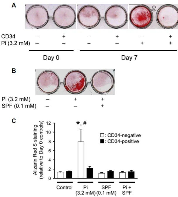
3.2. High phosphate-induced calcification of CD34-negative MSLCs MSLCs were cultured in the presence or absence of high inorganic phosphate (3.2 mM) for 7 days after reaching confluence. Marked calcification of CD34-negative, but not CD34-positive, cells occurred in the presence of high inorganic phosphate for 7 days.
Calcification was significantly suppressed by sodium phosphonoformate(SPF) [17], an inhibitor of inorganic phosphate transporter
1 (PiT-1) (Fig. 2A and B). Spectrophotometric quantification using the bound and released Alizarin Red S dye confirmed that calcification of CD34-negative, but not CD34-positive, MSLCs was significantly
accelerated by high inorganic phosphate, and this calcification was strongly suppressed in the presence of SPF (Fig. 2C).
3.3. Localization of MSLCs in calcified aortic valves
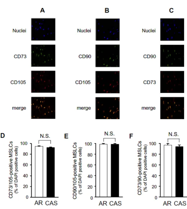
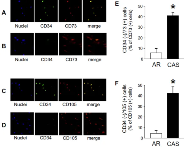
CD73-, CD90-, and CD105-positive MSLCs were present in both calcified (CAS) and non-calcified (AR) aortic valve specimens (Fig. 3A–C). The average DAPI-positive cell number from CAS aortic valve specimens (37.4 ± 2.7/1.4 mm2, n = 60) was not different from that of non-calcified AR specimens (32.1 ± 2.2/1.4 mm2, n = 60). There were no significant differences in the localization rates of CD73/105, CD90/105, and CD73/90-positive MSLCs between the CAS and AR specimens (Fig. 3D–F). We further investigated the localization of CD34-negative and -positive MSLCs. In the non-calcified AR aortic specimens, abundant CD34-positive MSLCs were found among the CD73- and CD105-positive cells (Fig. 4A and C). However, CAS aortic valve specimens rarely contained CD34-positive MSLCs (Fig. 4B and D). The localization rates of CD34-negative MSLCs to CD73- and CD105-positive cells were significantly higher in calcified CAS aortic valve specimens than in non-calcified AR specimens (Fig.
4E and F).
4. Discussion
We demonstrated for the first time that CD73-, CD90-, and CD105-positive MSLCs are localized in the aortic valve at both the cellular and tissue levels. These MSLCs comprised both CD34- positive and -negative cells, despite being CD45-negative.
Furthermore, high inorganic phosphate induced rapid calcification of the CD34-negative, but not CD34-positive, MSLCs. In addition, CD34-negative cells were abundant in CAS valves, but not AR valves. We showed that all passage 1 HAVICs exhibited positive CD73, CD90, and CD105 and negative CD45 expression, ensuring the International Society for Cellular Therapy’s recommended proportion as mesenchymal stem cells [18]. These cells were considered MSLCs because portions of the population expressed CD34, a marker of hematopoietic stem cells. We found that CD34-positive MSLCs were abundant in non-calcified aortic valve specimens, while CD34-negative MSLCs were significantly increased in calcified aortic valve specimens.
In addition, we found that during the culture of CD34-positive MSLCs for 1 week, approximately 10% of the proliferated cells changed to a CD34-negative phenotype (data not shown). From these results, we hypothesize that CD34-positive MSLCs may be transformed into CD34-negative MSLCs, resulting in augmented ectopic aortic valve calcification.
The cellular origin of ectopic aortic valve calcification has been unclear for a long time, though several hypotheses have been put forward. One suggestion was that aortic valve interstitial stromal cells, such as fibroblasts, differentiate into osteoblast-like cells, resulting in valve calcification. Although various molecular mechanisms concerning bone morphogenetic protein-2 and other osteogenic proteins have been proposed, an effective drug therapy to inhibit aortic valve calcification has not yet been developed [19]. Recently, Valgimigli et al. reported that aortic valve calcification is associated with ectopic ossification induced by CD34- and vascular endothelial growth factor receptor-2-positive endothelial progenitor (EP) cells [9]. Egan et al. proposed that ectopic calcification of aortic valve is induced by CD45- and osteocalcin-positive COP cells [13]. However, the precise definitions of EP and COP cells are yet undetermined.
On the other hand, Lin et al. demonstrated that CD34-negative cells in adipose tissue-derived mesenchymal stromal cells had a greater ability to differentiate into osteogenic lineages than CD34-positive cells [20]. In this study, we confirmed that calcification of CD34-negative MSLCs was rapidly induced using high inorganic phosphate (3.2 mM). Furthermore, large numbers of these cells were localized in calcified aortic valve specimens. From these results and previous reports, we propose that CD34-negative MSLCs are the cellular origin, or close to it, for ectopic aortic valve
calcification. In future studies, a more detailed elucidation of the origin of each CD34-negative and -positive MSLC, and identification of factors that trigger the transformation of MSLCs from CD34-positive to -negative may lead to novel therapeutic strategies for CAS.
High inorganic phosphate-induced calcification of CD34-negative cells was completely suppressed in the presence of PiT-1 inhibitor, SPF. Recent studies indicate that hyperphosphatemia is a major risk factor for vascular calcification [21]. Linefsky et al. reported that increased normal serum phosphate levels are associated with valve calcification [22]. These results also suggest that CD34-negative MSLCs are one of the cellular origins of aortic valve calcification, as evidenced by their high activity in phosphate transport through PiT-1. Further studies are expected to reveal the molecular mechanisms of high inorganic phosphate-induced calcification of CD34-negative MSLCs.
One limitation of this study is that although there was no difference in CD34-negative numbers between CAS and AR specimens as observed by cell isolation and culture, large differences were seen by immunohistochemical staining. This disparity is probably due to differences in cell proliferation rates in culture. Although we tried to measure the number of CD34-negative MSLCs immediately after harvest from both
CAS and AR specimens, no reliable data were obtained using FACS because of cell damage from the digestion, the existence of a lot of debris, and the detection limit of the apparatus for measuring a small volume of living cells. Another study limitation is the physiological relevance of the 3.2 mM inorganic phosphate used to induce calcification.
We investigated inorganic phosphate-induced calcification of both CD34-negative and -positive MSLCs using various concentrations of phosphate, ranging from 1.45 mM (physiological concentration) to 3.2 mM. However, inorganic phosphate concentrations up to 2.6 mM failed to induce calcification of these MSLCs.
In conclusion, CD73, 90, and 105-positive and CD45-negative MSLCs were successfully isolated from both calcified and noncalcified aortic valve specimens.
CD34-negative MSLCs are sensitive to high inorganic phosphate (3.2 mM), easily inducing calcification. Furthermore, large numbers of these MSLCs localize in calcific aortic valve specimens obtained from CAS patients. These results suggest that CD34-negative MSLCs are the cellular origin responsible for calcification of the aortic valve. These results should help to further clarify the mechanism of aortic valve calcification and contribute to the development of new therapies for CAS.
Acknowledgments
This work was supported by the Ministry of Education, Science, Sports and Culture of
Japan [Grant-in-Aid 24590310]. Financial support to Dr. Zaiqiang Yu from the Grants-in-Aid of The Cardiovascular Research Fund (Tokyo, Japan) is also acknowledged. We are grateful to the surgeons in the Department of Thoracic Cardiovascular Surgery, Hirosaki University Graduate School of Medicine for their support. We are also grateful to Ms. Izumi Miki, Hirosaki University Graduate School of Medicine, for technical support.
References
[1] J. Chikwe, A. Walther, J. Pepper, The surgical management of aortic valve disease, Br. J. Cardiol. 10 (2003) 453–459.
[2] J.J. Kaden, R. Kilic, A. Sarikoc, S. Hagl, S. Lang, U. Hoffmann, M. Brueckmann, M. Borggrefe, Tumor necrosis factor alpha promotes an osteoblast-like phenotype in human aortic valve myofibroblasts: a potential regulatory mechanism of valvular calcification, Int. J. Mol. Med. 16 (2005) 869–872.
[3] B. Jian, N. Narula, Q.Y. Li, E.R. Mohler III, R.J. Levy, Progression of aortic valve stenosis: TGF-beta1 is present in calcified aortic valve cusps and promotes aortic valve interstitial cell calcification via apoptosis, Ann. Thorac. Surg. 75 (2003)
457–466.
[4] J.J. Kaden, C.E. Dempfle, R. Grobholz, H.T. Tran, R. Kilic, A. Sarikoc, M.
Brueckmann, C. Vahl, S. Zagl, K.K. Haase, et al., Interleukin-1 beta promotes matrix metalloproteinase expression and cell proliferation in calcific aortic valve stenosis, Atherosclerosis 170 (2003) 205–211.
[5] L.L. Demer, Y. Tintut, Vascular calcification. Pathobiology of a multifaceted disease, Circulation 117 (2008) 2938–2948.
[6] X. Li, H.Y. Yang, C.M. Giachlli, BMP-2 promotes phosphate uptake, phenotypic modulation, and calcification of human vascular smooth muscle cells, Atherosclerosis 199 (2008) 271–277.
[7] G. Zhao, M.J. Xu, M.M. Zhao, X.Y. Dai, W. Kong, G.M. Wilson, Y. Guan, C.Y.
Wang, X. Wang, Activation of nuclear factor-kappa B accelerates vascular calcification by inhibiting progressive ankylosis protein homolog expression, Kidney Int. 82 (2012) 34–44.
[8] Z. Yu, K. Seya, K. Daitoku, S. Motomura, I. Fukuda, K.-I. Furukawa, Tumor necrosis factor-a accelerates the calcification of human aortic valve interstitial
cells obtained from patients with calcific aortic valve stenosis via the BMP2- Dlx5 pathway, J. Pharmacol. Exp. Ther. 337 (2011) 16–23.
[9] M. Valgimigli, G.M. Rigolin, A. Fucili, M.D. Porta, O. Soukhomovskaia
P. Malagutti, A.M. Bugli, L.Z. Bragotti, G. Francolini, E. Mauro, G. Castoldi R.
Ferrari, CD34+ and endothelial progenitor cells in patients with various degrees of congestive heart failure, Circulation 110 (2004) 1209–1212.
[10] E.R. Mohler, F.S. Kaplan, R.J. Pignolo, Boning-up on aortic valve calcification, J. Am. Coll. Cardiol. 60 (2012) 1954–1955.
[11] M. Gossl, S. Khosla, X. Zhang, N. Higano, K.L. Jordan, D. Loeffler, M. Enriquez- Sarano, R.J. Lennon, U. McGregor, L.O. Lerman, A. Lerman, Role of circulating osteogenic progenitor cells in calcific aortic stenosis, J. Am. Coll. Cardiol. 60 (2012) 1945–1953.
[12] R.K. Suda, P.C. Billings, K.P. Egan, J.-H. Kim, R. McCarrick-Walmsley, D.L.
Glaser, D.L. Porter, E.M. Shore, R.J. Pignolo, Circulating osteogenic precursor cells in heterotopic bone formation, Stem Cells 27 (2009) 2209–2219.
[13] P.K. Egan, J.H. Kim, E.R. Mohler III, R.J. Pignolo, Role for circulating osteogenic precursor (COP) cells in aortic valvular disease, Arterioscler. Thromb. Vasc. Biol.
31 (2011) 2965–2971.
[14] A. Shioi, Molecular mechanisms of vascular calcification, Clin. Calcium 20 (2010) 1611–1619.
[15] H. Puchtler, S.N. Meloan, M.S. Terry, On the history and mechanism of Alizarin and Alizarin Red S stains for calcium, J. Histochem. Cytochem. 17 (1969) 110–
124.
[16] C.M. Stanford, P.A. Jacobson, E.D. Eanes, L.A. Lembke, R.J. Midura, Rapidly forming apatitic mineral in an osteoblastic cell line (UMR 106-01 BSP), J. Biol.
Chem. 270 (1995) 9420–9428.
[17] M. Szczepanska-Konkel, A.N. Yusufi, M. VanScoy, S.K. Webster, T.P. Dousa, Phosphonocarboxylic acids as specific inhibitors of Na+-dependent transport of phosphate across renal brush border membrane, J. Biol. Chem. 261 (1986)
6375–6383.
[18] M. Dominici, K. Le Blanc, I. Mueller, I. Slaper-Cortenbach, F.C. Marini D.S.
Krause, R.J. Deans, A. Keating, D.J. Prockop, E.M. Horwitz, Minimal criteria for defining multipotent mesenchymal stromal cells. The International Society for Cellular Therapy position statement, Cytotherapy 8 (2006) 315–317.
[19] A. Parolari, C. Loardi, L. Mussoni, L. Cavallotti, M. Camera, P. Biglioli, E.
Tremoli, F. Alamanni, Nonrheumatic calcific aortic stenosis: an overview from basic science to pharmacological prevention, Eur. J. Cardiothorac. Surg. 35 (2009) 493–504.
[20] C.S. Lin, H. Ning, G. Lin, T.F. Lue, Is CD34 truly a negative marker for mesenchymal stromal cells?, Cytotherapy 14 (2012) 1159–1163
[21] E.L. Husseini, M.C. Boulanger, D. Fournier, A. Mahmut, Y. Boss, P. Pibarot P. Mathieu, High expression of the Pi-transporter SLC20A1/Pit1 in calcific aortic valve disease promotes mineralization through regulation of Akt-1, PLoS One 8 (2013) e53393.
[22] J.P. Linefsky, K.D. O’Brien, R.K. Dphil, I.H. de Boer, E. Barasch, N.S. Jenny, Association of serum phosphate levels with aortic valve sclerosis and annular calcification, J. Am. Coll. Cardiol. 58 (2011) 291–297
Fig. 1. MSLCs isolated from human aortic valves by collagenase digestion included
CD34-positive and -negative cells. CD markers for MSLCs were confirmed by flow cytometry. Furthermore, MSLCs were sorted by FACS into CD34-negative and -positive populations. (A–C) All MSLCs were CD73-(A), CD90-(B), and CD105-(C) positive. (D) Almost all MSLCs (more than 99%) were CD45-negative. (E) MSLCs included CD34-positive and -negative cells.
Fig. 2. CD34-negative MSLCs were easily calcified. After reaching confluence,
CD34-negative and -positive MSLCs were further cultured in the presence of high levels of inorganic phosphate (Pi, 3.2 mM) for up to 7 days. (A and B) Representative Alizarin Red S staining of cultured cells. CD34-negative MSLCs (A) were strongly calcified in the presence of inorganic phosphate, and calcification was completely
inhibited in the presence of sodium phosphonoformate, a selective PiT-1 inhibitor (SPF, B). Similar results were confirmed in 5 samples. (C) Quantification of Alizarin Red S staining at day 7 via extraction with cetylpyridinium chloride. The amount of released dye was quantified by spectrophotometry at 550 nm. CD34-negative (white bars) and CD34-positive (black bars) staining ratios were calculated relative to the day 0 control group. Each bar indicates the mean ± SEM; n = 5. ⁄p < 0.01 compared with CD34-negative MSLCs control; #p < 0.05 compared with CD34-positive MSLCs in the presence of inorganic phosphate (3.2 mM). (For interpretation of the references to colour in this figure legend, the reader is referred to the web version of this article.)
Fig. 3. MSLCs were present in aortic valve specimens. Sections of aortic valves
were incubated with various primary antibodies for stem cell markers and then detected by secondary antibodies with fluorescent dyes. (A–C) Typical double immunohistochemical staining of calcified aortic valve from CAS specimens with mesenchymal stem cell markers (CD73, CD90, and CD105). The merged figure is an overlay of the two panels. Cell nuclei were stained blue with DAPI. (D–F)
Localization rate of CD73/105-(D), CD90/105-(E), and CD73/90-positive (F) MSLCs in the calcified (CAS) and non-calcified (AR) aortic valve specimens. Bars represent mean ± SEM; n = 3.
Fig. 4. CD34-negative MSLCs were abundant in aortic valves of CAS specimens.
Representative immunohistochemical staining images of aortic valve specimen from AR (A and C) or CAS (B and D) by CD34 with CD73 (A and B) or with CD105 (C and D) are shown. The merged figure is an overlay of the two panels. Cell nuclei were stained blue with DAPI. Bar graphs show the localization rate of CD34(-)/CD73(+)
cells (E) and CD34(-)/CD105(+) cells (F) in the aortic valve. Each bar represents the mean ± SEM; n = 3.⁄p < 0.001 compared with AR.