Abstract
The objective of this study was to carry out epidemiological investigations in cows with dis-
placed abomasum (DA)and to evaluate the recov- ery of abomasal condition by determining serum pepsinogen and other biochemical values. One hundred twenty-three cows with DA were used in this study. The occurrence rates within 3 weeks after parturition were 80.9% (72
/89) for left dis-
placement of the abomasum (LDA) and 65.0%
(13
/20) for right displacement of the abomasum (RDA), whereas 42.9% (6
/14) of abomasal volvulus (AV)occurred after more than 4 weeks.
The percentages of dead and disused animal were higher for cows with AV (28.6%)and LDA (23.6%) than for those with RDA (5.0%). There were remarkable increases in nonesterified fatty acids
(NEFA)in all cows with DA at operation,where- as hyperglycemia was observed in cows with RDA and AV. The majority of cows with DA showed hypocalcemia and hypochloremia.
There were no significant variations in the serum and blood biochemical parameters between free and tie stall cows with DA. The pepsinogen
value was markedly decreased in all cows with DA. Pepsinogen values in cows with LDA,RDA and AV at 7-11 days after operation were still lower than the reference value, although most biochemical parameters returned to the normal ranges post-operation. This finding indicates that the mucosa of the abomasum did not recover even at that time.
Introduction
Displaced abomasum (DA) is a multifactorial disease. The disease is known to be associated with negative energy balance (NEB) and in- creased accumulation of triglycerides (TG)in the liver and is accompanied by other periparturient diseases (Muylle et al.1990 and Geishauser,1995).
The prevalence of cows with DA has been recog- nized to be increasing because cows are being fed intensively on large farms (Cameron et al. 1998,
Kelton et al. 1998 and Radostits et al. 2007).
Displaced abomasum results in economic losses due to treatment and reduced milk production.
In addition,it has been reported that 10% of cows with DA are culled or died (Bartlett et al. 1995 and Van Winden and Kuiper, 2003).
Nasser Z. A
BOUZEID, Eri S
AKUTA, Takahiro K
AWAI, Toshihiko T
AKAHASHI, Aya G
OTOH, Kazushige T
AKEHANA,
Garrett R. O
ETZELand Shin O
IKAWA(November 2007)
Assessment of Serum Pepsinogen and Other Biochemical Parameters in Dairy Cows with Displaced Abomasum or Abomasal Volvulus before
and after Operation
酪農学園大学獣医学部生産動物医療学教室
Department of Large Animal Clinical Sciences, School of Veterinary Medicine, Rakuno Gakuen University, 582 Bunkyodai-Midorimachi, Ebetsu, Hokkaido, 069‑8501, Japan
ザガジグ大学獣医学部臨床学教室
Department of Animal Medicine, Faculty of Veterinary Medicine, Zagazig University, Egypt 釧路地区農業共済組合厚岸支所
Akkeshi Veterinary Clinical Center, Kushiro Agricultural Mutual Aid Association, Akkeshi, Hokkaido, 088‑1125, Japan 酪農学園大学獣医学部獣医解剖学教室
Department of Veterinary Anatomy, School of Veterinary Medicine, Rakuno Gakuen University, 582 Bunkyodai- Midorimachi, Ebetsu, Hokkaido, 069‑8501, Japan
ウイスコンシン州立大学獣医学部臨床学教室
Department of Medical Sciences, School of Veterinary Medicine, University of Wisconsin, Madison WI 53706, USA
*連絡責任者
Corresponding author:Shin Oikawa
Pepsinogen is a proenzyme, generated by chief cells of the abomasal mucosa. Hydrochloride secreted from parietal cells changes pepsinogen to pepsin (Vianello et al. 1988 and Tanaka et al.
1991). It has been reported that the serum pep- sinogen value can be employed as a useful marker for assessing the pathogenesis of abomasal dis-
eases (Berghen et al. 1993, Zadnik and Mesaric, 1999 and Mesaric et al. 2002). The serum pep- sinogen value in healthy cows is not significantly changed with the lactating stage or age (Ohwada et al. 2002). An increase of pepsinogen reflects mucousal damage as a consequence of Ostertagia infection (Schaw et al.1997 and Scott et al.1999) and abomastitis in cows (Schillhorn, 1988). On the other hand, Ohwada et al. (2002) observed remarkable decreases of serum pepsinogen con- centrations in cows with left and right DA and those with abomasal volvulus (AV). These phe- nomena suggest the pathogenesis of atrophic gastritis. Although their report showed different pepsinogen values in each DA type at operation,
the changes after operation were not described.
Therefore, it could not be confirmed if the mucosal condition recovered.
The purpose of this study was to carry out an epidemiological investigation in cows with DA and to evaluate the recovery of abomasal condi- tion by determining serum pepsinogen and other biochemical values.
Materials and Methods
Animals
One hundred twenty-three Holstein cows with DA were used in this study. The animals were fed on 89 farms of the Kushiro region in Hok-
kaido, Japan from October 2004 through Febru- ary 2005. Cows with DA were divided into those with left displacement of the abomasum (LDA;
n
=89, 4.7
±1.8 yr), right displacement of the abomasum (RDA; n
=20, 5.0±2.2 yr) and
abomasal volvulus (AV; n
=14, 3.9±0.9 yr). Allcows were diagnosed based on physical examina- tion (ping sound and decreased appetite). The diagnosis was additionally verified surgically when an operation took place.
Epidemiological investigation
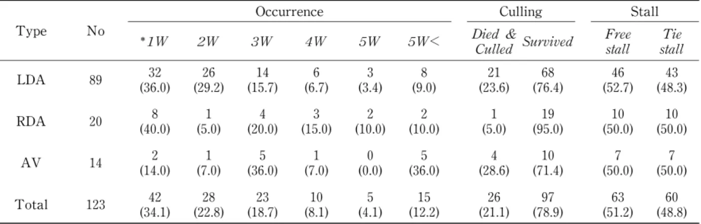
Occurrence time, culling within 2 months after operation and the stall system were investigated for all farms.
Blood Sampling
Ten milliliters of blood was collected from the jugular vein of each cow with DA into an evacuat- ed sterile tube without any anticoagulant three times (just before operation (day 0), days 1-3 and days 7-11 after operation). Samples were rapidly centrifuged at 3,000 rpm for 15 minutes. Sera were harvested and aliquots were stored at -20
℃until analysis. Moreover, 5 ml of blood was allowed to flow freely from the jugular vein of each cow into a tube containing sodium floride as an anticoagulant (Kelly, 1984).
Serum and blood biochemical analysis
Serum concentrations of nonesterified fatty acids (NEFA),chloride and calcium and the blood glucose value were measured at Kishimoto Clini- cal Laboratory, Sapporo, Japan.
Serum pepsinogen determination
The serum pepsinogen value was measured according to the method of Berghen et al. (1987).
Statistical analysis
The obtained data were compared and anal- yzed by Scheffeʼ s multiple comparison F test using SAS,2nd Ed,(Institute Inc.Cary,NC,USA).
Moreover the results were tested for significance using the t-test according to Selvin (1996).
Results
The occurrence rates of LDA, RDA and AV during the first three weeks after parturition were 80.9% (72
/89), 65.0% (13
/20) and 57.1%
(6
/14), respectively (Table 1). The percentages of dead and disused animals were higher for AV
(28.6%)and LDA (23.6%)than for RDA (5%).
The serum NEFA concentration in all cows
with DA at day 0 (1,338
±546 μEq
/L)was higher
than the reference value. Significant increases
of NEFA concentrations in cows with LDA were
observed compared to those with AV and RDA.
The serum NEFA concentration in all cows with DA returned to the reference value at 7-11 days.
The blood glucose level in cows with LDA was within the normal range, whereas those of cows with RDA and AV were higher than the reference value (hyperglycemia). At 7-11 days after opera- tion, the glucose value returned to the normal range. The majority of cows with DA had hypocalcemia and hypochloremia. Briefly, 6 (42.9%)of the cows with AV, 13 (65.0%)of those with RDA and 35 (39.3%)of those with LDA had calcium concentrations of less than 8 mg/ dL (sub-
clinical hypocalcemia). There were non- significant variations of calcium and chloride concentrations among cows with LDA, RDA and AV. Calcium and chloride levels were clearly recovered post-operation in all cows with DA,
especially in those with LDA. There were non- significant variations of serum and blood bio- chemical parameters between free stall and tied stall cows with DA.
The pepsinogen values in all cows with DA at day 0 (n
=123, 828±257 mU) remarkably de-creased compared to the reference range, but there were no significant difference among cows with LDA,RDA and AV (Table 2). The values in cows with LDA, RDA and AV at 7-11 days were still lower than the reference value. In addition, the pepsingen level was lower for dead and disused animals (n
=26, 790
±264 mU) than in healed animals (839±256 mU).
Discussion
It has been reported that the occurrence rates of LDA and RDA were within 6 weeks (Radostits et al. 2007)and 3-6 weeks after parturition (Con-
stable et al.1992 and Delgado-Lecaroz et al.2000) respectively. However, 80.9% of the cases of LDA and 65.0% of those of RDA happened within 3 weeks postpartum in this study. We could confirm the difference between the present and previous occurrences of DA. This phenomenon may be attributable to the changes of risk factors related to its occurrence. Briefly,it is presumed that cows with DA already fall into NEB before parturition and the close-up period. The path-
ogenesis induces severe conditions such as short- age of glucose and disturbance of calcium and ketone bodies metabolism (Muylle et al.1990 and Geishauser, 1995). On the other hand, 42.9% of AV occurred more than 4 weeks after parturition.
The direction of DA could be influenced predomi- nantly by the volume of the forestomach. Imme- diately after parturition, displacement occurs to the left because of a reduction in the size of the rumen volume. Several weeks later the dilated abomasum moves caudally and dorsally in the right abdomen because the volume of the fores- tomach is much larger, thereby providing an effective barrier (rumen barrier)(Radostits et al.
2007). With regard to the prognoses of different types of DA, the percentage of animals that died or became disused was higher for cows with AV (28.6%)and LDA (23.3%)than for those with RDA
Table 1 Occurrence time, culling and stall type in cows with displaced abomasum
Occurrence Culling Stall
Type No
1W 2W 3W 4W 5W 5W< Died &
Culled Survived Free stall Tie
stall
LDA 89 32 (36.0)
26 (29.2)
14 (15.7)
6 (6.7)
3 (3.4)
8 (9.0)
21 (23.6)
68 (76.4)
46 (52.7)
43 (48.3) RDA 20 8
(40.0) 1 (5.0)
4 (20.0)
3 (15.0)
2 (10.0)
2 (10.0)
1 (5.0)
19 (95.0)
10 (50.0)
10 (50.0)
AV 14 2
(14.0) 1 (7.0)
5 (36.0)
1 (7.0)
0 (0.0)
5 (36.0)
4 (28.6)
10 (71.4)
7 (50.0)
7 (50.0) Total 123 42
(34.1) 28 (22.8)
23 (18.7)
10 (8.1)
5 (4.1)
15 (12.2)
26 (21.1)
97 (78.9)
63 (51.2)
60 (48.8) LDA:left displacement of the abomasum. RDA:right displacement of the abomasum. AV:abomasal vovulus.
Weeks after parturation. Numbers in parenthesis indicate percent.
(5.0%). The case fatality rate of AV was consis- tent with previous reports(23.5%)(Radostits et al.
2007), whereas that of LDA was higher than that previously reported (5.6%)(Constable et al.1992).
This difference may be attributed to the involve- ment of risk factors related to the occurrence of LDA. Further researches are needed to eluci-
date this phenomenon.
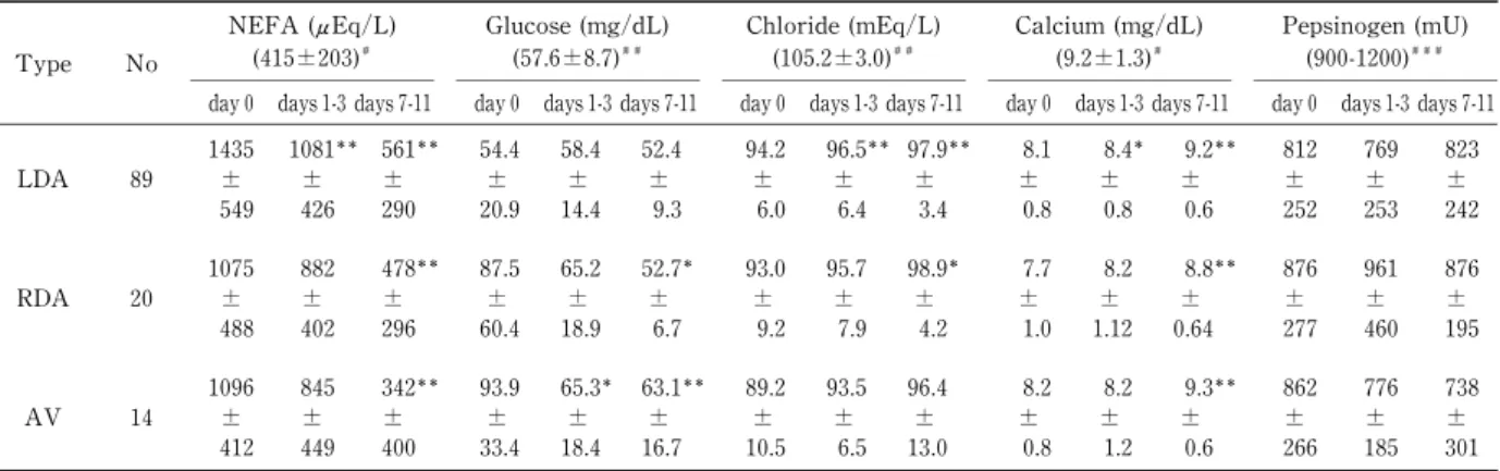
Regarding the serum biochemical parameters (Table 2), there were remarkable increases of NEFA concentrations in all cows with DA at day 0, compared with the reference value. The serum NEFA concentration in cows with LDA significantly increased compared with those for cows with AV and RDA. This may have been because LDA was strongly associated with NEB.
These results were in agreement with the report of Oikawa et al. (1997) that cows with LDA had higher NEFA concentrations. The odds for hav- ing LDA were 2 times greater for cows with plasma NEFA
>0.3 mEq/
L (cut point) between 3-35 days before calving than for cows that had NEFA below the cut point (Geishauser, 1995,
Cameron et al. 1998).
In this study hyperglycemia was observed in cows with RDA and AV. Previous authors simi- larly reported increased blood glucose values in cows with DA (Van Meirhaege et al.1988,Cupere et al.1991,Muylle et al.1990 and Itoh et al.1998).
On the basis of our results we hypothesized that hyperglycemia associated with RDA might be
associated with impaired outflow of pancreatic juice and disturbed blood circulation in the pan- creatic parenchyma because of changes of duodenal and omental position due to dislocation of the abomsum (Klucinski et al. 1988, Muylle et al. 1990, Kehrli and Goff, 1992). Therefore the elevated glucose level observed in RDA might be a secondary finding (Van Winden and Kuiper,
2003 and Van Winden et al. 2003).
The majority of the cows with DA showed subclinical hypocalcaemia (Table 2). Six (42.9%) of the cows with AV, 13 (65.0%) of those with RDA and 35 (39.3%) of those with LDA had calcium levels of less than 8 mg/ dL (subclinical hypocalcemia). This pathogenesis is considered to be an important factor to reduce the tone of the abomasal and ruminoreticular walls, which may induce DA. Briefly,previous researchers (Massy et al.1993,Delgado-Lecaroz et al.2000 and Rados-
tits et al. 2007)reported that cows with hypocal- cemia (serum calcium concentration
<7.9 mg/dL) 12 hrs before parturition had a 4-8 times greater risk of developing LDA than did normocalcemic cows. Hypochloremia was more pronounced in cows with AV especially (Table 2). Similar results were reported by Geishauser and Seeh
(1996). Moreover experimentally induced reduc- tion of the abomasal emptying into the cow duo- denum resulted in hypochloremia after eight hours. The hypothesis is that the ruminant fores- tomach compartments have the capacity to
Table 2 Changes of blood and serum biochemical parameters in cows with displaced abomasum during the experi mental period
-
NEFA (μEq/L) (415±203)
Glucose (mg/dL) (57.6±8.7)
Chloride (mEq/L) (105.2±3.0)
Calcium (mg/dL) (9.2±1.3)
Pepsinogen (mU) (900-1200) Type No
day0 days1-3 days7-11 day0 days1-3 days7-11 day0 days1-3 days7-11 day0 days1-3 days7-11 day0 days1-3 days7-11
LDA 89 1435
± 549
1081
± 426
561
± 290
54.4
± 20.9
58.4
± 14.4
52.4
± 9.3
94.2
± 6.0
96.5
± 6.4
97.9
± 3.4
8.1
± 0.8
8.4
± 0.8
9.2
± 0.6
812
± 252
769
± 253
823
± 242
RDA 20 1075
± 488
882
± 402
478
± 296
87.5
± 60.4
65.2
± 18.9
52.7
± 6.7
93.0
± 9.2
95.7
± 7.9
98.9
± 4.2
7.7
± 1.0
8.2
± 1.12
8.8
± 0.64
876
± 277
961
± 460
876
± 195
AV 14 1096
± 412
845
± 449
342
± 400
93.9
± 33.4
65.3
± 18.4
63.1
± 16.7
89.2
± 10.5
93.5
± 6.5
96.4
± 13.0
8.2
± 0.8
8.2
± 1.2
9.3
± 0.6
862
± 266
776
± 185
738
± 301 LDA:left displacement of the abomasum. RDA:right displacement of the abomasum. AV:abomasal vovulus.
Significant when compared with the value at day 0 (P<0.05; P<0.01).
Reference value according to Oikawa and Katoh 2002, Reference value according to Zadnik 2003. Reference value according to Ohwada et al. (2002).
absorb Na and Cl ,and the reflux and sequestra- tion of abomasal secretions that occur in an obstruction
/ligation model or naturally occurring obstructions may make these ions, to some degree,not accessible to the plasma by this route
(Gable and Martens, 1991 and Ward et al. 1994).
There are no published papers about the effect of stall type on biochemical parameters in cows. In this study we confirmed that there were non- significant variations in their parameters between free and tie stall cows with DA, which may indi-
cate that there is no difference of the path- ogenesis between the two stall systems for DA.
The pepsinogen value was markedly decreased in all cows with DA (n
=123, 828.3±257.1 mU)compared with the reference range. The results of this study were generally in agreement with a report that serum pepsinogen concentrations in LDA, RDA and AV cows were remarkably de- creased compared with healthy cows (Ohwada et al. 2002). The decreased value of serum pep-
sinogen in cows with DA is thought to be attribut- able to atrophy of the mucous membrane of the abomasum. Also in human beings,a decrease of pepsinogen in sera of patients with chronic atro-
phic gastritis was reported (Samloff et al. 1982, Dinis-Ribeiro et al. 2004 and Weck and Brenner 2006). Pepsinogen values in cows with LDA, RDA and AV at 7-11 days post-operation were still lower than the reference value, although almost biochemical parameters returned to the normal levels after operation. This finding indi- cates that the mucosa of the abomasum did not recover even at that time.
Acknowledgments
This study was supported by a Grant-in-Aid for Rakuno Gakuen Kyodo Kenkyu (2006). We are grateful to Mr. Kim Barrymore for his critical reading of the manuscript.
References
Bartlett,P.C.,M.Kopcha,P.H.Coe,N.K.Ames, P. L. Ruegg and R. J. Erskine. 1995. Economic comparison of the pyloroomentopexy vs the roll-and-toggle procedure for treatment of left displacement of the abomasum in dairy cattle.
J. Am. Vet. Med. Assoc. 206:1156
‑1162.
Berghen, P., H. Hilderson, J. Vercruysse and P.
Dorny. 1993. Evaluation of pepsinogen, gastrin and antibody response in diagnosing oster-
tagiasis. Vet. Parasitol. 46:175
‑195.
Berghen, P., P. Dorny and J. Vercruysse. 1987.
Evaluation of a simplified blood pepsinogen assay. Am. J. Vet. Res. 48:664
‑669.
Cameron, R. E. B., P. B. Dyk, T. H. Herdt, J. B.
Kaneene, R. Miller, H. F. Bucholtz, J. S. Lies- man, M. J. Vandehaar and R. S. Emery. 1998.
Dry cow diet,management,and energy balance as risk factors for displaced abomasum in high producing dairy herds.J.Dairy Sci.81:132
‑139.
Constable,P.D.,G.Y.Miller,G.F.Hoffsis,B.L.
Hull and D. M. Rings. 1992. Risk factors for abomasal volvulus and left abomasal displace-
ment in cattle. Am. J. Vet. Res. 53:1184
‑1192.
Cupere, F., E. Muylle, C. Van der Hende and W.
Oyeart. 1991. Metabolic profile tests in high yielding normal cows and in cows suffering from abomasal displacement. Bov. Pract. 26:
129‑ 130.
Delgado-Lecaroz,R.,L.D.Warnick,C.L.Guard, M. C. Smith and D. A. Barry. 2000. Cross- sectional study of the association of abomasal displacement or volvulus with serum electro-
lyte and mineral concentrations in dairy cows.
Can. Vet. J. 41:301
‑305.
Dinis-Ribeiro, M., G. Yamaki, K. Miki, A. Costa- Pereira,M.Matsukawa and M.Kurihara.2004.
Meta-analysis on the validity of pepsinogen test for gastric carcinoma, dysplasia or chronic atrophic gastritis screening.J.Med.Screen.11:
141
‑147.
Gable,G.and H.Martens.1991.Transport of Na and Cl across the forestomach epithelium:
mechanisms and interactions with short chain fatty acids.In:Tsuda T,Sasaki Y,Kawashima R, eds. Physiological Aspects of Digestion and Metabolism in Ruminants. Academic Press,
New York. pp.129‑ 151.
Geishauser, T. 1995. Abomasal displacement in the bovine-a review on character, occurrence aetiology and pathogenesis.J.Vet.Med.Ser.A.
42:229‑ 251.
Geishauser, T., C. Seeh. 1996. Duodeno-abomasal
reflux in cows with abomasal displacement.
Zentralbl Veterinarmed A. 43:445
‑450.
Itoh, N., M. Koiwa and A. Hatsugaya. 1998.
Comparative analysis of blood chemical values in primary ketosis and abomasal displacement in cows.Zentralbl Veterinarmed A.45,293
‑298.
Kelly, W. M. 1984. Veterinary clinical diagnosis.
6 Ed. pp.202
‑204, Bailliere, Tindall, London.
Kelton, D. F., K. D. Lissemore and R. E. Martin.
1998. Recommendations for recording and cal- culating the incidence of selected clinical dis- eases of dairy cattle.J.Dairy Sci.81:2502
‑2509.
Kehrli, M. E.,and J.P.Goff.1992.Periparturient hypocalcaemia in cows: Effects on peripheral blood neutrophil and lymphocyte function. J.
Dairy Sci. 72:1188
‑1193.
Klucinski, W., A. Degorski and E. Miernik- Degorska. 1988. Effect of ketone bodies on the mitogenic response of bovine milk lymphocytes, Zentralbl Veterinarmed A. 35:
626
‑631.
Massey,C.D.,C.Wang,G.A.Donovan and D.K.
Beede. 1993. Hypocalcemia at parturition as a risk factor for left displacement of abdomen in dairy cows. J. Am. Vet. Med. Assoc. 203:852
‑853.
Mesaric, M., T. Zadnik and M. Klinkon. 2002.
Comparison of serum pepsinogen activity between enzootic bovine leukosis (EBL) posi-
tive beef cattle and cows with abomasal ulcers.
Slov. Vet. Res. 39:227
‑232.
Muylle, E., C. Van den Hende, B. Sustronck and P. Deperez. 1990. Biochemical profiles in cows with abomasal displacement estimated by blood and liver parameters.Vet. Med. Assoc .37:259‑
263.
Ohwada, S., S. Oikawa, F. Mori, M. Koiwa, A.
Nitanai,T.Kurosawa and H.Sato.2002.Serum pepsinogen concentrations in healthy cows and their diagnostic significance with abomasal diseases. J. Rakuno Gakuen Univ. 26:289‑ 293.
Oikawa, S., N. Katoh. 2002. Decreases in serum apolipoprotein B-100 and A-I concentrations in cows with milk fever and downer cows.Can.J.
Vet. Res. 66:31
‑34.
Oikawa,S.,N.Katoh,F.Kawawa and O.Yasushi.
1997. Decreased serum apolipoprotein B-100
and A-I concentrations in cows with ketosis and left displacement of the abomasum.Am.J.Vet.
Res. 58:121
‑125.
Radostits,O.M.,D.C.Blood and C.C.Gay.2007.
Veterinary Medicine:a textbook of the diseases of cattle, sheep, pigs, goats and horses. 10 ed.
pp.353
‑367. Baillliere Tindall London.
Samloff, I. M., K. Varis, T. Ihamaki, M. Siurala and J. I. Rotter. 1982. Relationships among serum pepsinogen I, serum pepsinogen II, gas- tric mucosal histology. A study in relatives of patients with pernicious anemia. Gastroenter-
ology. 8:204
‑209.
Schaw, D. J., J. Vercruysse, E. Claerebout, J.
Agneessens and P. Dorny. 1997. Gastrointesti- nal nematode infections of first-season grazing calves in Belgium: general patterns and the effect of chemoprophylaxis. Vet. Parasitol. 69:
103
‑116.
Schillhorn van Veen, T. W. 1988. Evaluation of abomasal enzyme and hormone levels in the diagnosis of ostertagiasis. Vet. Parasitol. 27:
139‑ 149.
Scott, I., A. Dick, J. Irvine,M.J.Stear and Q.A.
Mckellar. 1999. The distribution of pepsinogen within the abomasa of cattle and sheep infected with Ostertagia spp. and sheep infected with Haemonchus contortus. Vet.Parasitol.82:145
‑159.
Selvin, S. 1996. Statistical analysis of epidemiological data. 2nd Ed. P.44
‑78. Oxford University Press, New York.
Statistical Analysis System “SAS”. 1996. SAS Userʼ s guide:Statistic 2 Ed. Institute Inc.
Cary, North Carolina.
Tanaka,Y.,K.Mine,Y.Nakai,N.Mishima,and T. Nakagawa. 1991. Serum pepsinogen I con- centrations in peptic ulcer patients in relation to ulcer location and stage. Gut. 32:849‑ 852.
Van Meirhaege,H.,P.Deperez,C.Ven den Hende and E. Muylle. 1988. Plasma glucose clearance and insulin response in cows with abomasal displacement. J. Vet. Med. A. 35:221
‑224.
Van Winden, S. C. L. and R. Kuiper. 2003. Left displacement of the abomasum in dairy cattle:
recent developments in epidemiological and
etiological aspects. Vet. Res. 34:47
‑56.
Van Winden, S. C. L., R. Jorritsma, K. E. Muller and J.P.T.M.Noordhuizen.2003.Feed intake, milk yield, and metabolic parameters prior to left displaced abomasum in dairy cows.J.Dairy Sci. 86:1465
‑1471.
Vianello,F.,M.Plebani and A.Piccoli.1988.Role of serum pepsinogen in detecting ulcer disease.
Clin. Chim. Acta. 172:335
‑340.
Ward, J. L., D. F. Smith, S. L. Fubini and D. N.
Deuel-Armando. 1994. Evaluation of abomasal outflow diversion as an experimental model of hypochloremic, hypokalemic metabolic al-
kalosis in lactating cows. Can. J. Vet. Res. 58:
13
‑19.
Weck,M.N.and H.Brenner.2006.Prevalence of chronic atrophic gastritis in different parts of the world.Cancer Epidemiol.Biomarkers Prev.
15:1083
‑1094.
Zadnik, T. 2003. A comparative study of the hemato-biochemical parameters between clinically healthy cows and cows with displace-
ment of the abomasums.Acta Veterinaria (Beo- grad). 53:297
‑309.
Zadnik, T. and M. Mesaric. 1999. Fecal blood levels and serum proenzyme pepsinogen activ-
ity of dairy cows with abomasal displacement.
Israel J. Vet. Med. 54:61
‑65.
要 約
本研究は,第四胃変位牛の発生に関する疫学調査を行うとともに,術後の回復状況を血清ペプシノーゲン値お よびその他の生化学値を測定することによって評価することを目的に実施された。供試牛として 123頭の第四胃 変位牛が用いられた。分娩後3週間以内の発生率は,第四胃左方変位(LDA)で 80.9%,第四胃右方変位(RDA)
で 65.0%であった。一方,第四胃捻転(AV)の 42.9%が分娩4週以上経過してから発生していた。AVの死廃率 は 28.6%,