Title
[症例報告]Carcinosarcoma (sarcomatoid carcinoma) of the
Gallbladder Presenting with a Cholecystocolic Fistula and a
Marked Leukocytosis : A Case Report
Author(s)
Hanashiro, Naoji; Tamaki, Satoshi; Naka, Tsunemori; Muto,
Yoshihiro
Citation
琉球医学会誌 = Ryukyu Medical Journal, 20(2): 77-80
Issue Date
2001
URL
http://hdl.handle.net/20.500.12001/3471
Carcinosarcoma ( sarcomatoid carcinoma ) of the Gallbladder Presenting
with a Cholecystocolic Fistula and a Marked Leukocytosis: A Case Report
Naoji Hanashiro", Satoshi Tamaki", Tsunemori Naka", and Yoshihiro Muto
'Division of Surgery, Naka Hospital, Kadena, Okinawa 904-0203 and
l The First Department of Surgery, Faculty of Medicine, University of the Ryukyus,
Nishihara, Okinawa 903-0215, Japan
(Received on July 18, 2000, accepted on October 24, 2000)
AB ST RACT
We herein report a case of carcinosarcoma (sarcomatoid carcinoma) of the gallbladder with a cholecystocolic fistula and evidence of marked leukocytosis in a 77-year-old Japanese man. The patient presented with episodes of right upper quadrant pain and fever in April 2000. The physi-cal examination revealed a painful, palpable mass in the right upper quadrant. Routine labo-ratory findings showed leukocytosis (18.000/mm3 ). Abdominal sonography showed an echogenic mass occupying the entire gallbladder lumen. Abdominal CT demonstrated a solid mass lesion measuring 8 × 7 cm in size with a clear, hypodense asterisk configuration, suggesting a
commu-nication between the gallbladder and the gastrointestinal tract. At exploratory laparotomy, a large solid gallbladder tumor showed direct invasion into the transverse colon and regional lymph
node metastasis. The tumor was evaluated to be Stage IV. With the intention of performing
cytoreductive surgery, a cholecystectomy together with a partial transverse colectomy was thus performed. The gallbladder tumor showed penetration into the transverse colon, forming a cholecystocolic fistula. On a cut section analysis, the tumor occupied the entire gallbladder lumen and measured 7 × 5 cm in size. No gallstones were observed. The tumor was composed of two different histologic features: an adenocarcinomatous feature that was composed of moder-ately differentiated adenocarcinoma and a sarcomatous feature that consisted primarily of pleomorphic, spindle cells. Immunohistochemically, the adenocarcinoma cells were positive forkeratin, EMA and CEA whereas the sacomatous spindle cells were positive for vimentin and CEA. However, both the carcinoma cells and sarcomatous cells were negative for G-CSF. Postoperatively, the patient's WBC counts increased from 26.9×10³/mm3 to 103.0×10³/mm3. Finally, the pa-tient died with clinical manifestations of peritonitis carcinomatosa one month after surgery.
Ryukyu Med. J. , 20(2)77-80, 2001
Key words: carcinosarcoma, gallbladder, cholecystocolic fistula, leukocytosis
INTRODUCTION
Carcinosarcomais a rare variant of the gallbladder It is characteized by invasive growth, a propen-sity of infiltrating into the neighboring organs and a worse prognosis compared to common adenocarcinoma. To our knowledge, this type of gallbladder cancer associated with malignant cholecystocolic fistula is extremely rare6-7)
This rarity has prompted us to report the present case. We
herein report a case of carcinosarcoma (sarcomatoid car-cinoma) of the gallbladder with associated cholecytocolic fistula and marked leukocytosis.CASE REPORT
A 77-year-old man presented with episodes of right
upper quadrant pain and fever in April 2000. Ti一e patient
was immediately admitted to our hospital. His medical history revealed that he had previously undergone an op-eration for a compression fracture of the lumbar verte-brae in 1997 and had also been treated with H2 receptor antagonists for a gastric ulcer in 1998. On admission, the patient appeared moderately ill. The physical examination re-vealed muscle guarding and tenderness over the entire abdomen and a palpable mass in the right upper quadrant. Routine labo-ratory findings showed leukocytosis (WBC 18,800/mm3 ) and CRP of 3.4mg/dl (normal, 0.27< mg/dl). The serum
ffl Malignant cholecystocolic fistula and Gallbladder carcinosarcoma
Fig. 1 Abdominal CT demonstrating a large solid mass lesion with air (arrow) measuring 8 cm in greatest dimen-sion of the gallbladder.
carcinoembryonic antigen (CEA) and carbohydrate anti-gen (CA 19-9 ) levels were not examined.
Abdominal ultrasonograms showed a heterogeneously echogenic mass occupying the entire gallbladder lumen. Computed tomograms demonstrated a solid mass lesion measuring 8 × 7cm in size with a clear, hypodense aster-lsk configuration similar to Benz sign which is ocassionally seen in gallstones, suggesting communication between the gallbladder and the gastrointestinal tract (Fig. 1).
Based on these findings, the patient was diagnosed to have advanced gallbladder carcinoma associated with an
interna一 biliary fistula. During an exploratory laparotomy,
a large solid gallbladder mass lesion showed direct invasion mainly to the transverse colon and the adjacent organs, re-suiting in the form of a large conglomerated mass lesion. The patient was determined to have Stage IV-gallbladder carci-noma. With the intention of performing cytoreductive sur-gery, a cholecystectomy together with a partial transverse colectomy was thus performed.
After surgery, the WBC counts were 26.9×10³ on May 6,53.1×10s on May 12,69.0×103 on May 15 and 103.0×10³ on May 16, respectively. About 60 to 70 percent of the WBCs
Fig. 2 The resected gallbladder revealing a large tumor lesion occupying the entire gallbladder lumen with a thickended wall (top) and the transverse colon showing the tumor invading into the oolonic lumen, thus forming a cholecystocolic fistula (bottom).
were composed of segmented neutrophils. During this pe-riod, ATL-like cells (adut T-cell lymphoma) were observed in the peripheral blood smears, but human T-cell lymphotropic virus type 1 (HTLV-1) (ELISA) was negative. Unfortu-nately, the serum G-CSF was not examined. Postoperatively his disease was extensively aggressive and he died while dem-onstrating chnica】 manifestations of peritonitis carcinomatosa one month after surgery.
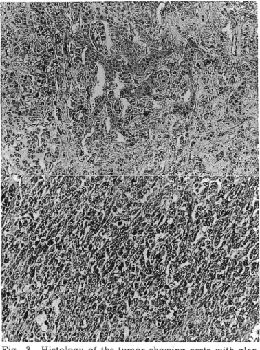
Grossly, the gallbladder was entirely occupied with carcinoma which had penetrated into the transverse colon, forming a cholecystocohc fistula measuring 2 cm in diame-ter. On bisection, the gallbladder tumor was solid, partially necrotic and measured 7 × 5cm in size. The gallbladder wall was diffusely thickened and extensively adhered to the trans-verse colon. The cystic duct of the gallbladder was oc-eluded by the carcinoma and no gallstones were found in the gallbladder (Fig. 2 ). Histologically, the tumor was composed of two different histologic features: an adenocarcinomatous feature that showed moderately to poorly differentiated carcinoma in the superficial portion of the tumor (ap-proximately l0% of the tumor) and a sarcomatous feature that consisted of pleomorphic, spindle cells with nuclear
pleomorphism (Fig. 3 ). Morphologically, a gradual
trans-Fig. 3 Histology of the tumor showing nests with glan-dular structures of round to oval tumor cells (top; HE
x50) and spindle-shaped cells (bottom; HEX50).
ltion was observed between these two components. Accord ing to an immunohistochemical examination, adenocarcinoma
cells were positive for keratin, EMA and CEA, but were
nega-tive for vimentin. Sarcomatous spindle cells were posinega-tive for vimentin and CEA, but were negative for keratin. Staining with anti-G-CSF monoclonal antibody did not demonstrated G-CSF in both the carcinoma and sacomatous
cells.
DISCUSSION
Internal biliary fistula (IBF) is an abnormal communi-cation between the biliary tract and adjacent organs, includ-ing the duodenum or colon9 10. The most frequent cause of IBF is cholelithiasis representing up to 90% of all cases while the second is a chronic duodenal ulcer, accounting for up to
6 %. The cause of gallbladder carcinoma comprises approxi-mately 4% of all cases. The frequency of IBF has been re-ported to be from 1.2 to 5.0% in biliary surgical caseslO Therefore, the frequency of IBF associated with gallbladder carcinoma may account for less than 0.2% in biliary
surgery12'.
On the other hand, approximately 40 gallbladder carcinosarcoma cases have been reported in the English
literature3 ". Furthermore, of more than 50 cases of G-CSF-producing tumors, only two cases of carcinosarcoma of the maxillary sinus and the esophagus have been reported18'. Based on these data reviewed in the literature, this case has attracted great interest because of the rare type of gall-bladder tumor, the formation of a malignant cholecystocohc fistula and a marked leukocytosis.
Carcinosarcoma is an unusual tumor which is char-acterized by a close admixture of carcinomatous and sarcomatous components21. Although there has been much confusion regarding the term, diagnosis and histogenesis of carcinosarcoma, it is now generally accepted that all of these various terms indicate the same tumor entity both clinically and morphologically…'. Regarding the treat-ment and prognosis of this tumor, the treattreat-ment of carcinosarcoma of the gallbladder is closely similar to that for adenocarcinoma of the gallbladder. Both tumors usually demonstrate advanced disease and thus have an extremely poor clinical outcome3). In general, the extent of tumor spread greatly influences the prognosis. In our experience, 3 cases of carcinosarcoma of the gallbladder including this case were clinically detected at a rather ad-vanced stage and the patient died within 1 t0 7 months after surgery4'5'
As mentioned previously, a malignant cholecystocohc fistula represents an uncommon complication of gallblad-der carcinoma. Gallbladgallblad-der tumors progressively grow, extend completely through the bowel wall and involve con-tiguous structures such as the duodenum or colon. Occa-sionally, central necrosis and ulceration of an intramural tumor causes a perforation or penetration into the adja-cent organs. Histologically, this gallbladder tumor showed necrosis of a tumor around the fistula. In this case, it is possible that the aggressive growth of the gallbladder tumor to the transverse colon could have caused the ne-crosis and ulceration of an lntramurally invaded tumor, thus leading to a cholecystocolic fistula.
Finally, a marked leukocytosis in this case was a unique feature. To our knowledge, there has been no previous clinical report of a marked leukocytosis of carcinosarcoma of the gallbladder in the literature. Although the mecha-nism of producing G-CSF by tumor cells is still unclear, leukocytosis is occasionally associated with a mahgant
tumor as a result of the production of growth factors that regulate proliferation, differentiation of myeloid cells and synergistic interactions among multiple cytokines, including granulocyte-colony stimulating factor (G-CSF), tumor ne-crosis factor a (TNF a ), and interleuken-1 (IL-1)16''". In our case, we did not extract and cultured the tumor tissues and examined serum G-CSF concentration for demonstra-tion of G-CSF activity. The immunohistochemical exami-nations of G-CSF in tumor cells were performed using the avidin-biotin complex method on parraffin-embedded sec-tions. A reaction for G-CSF was negative in both carcinoma and spindle cells. Unfortunately, the clinical implications of leukocytosis in tumors remains unresolved. If we
encoun-80 Malignant cholecystocolic fistula and Gallbladder carcinosarcoma
ter a cancer patient with a marked leukocytosis in fu-ture, increased serum G-CSF activity, demonstration of G-CSF activity in the tumor extract solution or tumor tissue culture solution or immunostaining of the tumor tissues with anti-G-CSF antibody should be examined.
REFERENCES
1 ) Edmondson H. : Tumors of the gallbladder and extraheaptic bile ducts. In: Atlas of tumor pathology. Washington D.C., Armed Forces Institute of Pathology, pp.61-67. 1976.
2 ) Lopez G.E‥ Strimel W. and Herrera-Ornelas L∴
Carcinosarcoma of the gallbladder: Report of a case.
J. Surg. Oncol. 29: 224-226, 1985.
3 ) Iezzoni J. C. and Mills S.E.: Sarcomatoid
carcin0-mas (carcinosarcocarcin0-mas) of the gastrointetinal tract: A review. Semin. Diagn. Pathol. 10: 176-187, 1993. 4 ) Shimabuku M., Yamada M., Shiraishi M., Yamazato
M.. Kusano T.. Muto Y. and Toda T.: Carcinosarcoma of the gallbladder: a case report. Asian J. Surgery 21: 315-318, 1998.
5) Samura H., Isa T., Kuniyoshi S., Shiraishi M.,
Kusano T. and Muto Y.: Sarcoma-toid carcinoma
(so-called carcinosarcoma) of the gallbladder, a case
report and review of the literature. Ryukyu Med. J.
19: 39-42, 1999.
6 ) Khaira H.S., Awad R.W. and Thompson A.K.: Squamous
cell carcinoma of the gallbladder presenting with a biliary-colic fistula. Eur. J. Surg. Oncol. 21: 581-582, 1995.7 ) Ercolani G., Nagino M., Kamiya J., Kondo S., Kanai
M., Nishio H. and Nimura Y. Advanced adenosquamous
carcinoma of the gallbladder with bilio-biliary fistula: an uncommon case treated by hepatopancreatoduodenectomy. Hepatogastroenterology 46: 1650-1654, 1999.8 ) Okabe T., Ohwada S., Ogawa T., Takeyoshi I., Kamoshita N., Kon Y., Fukusato T., Ohhara K. and Morishita Y.: Gallbladder carcinoma with choledochoduodenal fistula: a case report with surgical treatment. Hepatogastroentero-logy 46: 1660-1663, 1999.
9 ) Shah M. and Mori W: A clinico-pathological study of spontaneous internal biliary fistula. Acta Pathol. Jpn. 23: 349-358, 1973.
10) Safaie-Shirazi S., Zike W.L. and Printen K. J. Spon-taneous enterobihary fistulas. Surg. Gynec. Obstet. 137: 769-772, 1973.
ll) Sameshima Y., Uchimura M., Muto Y., Waki S.,
Hayashi T., Nakayama M. and Yamada M.: Internal
biliary fistulas due to gallstone. Nippon Rinshogeka
Gakkai Zasshi 43: 960-966,1982.
12) Hutchings V.Z., Wheeler J.R. and Puestow C.B. :
Choledochoduodenal fistula complicating duodenal
ulcer: A report of five cases and a review of the
lit-erature. Arch. Surg. 73: 598-605, 1956.
13) Ota S., Kato A., Kobayashi H., Yonezumi M., Musashi
M., Imamura M. and Asaka M.: Monoclonal origin
of an esophageal carcinosarcoma producing granulocyte-colony stimulating factor, a case report. Cancer 82: 2102-2111, 1998.14) Taniyama K., Sasaki N., Mukai T., Uemura N., Miyoshi N., Nakai H., Nakayama H. and Tahara E.: Carcinosarcoma of the esophagus. Pathol. Int. 45: 297-302, 1995.
15) Iascone C. and Barreca M. : Carcinosar∞ma and pseudosarcoma of the esophagus: Two names, one
dis-ease - Comprehensive review of the literature. World
J. Surg. 23: 153-157, 1999.
16) Suzuki A, Takahashi T, Okuno Y, Tsuyuoka R, Fukumoto M. and Nakamura K.: IL1 production as a regulator of G-CSF and IL-6 production in G-CSF pro-ducing cell lines. Br. J. Cancer 65: 515-518, 1992. 17) Koeffler H.P., Gasson J., Ranyard J., SouzaL.,
Shepard M. and Munker R.: Recombinant human TNF a
stimulates production of granulocyte colonystimulating factor. Blood 70: 55-59, 1987.
A CKNOW LEDGEMENTS
We are grateful to the pathologists at the local
hospi-tal described above who contributed to this case study and