Clinical and Hematological Observations on Experimental Immunohemolytic Anemia in Horses
Mitsuo SoNODA* and Kiyokazu MORI* *
Two groups of 3 healthy horses each were injected intravenously with 1.0 ml of normal ovine serum (control group) and 1.0 ml of anti-horse red cell ovine serum (experimental group) per kg of body weight, respectively. Clinical and hematological observations were carried out on them for 6 and 21 days after injection, respectively. In the control group, no clinical signs were noted at all. Prominent leukopenia appearing 3 hours after injection was the only significant blood change throughout the experimental period. In the experi mental group, the main clinical signs observed were accelerated breathing immediately after injection, pyrexia, depression, anorexia, irregular heart sounds, accelerated peristalsis, anemia, icterus, marked hemoglobinuria and albuminuria in the initial stage, slight depres sion and anemia and/or icterus in the middle stage, and slight depression and anemia in the terminal stage of the experimental period. In the hematological observation, the main findings after injection were slight diphasic anemia, leukocyte count decreasing 3 hours after injection and increasing thereafter, reduction in the minimum and maximum resistance of red cells, slight increase in the rate of appearance of red cells with Jolly's body and marked anisocytosis on blood films, appearance of sideroleukocytes and erythrophages, large value of indirect bilirubin, and positive Coombs' test.
It is well known that there has been a sporadic occurrence of hemolytic icterus of newborn foals, which is a type of immu nohemolytic anemia, in the Hidaka dis trict of Hokkaido since many years ago.
In their previous paper,l) the authros reported the clinical and hematological
observations of the disease. The onset of the disease was so abrupt and the course so acute in general that it was very
difficult to carry out detailed clinical and hematological examinations in most spon taneous cases of the disease, especially in the early stage.
Therefore, an experimental study on immunohemolytic anemia was conducted
by the use of anti-horse red cell ovine serum to compare with the clinical and
hematological findings of the spontaneous hemolytic icterus of newborn foals re-ported already."2)
Received for publication June 30, 1976.
*Department of Veterinary Internal Medicine , School of Ve terinary Medicine, Rakunogakuen College, Ebetsu, Hokkaido 069-01, Japan.
**Shintoku Animal Husbandry Experiment Station , Hokkaido. This work was supported by a grant-in-aid from the Equine Health Laboratory, Japan Racing Association.
The outline of this paper was read before the 73rd Meeting of the Japanese Society of Veterinary Science held in Tokyo in April, 1972.
Immunohemolytic Anemia in Horses
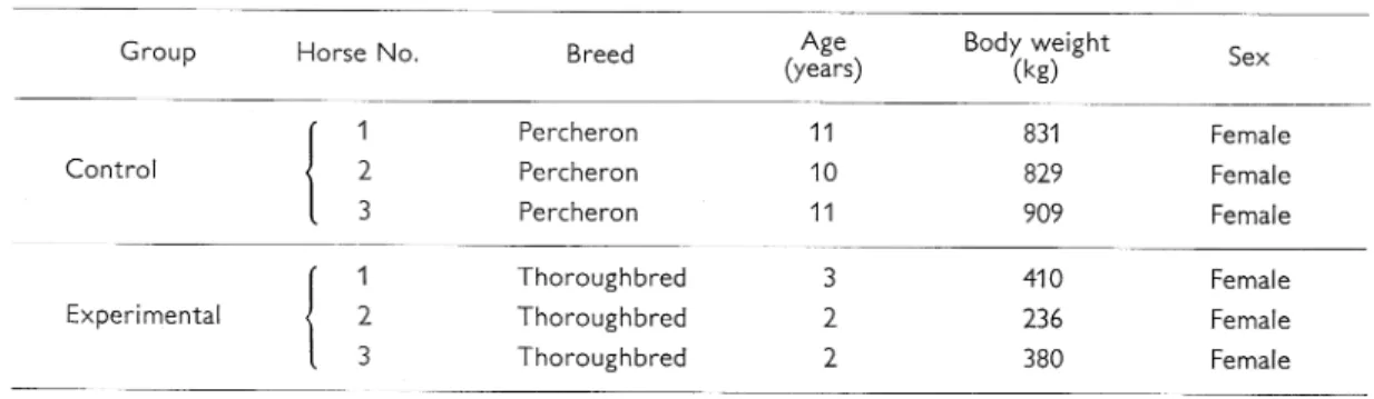
Materials and Methods Horses used
Six clinically and hematologically heal-thy female horses ranging from 2 to 11 years of age were used for the experiment. They were divided into two groups of three horses each. One group was in jected with normal ovine serum (control
group) and the other with ovine antiserum (experimental group).
Sera used and amounts injected
1. Normal ovine serum. It was obtained from the blood of a clinically healthy male sheep by centrifugation. The hemo lysin and agglutinin titers of the serum for horse red cell were X 4 and x 1, respective-ly.
2. Anti-horse red cell ovine serum. Fresh anticoagulative horse red cells were wash-ed 5 times with isotonic saline solution . A sheep was injected intravenously with 8 ml of 50% red cell suspension in saline solution 7 times every other day . On the 7th day after the last injection, his serum was checked for hemolysin and agglutinin titers for horse red cells. Then he was sac rificed by total bleeding. The serum was separated by centrifugation and stored at -20°C in the refrigerator until use. It was proved to have hemolysin and agglu
tinin titers of 1:128 and 1:2,048, respec tively.
These normal and anti-horse red cell ovine sera were injected intravenously once into the horses of the control and ex perimental groups in a dose of 1 ml per kg of body weight.
Methods of observation 1. Clinical observation
Clinical observation was carried out on the horses of the control and experimental groups for 6 and 21 days after injection with serum, respectively. Urinalysis was performed on urine samples obtained by catheterization for pH, protein, glucose
and hemoglobin by a hemacombistix at
intervals of 1-7 days. Especially during the first 24 hours, it was done 3, 6, and 24 hours after injection with serum.
2. Hematological observation
Hematological observation was carried out on the horses of the 2 groups at the same time with the urinalysis. It covered the following items: red and white cell counts by the Thoma-Zeiss method, he matocrit reading by the micro-hematocrit method, hemoglobin value by the cyan methemoglobin method, osmotic fragility of red cells by a descending series of saline solution, morphology of red cells in blood films stained with Giemsa stain,
leukocyte count in thick smear films of leukocytes fixed in formaldehyde gas and stained with Prussian blue and carbol fuchsin solution, erythrophage in thick smear films of leukocytes stained with Giemsa stain, serum bilirubin value by Evelyn-Malloy method, and agar gel immunodiffusion test for equine infec tious anemia by Nakajima and Ushimi's method.3)
Results Clinical findings
1. Control group
No abnormal clinical signs were ob-served in any horse. Urinalysis revealed no abnormal data at any time during the period of observation.
2. Experimental group
Horse No. 1. Soon after injection with antiserum, she exhibited depression, in-creased pulse, and accelerated breathing rate. All the signs, except depression, dis appeared completely in 60 minutes. Ex amination 3 and 6 hours after injection revealed pyrexia (39.0 and 38.9°C), ano rexia, increased pulse rate (82 and 84), accelerated peristalsis, dullness in action, marked albuminuria and hemoglobinuria. When examined 24 hours after injection, the former 3 signs disappeared, but she was still slightly depressed and dull in action. Albuminuria and hemoglobinuria were persisted until the 3rd day after in jection. Anemic discoloration and icterus began to be seen on the mucous memb ranes 6 hours and 3 days after injection, respectively. The former sign persisted over the entire experimental period, but the latter disappeared on the 21st day of observation.
Horse No. 2. Accelerated breathing and
depression were noted immediately after injection, but disappeared soon. In the ex aminations 3 and 6 hours after injection, mild pyrexia, depression, anorexia, irre gular heart sounds, marked albuminuria and hemoglobinuria were presented. Dep ression became quite clear on the 3rd day and anorexia was very severe during the 6th and 10th days of observation. Anemia and icterus began to be noted on the 2nd day and aggravated on the 4th and 5th day of observation, respectively. At the end of the observation period, the mare was still anemic, but not icteric on the mucous membranes. Irregular heart sound was noted again during the 4th and 8th days. Peristalsis was accelerated and the action became dull 24 hours after in jection. The former began to be more
serious on the 6th day and returned to a normal state on the 14th day of the ob servation. The latter, however, persisted even after the period of observation.
In urinalysis, albuminuria and hemo globinuria were seen continually until the 8th day and transient glucosuria was posi tive when tested on the 2nd and 3rd days of observation.
Horse No. 3. Accelerated breathing was the only sign which appeared immediately after injection. It disappeared within 30
minutes. When examined 3 hours after
injection, the horse showed mild depres sion, slight anorexia and dullness in action.
In urinalysis, marked albuminuria and
hemoglobinuria were positive. Irregularity in heart sounds appeared 6 hours after injection. Anemic coloration was seen on
the mucous membranes 24 hours after
injection. In addition, icterus was noted for 3 days after the 4th day of observation.
Immunohemolytic Anemia in Horses
Fig. 1. Clinical findings of horse No. 2
Remarks. B; Before injection.
disappeared on the next day. An increased pulse rate was clear on the 5th day. The signs of depression and anemia persisted until the end of observation period. Dull-ness in action became very conspicuous 24 hours after injection, but dissipated on the 8th day of observation.
Marked albuminuria and hemoglobi nuria persisted from 3 hours to the 6th and 8th days of observation, respectively.
They became negative on the 14th day. On the 21st day, or the last day, of ob servation, the horse exhibited no signs, except slight depression and anemic colo ration on the mucous membranes. Hematological findings
1. Erythrocyte count. In the control and experimental groups the erythrocyte count was 7.45 and 7.43 millions /mm3 on the average, respectively, before injection.
Fig. 2. Findings of erythrocytes, hematocrit and hemoglobin
Remarks. B; Before injection.
•¬: Mean value of experimental group .•¬: Mean value of control group.
In the control group, the count varied be tween 7.24 and 8.30 millions/mm3, or in the nomal range of horse, after injection. In the experimental group, it began to decrease gradually 3 hours after injection
and reached 2.72 millions/mm3, or the
smallest mean value, on the 8th day . Then it increased to 3.65 millions/mm3 on the average, or almost a half of the initial count, on the last day of observa tion.
2. Hematocrit reading. The mean values of the hematocrit reading were 36.3 and 37.0% in the two groups , respectively, before injection. In the control group , it
fluctuated between 35.8 and 39.2% in
the course of experiment. On the other hand, in the experimental group, it began to decrease after injection and showed 15%, or the lowest mean value, on the 10th day of experiment. Then it increased gradually and was 22% on the last day of observation.
3. Hemoglobin value. The hemoglobin value was 13.0 g/ 100 ml on the average in the control group and 12.7 g/ 100 ml on the average in the experimental group before injection. After injection, the values of the two groups fluctuated in almost parallel with those of the hematocrit reading described above. That is, the value of the control group was always in a normal range and those of the experi mental group the smallest on the 10th day of observation.
4. Leukocyte count. In the control and experimental groups, the leukocyte count was 7,133 and 8,867 on the average, respectively, before injection. It decreased to 5,733 and 6,600, respectively, 3 hours after injection. It returned to 7,567 and 8,467, respectively, at 6 hours. Since then,
Fig. 3. Findings of leukocytes Remarks. See the footnote of Fig, 2,
Immunohemolytic Anemia in Horses
in the control group, it was within a nor mal range of the horse. In the experi mental group, it increased to reach 17,600, or the highest count, on the 8th day . On the last day of observation, it was 7,333 on the average.
5. Erythrocyte fragility. Before injection , the minimum and maximum resistances
Fig. 4. Findings of erythrocyte fragility Remarks. See the footnote of Fig. 2,
of erythrocytes were 0.65 and 0.46%,
respectively, in the control group and
0.67 and 0.47% in the experimental
group. In the control group, they ranged from 0.65 to 0.67% and from 0.45 to 0.48 , respectively, after injection.
In the experimental group, both mini-mum and maximini-mum resistances began to be reduced markedly 3 hours after in jection. The minimum resistance was
within 0.85 and 0.87% during the 2nd and 8th days of observation. The maxi-mum resistance was within 0.56 and 0.58% from 3 hours to the 4th day after injection. Then it increased gradually. In the terminal stage of the period of ob servation, the minimum resistance was still reduced, but the maximum rather in-creased to be higher than that before in jection.
6. Findings of erythrocytes. In the 3
horses of the control group, neither
juvenile nor abnormal red cells, except a very few cells with Jolly's body, were seen in any stage of the experimental period. On the other hand, in the horses of the experimental group, red cells with Jolly's body increased slightly in number
on the 3rd or 8th days. Anisocytosis was marked on the 3rd or 6th days of ob
servation. These changes of red cells,
however, were hardly noticed on the last day of observation.
7. Sdderoleukocytes. No sideroleukocytes appeared in any horse of the control group at any time of observation. In the experimental group, the cells appeared in all the 3 horses for the first time 3 hours after injection. They were all granular in type. The average count was 7 per 10,000 leukocytes. Nine hours after injection, it was 33 per 10,000 leukocytes, or the highest. Then it decreased gradu ally. A few sideroleukocytes were still seen on the last day of observation in all the 3 horses. The cells appearing 9 hours after injection were granular, diffuse, or mixed in type. The latter two types were always very few.
8. Erythrophage. No erythrophages were noted in the control group at any time
Fig. 5. Findings of sideroleukocytes Remarks. See the footnote of Fig. 2.
of observation. In the experimental group, they appeared in all the 3 horses 3 hours after injection, viz., 10%0 on the average in number. Thereafter, they fluctuated between 0.3 and 19% on the average until the 14th day of observation. On the 21st, or the last day of observation, however, they were not found at all in any horse. 9. Serum bilirubin value. Before injec tion, the direct and indirect bilirubin values were 0.21 and 0.22 mg/ 100 ml on the average, respectively, in the con trol group. In the examination after injec tion, both of them were always in a normal range. The former fluctuated from 0.11 to 0.30 mg/ 100 ml and the latter from 0.02 to 0.42 mg/ 100 ml.
In the experimental group, the direct and indirect bilirubin values were 0.22 and 0.39 mg/ 100 ml on the average, res pectively, before injection. In the exami nation after injection, the direct bilirubin increased slightly (0.59-1.05 mg/ 100 ml), except that in the terminal stage of ob servation. On the other hand, the indirect one began to increase markedly 3 hours
after injection and reached 7.91 mg/ 100 ml, the highest mean value, on the
6th day. After that it declined promptly. The fluctuation of the total bilirubin value was perfectly identical with that of the
indirect bilirubin value.
10. Direct Coombs' test. The direct Coombs' test was negative in the 3 horses of the control group at any time of ob servation. In the experimental group, it was negative in all the 3 horses before injection. It turned, however, to be posi tive and remained in this state for 1-12 days, or 5 days on the average.
11. Agar gel immunodiffusion test. The
agar gel immunodiffusion test revealed
that sera collected from all the horses of
both control and experimental groups
before injection and on the last day of the
observation period were negative for
equine infectious anemia. Discussion
In the present study, no clinical signs were noted in the group injected with normal ovine serum. Clear clinical signs, however, were recognized in the group injected with ovine antiserum soon after injection and in the initial and middle stages of observation. They may have been
caused by the antigen-antibody reaction
and the subsequent hemolysis.4)
In the hematological examination, mild diphasic anemia was seen only in the ex perimental group on the 2nd and 8th days. A finding like this in experimental im munohemolytic anemia was already re-ported in rabbits by Toki5) and in dogs by the authors.6) It was assumed7'8) that anemia in the first phase might be mainly due to intravascular hemolysis caused directly by hemolysin and complement in the blood, and that anemia in the second phase might be due to extravascular
Fig. 6. Findings of bilirubin Remarks. See the footnote of Fig, 2.
Immunohemolytic Anemia in Horses
hemolysis caused by agglutinin or im perfect antibody. In the present study, anemia may have been caused by a simi lar mechanism.
Leukocytes decreased clearly in count in both groups 3 hours after injection and in-creased thereafter only in the experi mental group. The decrease in the leuko cyte count was supposed to be due to ana phylactic shock by injection with
hetero-antiserum, and the following increase to an increase in the function of bone marrow following acute hemolysis. The slight in-crease in the rate of appearance of red cells with Jolly's body and marked ani socytosis noted on blood films in the experimental group were thought to reflect the increased regeneration of ery throgenesis in bone marrow.
A marked reduction was seen in the osmotic resistance of red cells only in the experimental group 3 hours after injection. It indicated that the red cells might have been sensitized by antibody against horse
red cells injected and/or incomplete
antibody appearing in the blood after
injection, and that they might have been weakened by the osmotic pressure of saline. In addition, it might be thought that a large number of sensitized red cells were
phagocytized by monocytes and neutro
phils, which changed into erythrophages. In the 3 horses of the experimental group, it was clarified that the injection of antiserum had resulted in the appear ance of sideroleukocytes in the blood. The cells began to appear 3 hours and reached the highest count 6 hours after injection. They decreased gradually thereafter. Al-most all the sideroleukocytes found in the experimental group were granular in type. The diffuse and mixed types were a very
few throughout the entire period of obser vation. The authors presumed that the cells of the former type might come from phagocytic cells in the blood containing hemosiderin in which damaged red cells had been phagocytized and digested, and that the latter two types might come from
cells containing hemosiderin and/or fer
ritin released from the reticuloendothelial
system9' 10) which have phagocytized a
number of damaged red cells. From the negative results of the agar gel immuno diffusion test, it could be said that the ap
pearance of sideroleukocytes in these
horses had nothing to do with equine in fectious anemia.
Serum bilirubin value fluctuated an
tagonistically with the red cell count. It began to increase 3 hours after injection, reached a maximum on the 6th day, and then decreased rapidly. In this instance, most part of the bilirubin was the indirect one. From this finding it was certain that the anemia observed in the experimental group was due to the hemolytic destruc tion of red cells.
In the present study, detailed clinical and hematological observations were car ried out in the course of experiment, es pecially in the initial stage. The results obtained were essentially the same as the actual state of hemolytic icterus of foals reported already."2) Therefore, it may be reasonable to say that the findings of the present experiment are very useful for understanding the mechanism of occur rence of hemolytic icterus of foals. It is natural that there are some differences in the type of antibody used and the route of its entrance into the blood stream between hemolytic icterus of foals and the present experimental hemolytic anemia.
Literature Cited
1) Sonoda, M., H. Noda, K. Kobayashi and Y. Maede. 1972. Clinical and hematological studies on hemolytic icterus of foals. Exp. Rep. Equine Hlth Lab. No. 9, 103-111.
2) Doll, E. R. 1952. Observations on the clinical features and pathology of hemolytic icterus of newborn foals. Amer. J. Vet. Res. 13, 504-508. 3) Nakajima, H, and C. Ushimi. 1972. Detection of
precipitating antibody in equine infectious anemia by concentrated virus antigen. Nat. Inst. Anim. Hlth Quart. 12, 47-53.
4) Shibusawa, K., T. Fukuda and G. Otani. 1963. Yuketsu fukuhanno [Side reaction of blood trans
fusion], p. 325-354, In Rinsho Yuketsugaku. Igaku Shoin, Tokyo [in Japanese].
5) Toki, K. 1957. Studies on the experimental immunohemolytic anemia with special reference to the mechanism of hemolysis in vivo. Tokyo J . Med. Sci. 65, 415-431 [in Japanese with English
summary].
6) Mori, K, and M. Sonoda. 1974. Hematological observations of experimental immuno-hemolytic anemia in the dog. Jui Chikusan Shinpo (J. Vet. Med.) No. 610, 241-247 [in Japanese with English
summary].
7) Kuroyanagi, T., A. Kurusu and N. Mimura. 1955. The significance of univalent antibody in the mechanism of experimental immunohemolytic anemia. Sogoigaku 12, 755-766 [in Japanese]. 8) Dameshek, W. and S. 0. Schwarz. 1939. Hemolysis as the cause of clinical and experimental
hemolytic anemias. Amer. J. Med. Sci. 196, 769-792. 9) Ishii, S. 1942. On the histopathological studies
of infectious anemia in horses. IV. Observations on tissue iron and siderocytes. Juekichosajo Kenkyu hokoku No. 19, 294-317 [in Japanese].
10) Ishi.i, S., K. Nobuto and K. Tanaka. 1940. On the histopahtological studies of infectious anemia in horses. On the detection of siderocytes in the blood of vena jugularis and its clinical-diagnostic value. Jap. J. Vet. Sci. 2, 555-557.
Immunohemolytic Anemia in Horses 馬の実験的免疫性溶血性貧血の臨床学的ならびに血液学的観察 其 田 三 夫*・ 森 清 一** 6頭 の健 康 馬 を3頭 ず つ2群 に 分 け ,正 常緬 羊 血 清 (対 照 群)お よび抗 馬赤 血 球 緬 羊 血 清(実 験 群)を い ず れ も1ml/kgず つ 静 脈 内 に1回 注 射 し,そ の後 そ れ ぞ れ6お よび21日 間,臨 床 学 的 お よび 血 液 学 的 に 観 察 した 。 そ の結 果,対 照 群 では 何 らの異 常 症 状 もみ られ ず,ま た 血 液 学 的 には 注 射 後3時 間 目に 発 現 した 白血 球 の減 少 の ほか 著 変 は なか った 。 実験 群 で は,注 射 直 後 に 呼 吸 の速 迫 が 発 現 した 。 そ の 後,引 き続 い て観 察 の初 期 に は 発 熱,元 気 の 沈衰 ,食 欲 の減 退,心 音 の不 整,腸 蠕 動 の 亢 進,貧 血,黄 疸 お よび 著 明 な血 色 素尿 と蛋 白尿,観 察 の 中 期 に は 軽 度 の 元 気 沈衰 お よび貧 血 と黄 疸,さ らに 末 期 に は 軽 度 の 元気 沈衰 お よび貧 血 が 主 な 症 状 で あ っ た 。血 液 学的 に は,2相 性 の貧 血,3 時 間 後 の 白血 球 の一 時 的 減 少,そ れ に 引 き続 いた 白血 球 の 増加,赤 血 球 の最 小 お よ び 最 大 抵 抗 の減 弱 化, Jolly小 体 を 有 す る赤 血 球 の軽 度 の増 加 と著 明 な大 小 不 同症,担 鉄 細 胞 お よび赤 血 球 貪 食 細 胞 の 出現,間 接 ビ リル ビ ンの増 加 お よびCoombs試 験 の陽 性 化 が 認 め られ た 。 昭和51年6月30日 受付 *酪農学 園大学獣 医学科家 畜内科学教室 **鼎 北海 道新得畜産試験場衛生課 本研究 は日本 中央競 馬会競走馬保健研究所 の依託研究費に よって なさ れ たものである。 59