Paenibacillus sp. W-61のキシラン分解・資化にお
ける菌体表層キシラナーゼの機能解明
著者
Fukuda Mutsumi
学位授与機関
Tohoku University
Chapter I
Cell-Surface Xylanase 5 is Essential for the Expression of the Xylanase Genes in
Paenibacillus sp. W-61
Introduction ……….1
Materials and methods………..3
Results……….10
Discussion………15
Refereneces………..20
Chapter II
Membrane location of Paenibacillus sp. W-61 LpX lipoprotein and its function as a chaperon in Xylanase 1 synthesis Introduction ………32Materials and methods……….32
Results……….38
Discussion………43
Refereneces………..44
Chapter I
1
Cell-Surface Xylanase 5 is Essential for the Expression of the
2Xylanase genes in Paenibacillus sp. W-61
34 5
Introduction
6
To prevent facing global warming, we need to shift our energy 7
sources from fossil resources to biological ones that do not increase 8
greenhouse gas. Cellulose and hemicellulose are the major components of 9
plant cell walls and are most abundant carbohydrate resources on the 10
earth (Wyman, 2007). Because these carbohydrates are rich in none-edible 11
parts of plants, such as rice husk, rice straw, and wheat bran, they do not 12
compete with foods (e.g. starch in grains), making them most promising 13
biological resources alternative to fossil fuels. Glucose and xylose, the 14
hydrolysis products of cellulose and hemicellulose respectively, can be 15
converted into ethanol (a fuel) and other chemicals by fermentation 16
(Wong et al., 1988). Many researches have been focusing on enzymatic 17
degradation of cellulose and hemicellulose into the sugars. Such 18
bioconversion processes should also help to reduce agricultural and 19
forestrial residues and wastes. 20
Beta-1,4-xylan (xylan) is a backbone polysaccharide of 21
hemicellulose (Whistler et al., 1970) and β-1,4-xylanases (EC 3.2.1.8) 22
hydrolyzes the xylan backbones into xylo-oligosaccharides and xylose 23
(Wong et al., 1988; Gilbert et al.,1993; Sunna & Antranikian, 1997). 24
Some microorganisms, which utilize xylan as a carbon source, produce 25
several xylanases to effectively hydrolyze recalcitrant β-1,4-xylan 26
(Gilbert et al., 1993). Apparently several xylanases with different 27
catalytic properties are required to achieve efficient hydrolysis of the 28
polysaccharide. Understanding of catalytic properties and roles in xylan 29
hydrolysis of xylanases produced by xylan-utilizing microorganisms 1
would provide a clue for development of an efficient system for 2
enzymatic xylan degradation. 3
An aerobic β-1,4-xylanolytic bacterium, Paenibacillus sp. W-61, 4
formerly classified as Aeromonas caviae W-61, efficiently degrades 5
xylans via five extracellular xylanases, (designated Xylanases 1 [Xyn1], 2 6
[Xyn2], 3[Xyn3], 4[Xyn4], and 5 [Xyn5], of 22, 41, 58, 120, and 140 7
kDa, respectively) (Nguyen et al., 1993). Xyn1, Xyn2, Xyn4 and Xyn5 8
hydrolyze oat spelt xylan to yield xylobiose (X2), xylotriose (X3), 9
xylotetraose (X4), xylopentaose (X5), and xyloheptaose (X6) (Nguyen et 10
al., 1991; Nguyen et al., 1993; Roy et al., 2000; Ito et al., 2003), whereas
11
Xyn3 produces xylo-oligosaccharides larger than xyloheptaose (X7) 12
(Okai et al., 1998). Strain W-61 has three xylanase genes, xyn1, xyn3, and 13
xyn5; xyn1, xyn3, and xyn5 encode xylanases of the glycoside hydrolases
14
family 11, family 5, and family10, respectively (Okai et al., 1998; Ito et 15
al., 2003; Watanabe et al., 2008). Xyn2 and Xyn4 are derivatives of Xyn3
16
and Xyn5 respectively that are truncated at the C-terminals by proteolysis, 17
(Okai et al., 1998; Ito et al., 2003). 18
In 2003, Ito et al. cloned the xyn5 gene. The product Xyn5 is a 19
polypeptide of 1,326 amino acid residues having five domains. An N-20
terminal domain contains two family 22 carbohydrate-binding modules 21
(CBMs), followed by the domains of family 10 glycoside hydrolase, 22
family 9 CBM, a region homologous to the lysine-rich region of 23
Clostridium thermocellum SdbA (Leibovitz et al., 1996; Leibovitz et al.,
24
1997), and a domain of three S-layer-homologous (SLH) motifs (Fig. I-1). 25
The SLH domains would anchor Xyn5 to the cell surface of strain W-61, 26
and CBM9 could combine cellulose microfibrils of the plant cell wall (Ito 27
et al., 2003). Newly synthesized Xyn5 can be exclusively found in the
28
cell envelopes and afterwards its small portion is released into the 29
medium (Ito et al., 2003). 1
In 1983, Lamed et al reported that anaerobic cellulose degraded 2
bacterium Clostridium thermocellum forms a large extracellular 3
polysaccharolytic complex called cellulosomes. This complex consists of 4
a scaffolding protein and many bound cellulases and which play a key 5
role in effective cellulose degradation (Koguchi et al., 2002). 6
Cellulosome has only in anaerobic bacteria. In contrast, it has not been 7
reported that cell surface localized xylanases in aerobic bacterium, and its 8
role of cell-surface Xylanase in xylan degradation is poorly understood. 9
Functional analysis of Xyn5 will elucidate the role of this enzyme in 10
xylan degradation by the xylanolytic bacterium. Functions of the Xyn5 11
domains, except for the catalytic domain, in xylan hydrolysis, as well as 12
how and what cell-envelope component Xyn5 interacts with. In this 13
chapter, I show that Xyn5 associates with the cell surface via the C-14
terminal SLH domain. I also show that Xylobiose from Xyn5 hydrolytic 15
products of xylan act as inducers for the expression of xyn1, xyn3, and 16
xyn5 genes.
17 18
Materials and methods
19
Bacterial strains, plasmids, and culture media.
20
Paenibacillus sp. W-61 was isolated and stocked in our
21
laboratory (Nguyen et al., 1991). Bacterial strains and plasmids used in 22
this study are listed in Table I-1. Paenibacillus sp. W-61 was aerobically 23
grown at 30ºC in medium I (0.2% yeast extract, 0.25% NaCl, 0.5% NH4Cl,
24
1.5% KH2PO4, 3% NaHPO4, 0.025% MgSO4䡗7H2O, and 0.7% xylan; pH 25
7.0) as previously described (Roy et al., 2000). Escherichia coli DH5α 26
and BL21 (DE3) were cultivated in Luria-Bertani (LB) medium 27
(Sambrook et al., 1989). 28
Construction of a xyn5 knockout mutant.
1
Standard methods (Sambrook et al., 1989) were used for DNA 2
manipulations. A 2.5-kbp DNA fragment encoding the family10 catalytic 3
domain of Xyn5 was amplified by PCR using plasmid pUX5S-22 (Ito et 4
al., 2003) as a template and an oligonucleotide primer pair of
xyn5-KO-5
fw (5’-CAGGTCGTTTCCGGGTTACTC-3’) and xyn5-KO-rv
(5’-6
CTCAACGGAATCCTGCTCCC-3’). The resultant PCR product was 7
blunt- ended and cloned into the SmaI site of plasmid pUC119 (TaKaRa 8
Bio, Kyoto, Japan). The internal 902-bp HindIII fragment was replaced 9
with a cat gene cassette (Kato., 2005). A resultant xyn5::cat fragment was 10
transferred into the thermo-sensitive shuttle vector pKAF (Kato., 2005). 11
The resultant plasmid, pX5K01, was then transformed into Paenibacillus 12
sp. W-61 by electroporation. Transformants were cultivated at 42˚C, a 13
none-permissive temperature for the vector plasmid. Knockout mutants of 14
xyn5 generated by a single crossover were selected on LB plate containing
15
10 µg chloramphenicol/ml and one of the knockout mutant, designated as 16
PW101, was selected for further studies. 17
A plasmid carrying xyn5 was constructed for use in 18
complementation experiments of PW101 as follows. The entire region of 19
xyn5 was amplified by PCR using plamid pUX5S-22 as a template and a
20
primer pair, X5-pro-Fw (5’-CTTCCCGGGAGTGGTATTATCTGGTGAG 21
AAAGG-3’) and X5-ter-RV (5’-GAAGGATCCTTGTTGAGCTGCAAATGGAAA 22
CGGTTG-3’). A resultant full-length xyn5 DNA fragments were blunt-23
ended and inserted the SmaI site of plasmid pHY300PLK, to obtain 24
plasmid pX5K02. Strain PW101 harboring pX5K02 was designated as 25
PW102. I also constructed a plasmid harboring Xyn5 lacking the C-26
terminal SLH domain. DNA fragments of xyn5 (nt 2853 to 4140) without 27
the SLH coding-region were amplified by PCR using plasmid PUX5S-22 28
as a template, and primer pair X5-pro-Fw
CTTCCCGGGAGTGGTATTATCTGGTGAGAAAGG-3’) and Xyn4-RV 1
New (5’-GGGTTAAGACTTGGTTACGTAGGCTACGG-3’). DNA
2
fragments thus amplified were inserted into plasmid pHY300PLK at SmaI 3
site, to obtain pX5K03. Strain PW101 harboring plasmid pX5K03 was 4
designated as PW103. 5
6
Preparation of cell proteins.
7
Strains W-61 (wild type), PW101 (xyn5::cat), and PW102 8
(xyn5::cat, xyn5 on pX5K02) were grown at 37˚C for 24 h in 4 ml medium I 9
containing 0.7% glucose as a carbon source. Cells were harvested by 10
centrifugation and suspended in 4 ml medium I without carbon source. 11
After shaking at 30˚C for 1 h to deplete intracellular glucose, cells were 12
sedimented by centrifugation and resuspended in 4 ml of medium I 13
containing 0.7% xylan as a carbon source at final cell density of 1.2 x 10⁹ 14
cells/ml. One-fifth ml of the cultures were withdrawn at indicated time of 15
incubation period, then cells and extracelluar proteins in the samples 16
were precipitated by adding trichloroacetic acid (TCA) to a final 17
concentration of 10%. Precipitates were collected by centrifugation, 18
washed with cold acetone and dried. Then the dried protein samples were 19
suspended into 100 µl of 1 x SDS-PAGE sample buffer. 20
21
SDS-PAGE, Western blotting and zymography.
22
SDS-PAGE, Western blotting and zymography were done as 23
previously described (Ito et al., 2003). Briefly, SDS-PAGE was performed 24
according to Laemmli (Laemmli., 1970). Proteins resolved by SDS-PAGE 25
were electroblotted onto HybondT M-ECLT M membranes (GE Healthcare 26
Bio-Science KK, U. S. A.). Blotted proteins were immuno-stained with 27
antiserum against Xyn5 and alkaline phosohatase-conjugated anti-rabbit 28
immunoglobulin G (Promega, Madison, Wis.). Immuno-complexes of 29
Xyn5 on the membranes were visualized using nitroblue tetrazolium 1
(Wako Pure Chemicals, Osaka, Japan), and 5-bromo-4-chloro-2
indolylphosphate (Wako Pure chemicals). Zymography of xylanases were 3
done using Remazol Brilliant Blue-stained xylan as a substrate (SIGMA, 4
U. S. A.) as previously described (Roy et al., 2000). 5
6
Immuno-gold labeling of Xyn5 on whole cells and electron microscopic
7
observation.
8
Immuno-gold labeling of Xyn5 on the cell surface was performed 9
by the method of Egelseer et al. (1995, 1996) (Ghitescu et al., 1990, 10
Balslev et al., 1990). Strains W-61 and PW103 were cultivated with 11
shaking to the early stationary phase (9 h of incubation) in 4 ml medium I 12
containing 0.7% soluble xylan. Cells from a 1 ml culture were harvested 13
by centrifugation, washed once phosphate-buffered saline (PBS) and 14
suspended in 250 µl of PBS. The cell suspensions were incubated with 15
antiserum against Xyn5 (diluted ten fold with PBS) for 10 h at 4˚C. After 16
harvesting and washing as above, cells were subsequently treated with 10 17
µl of concentrated protein A-colloidal gold conjugate (10 nm in diameter; 18
GE Healthcare). After 1 h of incubation at room temperature, free protein 19
A-colloidal gold conjugates were removed by three times of 20
centrifugation in 250 µl of PBS. Immuno-gold stained cells were then 21
suspended in 10 µl of PBS and immediately applied onto glow-discharged 22
carbon-coated copper grids, followed by observation under a HITACHI 23
Z-8100 electron microscope, operated at 75 kV. 24
25
Purification of the SLH domain.
26
A 1.5 kbp DNA fragment encoding the SLH domain of Xyn5 was 27
amplified by PCR using plasmid pUX5S-22 (Ito et al., 2003) as a template 28
and an oligo-nucleotide primer pair K-slh-Bam-fw
GATGGATCCGGTCAAGGTTACCAGGATACG -3’) and K-slh-rv (5’-1
GGCCCCGGGATTTGAAAAAAGCTGCCGTCTG –3’). PCR products 2
were digested with BamHI and SmaI (underlined), and ligated to plasmid 3
pGEX4T-1 (GE Healthcare) digested with the same restriction enzymes, 4
to fuse in-frame the SLH-coding sequence to the gst gene on the plasmid. 5
The resultant plasmid containing the SLH domain was designated as 6
pX5K05 (gst::slh). The plasmid was introduced into E. coli BL21 (DE3) 7
by transformation, and a recombinant harboring pX5K05 was designated 8
as PE101. E. coli cells were grown in 200 ml LB broth containing 9
ampicillin (50 µg/ml) at 30˚C. When A6 0 0 reached 0.5, isopropyl-β-D-10
thiogalactopyranoside (IPTG) was added the culture to a final 11
concentration of 1 mM. After 4 hrs, cells were harvested and suspended in 12
10 ml of 50 mM sodium phosphate buffer (pH 7.3), then disrupted by 13
passage through a French pressure cell at 4000 psi. After unbroken cells 14
and large debris were removed by centrifugation at 3,500 x g for 10 min, 15
clear supernatant was centrifuged at 200,000 x g for 60 min at 4˚C, to 16
remove the cell envelopes. GST-SLH fusion in the supernatant was 17
purified using a GSTrap FF column (1 ml, GE Healthcare) according to 18
the manufacture’s protocol. 19
20
Preparation of peptidoglycan and secondary cell wall polymers from a
21
xyn5 knockout mutant.
22
Peptidoglycan or secondary cell wall polymers were prepared 23
from PW101 cells (xyn5::cat) as described by Ries (Ries et al., 1997). 24
PW101 cells were harvested from 1-liter culture in medium I containing 25
0.7% glucose and disrupted using a French Pressure Cell as described 26
above. Cell envelopes were collected by centrifugation at 4˚C at 100,000 27
x g for 1 hr. The pellets were suspended in 50 mM Tris-HCl buffer (pH 28
7.4) containing 0.5% (w/v) Triron X-100 and 5 M guanidine-HCl, and 29
incubated at 20˚C for 1 hr. After centrifugation at 40,000 x g for 30 min at 1
4˚C, resultant pellets were washed 4 times with 50 mM phosphate buffer 2
(pH 7.4). Washed pellets were resuspended and incubated in 50 mM 3
phosphate buffer (pH 7.4) containing 1% SDS at 100˚C for 1 hr. 4
Precipitates obtained by centrifugation at 40,000 x g for 30 min at 4˚C 5
were washed 6 times with distilled water. Prepared cell walls were 6
resuspended in 50 mM phosphate buffer (pH 7.4). Secondary cell-wall 7
polymers were prepared from the cell walls through following procedures. 8
The cell walls were incubated with 48% hydrofluoric acid (HF) at 4˚C for 9
48 h. Pellets obtained by centrifugation conditions were washed once with 10
48% HF and subsequently 5 times with distilled water. The resultant 11
secondary cell-wall polymers were dissolved in 50 mM phosphate buffer 12
(pH 7.4). 13
14
Binding assays of the SLH domain to peptidoglycan and to secondary
15
cell-wall polymers.
16
Ten µg of recombinant GST-SLH polypeptides were added to 50 17
µl of the secondary cell-wall polymer solution in 50 mM sodium 18
phosphate buffer (pH 7.4) or peptidoglycan suspension, and mixed for 1 19
hr with rotating at 4˚C. Then, cell-wall polymers and peptidoglycan were 20
recovered by centrifugation. The fusion proteins in the supernatants and 21
in cell-wall polymers or peptidoglycan were resolved by SDS-PAGE and 22
quantified by Western blotting using anti-GST antibody. 23
24
Xylanase assay.
25
Xylanase activity was measured as described previously (Nguyen 26
et al., 1991). One unit of enzyme was defined as the amounts of enzyme
27
that liberates 1 µmol of the reducing sugar from oat spelt xylan. 28
Isolation of RNA and real time RT-PCR analysis.
1
Strain W-61 and PW101 cells were grown in 4 ml of medium I 2
containing 0.7%(w/v) glucose at 37˚C for 24 h. 100 µl of the cultures 3
were inoculated into 100 ml medium I containing 0.7%(w/v) xylan, xylo-4
oligosaccharide, or glucose, to O.D6 6 0 of 0.035. The cultures were 5
incubated at 30˚C and 2 ml portions of the culture were centrifuged to 6
harvest cells at indicated time. Total RNA was prepared by the method 7
described by Aiba et al. (1981). The amounts of xyn1, xyn3, and xyn5 8
mRNA were determined by real-time reverse transcription PCR (real-time 9
RT-PCR). cDNAs of xyn1, xyn3, and xyn5 were synthesized using TaKaRa 10
RNA PCR Kit (AMV) Ver.3.6 (TaKaRa bio, Kyoto, Japan) in reaction 11
mixtures containing 100 ng of total RNA and primers xyn1-RT (5’-12
CCCAGTTGTCCACCACGTAG-3’), 䚭 xyn3-RT
(5’-13
GTTTCGACCATGTCGCTTGGC-3’), or xyn5-RT
(5’-14
CAGCTGCAGATGGATTAACATCC-3’). Resultant cDNAs (5 ng) were 15
then used as templates in real time RT-PCR using specific primer pairs, 16
Xyn5 freal (5’ AGGGCAAAGCAACTCAATCC3’) Xyn5 rreal (5’ -17
GCAATATCCACGCCATCATAG-3’) for xyn5, Xyn1 f-real
(5’-18
GCAGAACTGGACAGATGGAG-3’) Xyn1 r-real
(5’-19
CGTCAAATATCCGTTGCCAGATG-3’) for xyn1, Xyn3 f-real (5’-20
GCCTCTCATGTTAACGCCGAC-3’) Xyn3 r-real
(5’-21
CGCACCTTGCTCTATGGCTC-3’) for xyn3, LightCycler (Roche) and 22
detected LightCycler-Faststart DNA Master SYBR Green I kit (Roche). 23
RT-PCR was performed in triplicates. 24
25
Luciferase assay.
26
A 2.0 kbp fragment of the luciferase gene was amplified by PCR 27
using PicaGene Basic Vector 2 (TOYO B-Net) as a template and an 28
oligonucleotide primer pair of fw
TCGATAGTACTAACATACGCTCTCC-3’) and rv (5’-1
CTCATCAATGTATCTTATCATGTCTGC-3’). Amplified PCR products 2
were blunt-ended and cloned into the blunted HindIII and EcoRI sites of 3
plasmid pHY300PLK to obtain pX5K06. Putative xyn5 promoter region 4
was PCR-amplified using W-61 chromosomal DNA as a template and 5
primers, Xyn5-luci-fw (5’-CTAGGTACCACTGCCTTATCTTCGGACG-6
3’) and Xyn5-luci-rv
(5’-7
TAACCCGGGAACGACCTGCTTAAATGATTTCC-3’). Resultant PCR 8
products were digested with KpnI or SmaI, and cloned into the same 9
restriction enzymes site of plasmid pX5K06. PW101 cells carrying the 10
resulting plasmid pX5K07 (Px y n 5-luciferase) were grown in the medium I
11
containing 0.7% xylan. Cells were collected by centrifugation and washed 12
in 50 mM sodium phosphate buffer (pH 7.0). Then cells were suspended 13
in 1 ml of same buffer containing 300 µg/ml lysozyme and incubated at 14
37˚C for 10 min. The cell lysate was then centrifuged for 10 min at 5,000 15
x g at 4 °C, and the resultant supernatant was incubated at 30 °C with 16
agitation for different post-irradiation incubation times, Luciferase 17
activities in cells lysate (100 µl) were measured using a PicaGene assay 18
system (TOYO B-Net). Chemiluminescent signals were detected using a 19
Luminescencer PSN AB-2200 (ATTO, Tokyo, Japan). Induction was 20
calculated as follows: relative light units (RLU) for sample culture/RLU 21
for medium-only control culture, if RLU for sample > RLU for control. A 22
decrease in luciferase activity of the sample culture compared to the 23
control culture was calculated as RLU for control/RLU for sample. 24
25
Results
26
Xyn5 is located on the cell surface.
27
Newly synthesized Xyn5 was exclusively detected in the cell 28
envelopes by immunological and enzymatic assays and small portion of 29
the cell envelope Xyn5 is released into the medium as a free form (Ito et 1
al., 2003). W-61 cells, which were grown for 9 hr in medium I containing
2
0.7% (w/v) xylan, were treated with anti-Xyn5 antibodies and immuno-3
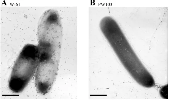
gold particles to observe immuno-gold stained Xyn5 the cell surface 4
using a transmission electron microscopy. As shown in Fig. I-2A 5
immuno-gold particles were observed on the cell surface. On the other 6
hand, no immuno-gold was observed with the W-61 cells grown in 7
medium I containing 0.7% glucose, or in medium I supplemented with 8
both 0.5% xylan and 0.5% glucose (data not shown). These results showed 9
that Xyn5 synthesis was inducible by xylan and subjected to catabolite 10
repression by glucose. On the other hand, no immuno-gold was observed 11
on the cell surface of strain PW103 that produces truncated Xyn5 lacking 12
the C-terminal SLH domain (Xyn5∆SLH) (Fig. I-2B). From these results, 13
I assumed that Xyn5 is anchored onto the cell surface via the C-terminal 14
SLH domain. To examine this hypothesis, I employed Western blotting to 15
detect Xyn5 and Xyn5∆SLH in the cell envelopes and the culture 16
supernatants. These strains were cultivated for 12 h in xylan and 17
harvested by centrifugation to separate the cells from the culture fluids. 18
The cells were then disrupted by French Pressure Cell to obtain cell 19
envelopes. Proteins of 140 kDa and 180 kDa were found in the cell 20
envelopes, but not in the culture supernatant, of the wild type cells (Fig. 21
I-3A) On the contrary, a protein of 120 kDa, which corresponds to 22
molecular mass of Xyn5∆SLH, was detected in the culture supernatant, 23
but not in the cell envelopes of PW103 (Fig. I-3B). These results 24
substantiate the notion that the C-terminal SLH domain anchors Xyn5 on 25
the cell surface. 26
27
Binding of the C-terminal SLH domain to peptidoglycan and secondary
28
cell wall polymers.
Binding of purified recombinant SLH (rSLH) domain (see 1
Materials and Methods) to the cell envelope components of strain W-61 2
was examined in vitro. The SLH domain was expressed and purified as a 3
GST fusion polypeptide in strain PE101. Peptidoglycan and secondary 4
cell wall polymers were prepared from a xyn5 knockout mutant PW101 5
and incubated with various amounts of GST-SLH. When GST-SLH was 6
incubated with peptidoglycan, the amounts of GST-SLH co-precipitated 7
with peptidoglycan increased as the amounts of the protein in the reaction 8
mixtures increased, with concomitant decrease in the amounts of free 9
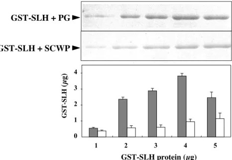
GST-SLH in the supernatants (data not shown). GST-SLH also bound to 10
the secondary cell wall polymers as to peptidoglycan (Fig. I-4). SLH 11
domain of Xyn5 thus has ability to bind to both peptidoglycan and 12
secondary cell-wall polymers of strain W-61. Binding affinity of GST-13
SLH to the cell envelopes was comparable to those to peptidoglycan and 14
to secondary cell-wall polymers (data not shown). 15
16
Binding of Xyn5 to oat spelt xylan.
17
Previously, it was demonstrated that purified rXyn5 binds to 18
crystalline cellulose, and Avicel PH-101, but not to oat spelt xylan (Ito et 19
al., 2003). It appears that CBM9 domain of Xyn5 has a binding ability
20
specific to cellulose, but CBM22 has no binding activity to cellulose and 21
xylan (Ito et al., 2003). However, Xyn5 should bind to xylan particles to 22
degrade them. The molecular particle size of oat spelt xylan (500-710 µm) 23
and Avicel PH-101 (40 µm) are different (Obae et al., 1999). So, I 24
predicted that Xyn5 binds to small size xylan particles. Oat spelt xylan 25
was homogenized by a mortar and pestle before being used for binding 26
assay. As shown in Fig. I-5, rXyn5 co-sedimented with the homogenized 27
oat spelt xylan (Fig. I-5A) as well as with Avicel PH-101 (Fig. I-5B), 28
showing that the SLH domain can bind to both cellulose and xylan, 29
although binding affinity to cellulose was significantly higher than that to 1
xylan. 2
3
Xyn5 is essential for the utilization of insoluble xylan.
4
To elucidate a role of Xyn5 in xylan utilization, I constructed a 5
xyn5 knockout mutant (PW101). This mutant poorly grew in minimal
6
media containing insoluble or even soluble xylan. However PW101 grew 7
well in minimal medium supplanted with xylo-oligosaccharides 8
(xylobiose to xyloheptaose) as a carbon source (data not shown). Since 9
the mutant assimilated all kinds of xylo-oligosaccharides, as confirmed 10
by the absence of the oligosaccharides in the culture, it may produce a 11
xylanase (Xyn1 or Xyn3) capable of hydrolyzing the oligosaccharides. 12
PW103 harboring plasmid pX5KO3 (xynΔslh) secreted Xyn5ΔSLH (Fig. 13
I-3). When strain W-61 and PW103 were cultivated in 0.7% (w/v) 14
insoluble xylan medium, growth of PW103 was significantly retarded, 15
while soluble Xyn5 was produced. After cultivation for 24 hr, xylanase 16
activity in the culture of PW103 was 0.4-fold lower than wild type strain 17
(Fig. I-6). In contrast, PW103 grew similarly to wild type W-61 in 0.7% 18
(w/v) soluble xylan medium (Fig. I-6). These results suggest that not only 19
production of Xyn5 but also its association with the cell surface are 20
important for the utilization of insoluble xylan by strain W-61. 21
22
Involvement of Xyn5 in the expression of xyn1 and xyn3.
23
To further study the roles of xyn5 in xylanase synthesis, I 24
compared total xylanase activity and xylanase molecules (Xyn1 through 25
Xyn3) produced between the wild-type W-61 and the xyn5-knockout 26
mutant PW101. Producing of five xylanases were compared by using 27
zymography. None of the xylanases was present in the culture of PW101 28
in the 0.7% (w/v) xylan medium (Fig. I-7). Strain PW102, carrying 29
plasmid pX5KO2 (xyn5), grew well in medium I in 0.7%(w/v) xylan and 1
produced all xylanases, whose amounts were comparable to those of 2
wild-type strain (Fig. I-7). Thus, xyn5 appears to play a crucial role in the 3
synthesis of both Xyn1 and Xyn3. 4
When PW101 (xyn5::cat) was cultivated in medium I containing 5
the xylo-oligosaccharides, it produced both Xyn1 and Xyn3 (Fig. I-9A), 6
suggesting that the xylo-oligosaccharides serves as inducers for Xyn1 and 7
Xyn3 synthesis. To substantiate the induction effects of the xylo-8
oligosaccharides and to examine their effects on xyn1 and xyn3 9
transcription, I measured the amounts of xyn1 and xyn3 mRNA in PW101 10
cells grown under inducible (in xylo-oligosaccharide medium) and none-11
inducible (in glucose medium) conditions using real-time RT-PCR. When 12
PW101 cells were cultivated in xylan medium, no xyn5 mRNA was
13
detected (data not shown) and the amounts of xyn1 and xyn3 mRNAs were 14
very low (Fig. I-8B). Strain PW101 started xyn1 and xyn3 transcription 15
within 3 hrs after being transferred from glucose medium into xylo-16
oligosaccharide medium (Fig. I-8C). The amounts of xyn5 mRNAs 17
increased in the wild-type cells cultivated in xylan medium. Transcription 18
of the xylanase genes initiated at different time of cultivation. In xylan 19
medium, xyn5 transcription began around in 3 h and reached maximum 20
levels during 4.5 and 7.5 h, then decreased after 9 h. Under the same 21
condition, xyn1 and xyn3 mRNA synthesis started in 6 and 7.5 h, 22
respectively (Fig. I-8A). Very few amounts of xyn1, xyn3, and xyn5 23
mRNAs were syntheized when the wild type W-61 was cultured in glucose 24
medium (data not shown). Xylobiose and xylotriose induced xyn1 and 25
xyn3 expression (Fig. I-9A). As measured by luciferase activity of a
26
Xyn5-luciferase fusion, expression of the xyn5-luc (luciferase gene) was 27
enhanced 30-fold in the presence of the xylo-oligosaccharides (Fig. I-10). 28
Taken together, xylobiose and xylotriose resulted from xylan by the 29
action of Xyn5 appear to act as the inducers of the xyn1, xyn3 and xyn5 1
genes.These findings suggest that expression of xyn5 is also induced by 2 xylo-oligosaccharide. 3 4 Discussion 5
In this chapter, I showed that Xyn5 of Paenibacillus sp. W-61 is 6
anchored onto the cell surface via its C-terminal SLH domain, that the 7
cell-surface Xyn5 plays a key role in an initial stage of xylan degradation, 8
and that xylo-oligosaccharides, possible products of xylan hydrolysis by 9
Xyn5, induce the expression of xyn1, xyn3, and xyn5. 10
䚭 䚭 䚭 䚭 As revealed by electron-microscopic observation of the W-61 11
cells labeled with immuno-gold particles using anti-Xyn5 antibodies (Fig. 12
I-2A), Xyn5 appear to cluster on the surface and the peripheral of the 13
cells. Xyn5 does not uniformly distribute on the cell-surface. Gold 14
particles can also be observed around the PW103 cell (Fig. I-2B), 15
although cells are washed with 250 µl of PBS. Xyn5 lacking the C-16
terminal SLH domain (produced by PW103) seems to freely defuse into 17
PBS buffer, unlike intact Xyn5 that is anchored on the cell surface. Xyn4, 18
a truncated form of Xyn 5, as well as intact Xyn5 are released in lesser 19
amounts compared to Xyn5ΔSLH. Some gold particles appear directly 20
attach on but not veil the W-61 cells (Fig. I-2A). Bacillus 21
stearothermophilus DSM2358 and B. stearothermophilus ATCC12980,
22
which produce high molecular-weight amylases associated with the cells, 23
have the surface layers similar to that of W-61 (Egelseer et al., 1995 and 24
Egelseer et al., 1996). The cell-associated amylases cover the whole cell 25
surface. The structure of bacterial cell surface, its components, and cell 26
wall polymers were not different among species (Schaffer, C and P, 27
Messner., 2005). Thus, cell surface structure has high diversity even 28
among Bacillus species. 29
Xyn5 is associated with the cell surface via its C-terminal SLH 1
domain (Fig I-3). Cell surface proteins of Gram-positive bacteria have a 2
domain necessary for targeting them onto the cell walls (Fujino et al., 3
1993; Lupas et al., 1994). Such a domain (ca. 55 residues) contains 4
modules of 10-15 converted amino acids, which is referred to the surface 5
layer homologous (SLH) domain. SLH domains, which is composed of 6
one or three modules, have been identified in over 40 proteins of Gram-7
positive bacteria (Engelhardt et al., 1998). The 180-kDa xylanolytic 8
protein, which cross-reacts with anti-Xyn5 antibodies (Fig I-3), appears 9
after the cell-surface Xyn5 is produced (data not shown). This 180-kDa 10
protein is susceptible to trypsin digestion (Ito et al., 2003). In contrast, 11
PW103 (xyn5∆slh) cells that were cultivated for 12 h in 0.7% xylan 12
medium had no 180-kDa protein on the cell surface (Fig I-3). Thus, Xyn5 13
may have an interaction with a cell-surface protein(s) via the SLH domain 14
of Xyn5 to make the 180-kDa complex, which is not dissociated by SDS, 15
implying that Xyn5 bounds covalently to this cell surface protein. I 16
determined the N-terminal amino acid sequence of the 180-kDa complex. 17
The determined sequence was Asp-Thr-Ala-Thr-Ser-Pro-Gln-Gln-Gln-18
Phe-Asp-Ala. This sequence is identical to the N-terminal sequence of the 19
100-kDa major S-layer protein of strain W-61 (Ito et al., 2003). The 20
counterpart of Xyn5 in the complex therefore should be the S-layer 21
protein. A molar ratio of Xyn5 to 180-kDa S-layer protein on the cells was 22
4:1 (data not shown). Further study to understand the molecular nature of 23
the Xyn5 complex is way under. 24
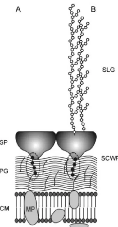
䚭 䚭 The GST-SLH domain of Xyn5 interacts with both peptidoglycan 25
and secondary cell wall polymers (Fig. I-4). Secondary cell wall polymers 26
(SCWP), which mainly consist of N-acetylglucosamine and
N-27
acetylmannnosamine, mediate non-covalent attachment of S-layer proteins and 28
SLH domain to under-layer peptidoglycan (Schaffer, C and P, Messner., 29
2005) (Fig. I-11). The binding affinity of GST-SLH to the secondary cell-wall 1
polymers is about one-third of that to the peptidoglycan (Fig. I-4). The 2
decreased in interaction between 5 µg GST-SLH and peptidoglycan is predicted 3
to be saturated GST-SLH protein. So, saturated cell surface localized Xyn5 4
could be easy to peel off the peptidoglycan, and excreted into the medium. Total 5
sugars of the secondary cell-wall polymers and the peptideglycan, as measured 6
by the phenol-sulfuric method (Hodge, J. E. and B. T, Hofreiter. 1962), were 7
57.8 µg and 14.6 µg per 100 µg dry weight, respectively. The SLH domain of 8
Xyn10B from an anaerobic bacterium Clostridium stecorarium binds to 9
peptidoglycan but not to secondary cell-wall polymers (Feng et al., 2000). 10
Ito et al, reported that recombinant Xyn5 binds to crystalline cellulose 11
but not to oat spelt xylan (Fig. I-5). However, it could bind to homogenized oat 12
spelt xylan with a smaller particle size with two-times lower affinity that to 13
Avicel (Fig. I-5A and I-5B). As xylan usually co-exists with cellulose, binding 14
of Xyn5 to cellulose would allow its access to the substrate xylan. CBM9 has 15
been shown to bind to cellulose (Ito et al., 2003). Xyn5 has another 16
cellulose-binding domain of family 22 (CBM22, Fig. I-1). Further study 17
is required to elucidate the roles of these CBMs in Xyn5 binding to 18
cellulose and xylan. 19
Xyn5 plays a crucial role in the utilization of insoluble xylan, but 20
not soluble xylan, by strain W-61 (Fig. I-6). Anaerobic bacteria that 21
efficiently degrade cellulose and xylan, including Clostridium 22
thermocellum, Clostridium cellulovorans, Ruminococcus flavefaciens,
23
Acetivibrio cellulolyticus and, have a super-molecular complex, termed
24
the ‘cellulosome’, on their cell surface (for reviews see: Doi & Kosugi., 25
2004; Beguin et al., 1996; Shoham et al., 1999; Bayer et al., 2004., 26
Demain et al., 2005). In contrast, most xylan-utilizing aerobic bacteria 27
secreted xylanases into the extracellular milieu, without localizing them 28
on the cell surface (Tomme et al., 1995; Warren et al., 1996). Scaffoldin 29
is a major component of the cellulosome and has ternary functions that 1
include binding to cellulosomal enzymes, binding to substrate cellulose, 2
and binding to cell-surface proteins (Doi & Kosugi., 2004). Cell-surface 3
Xyn5 of strain W-61 resemble the scaffoldin system of anaerobe 4
bacterium. Cellulosomes have many potential biotechnological
5
applications in the conversion of cellulosic biomass into sugars for the 6
production of valuable products such as ethanol or organic acids (Doi & 7
Kosugi., 2004). Strain W-61 represents aerobic xylanolytic bacteria that 8
have potential for application in biomass conversion. 9
䚭 䚭 䚭 䚭 I found that xyn5 knockout mutant grows poorly in media
10
containing insoluble xylan as a sole carbon source and that it produces 11
little amount of Xyn1 and Xyn3 (Fig. I-7). When xylo-oligosaccharides, 12
such as xylobiose and xylotriose, are present in the medium, the mutant 13
became able to produce Xyn1 and Xyn3 (Fig. I-9B). The cell-surface 14
Xyn5 appears to play a major role in the expression of xyn1 and xyn3 15
genes by generating their inducers, xylobiose and xylotriose, from xylan. 16
When the xyn5 mutation is complemented in trans by xyn5 cloned into 17
pHY300PLK, the xyn5 knockout mutant becomes able to produce the 18
xylanases (Fig. I-7), confirming that xyn5 is responsible for xylan-19
utilization and xylanase synthesis by strain W-61. 20
Real time RT-PCR revealed that the xylanase genes in strain W-21
61 are expressed in the order of xyn5, xyn1, and xyn3 (Fig. I-8A). 22
Regulatory mechanisms underlying the xyn gene expression are presently 23
poorly understood. In Prevotella bryantii B14, large
xylo-24
oligosaccharides (approximately 30-40 degree of polymerization) are 25
responsible for the induction of the xylanase genes (Miyazaki et al., 26
2005). The xylanase gene expression in Bacillus stearothermophilus No. 27
236 and B. subtilis is subjected to catabolite repression (Cho et al., 1999; 28
Jeong et al., 2006). However, it is not known how multiple xylanase genes 29
are regulated. A remarkable characteristic of the xyn5 knockout mutant is 1
that other xylanase genes (xyn1 and xyn3) are not induced even xylan is 2
present in the medium (Fig. I-8B). In this context, it should be noted that 3
xylo-oligosaccharides can induce the expression of the xylanase genes in 4
the xyn5 mutant and that xyn1 and xyn3 are expressed earlier in xylo-5
oligosaccharide medium than in xylan medium (Fig. I-8C). In conclusion, 6
cell-surface Xyn5 acts in the initial stage of xylan degradation to xylo-7
oligosaccharides, which induce xyn1 and xyn3 expression. Further study 8
is needed to elucidate the mechanism involved in the induction of the 9
xylanase gene expression in strain W-61. 10
Xylobiose most strongly activates xyn1 and xyn3 transcription 11
(Fig. I-9A). Xylotriose can also induce expression of these genes but 12
xylose is inert to do so. These results suggest that xylobiose and 13
xylotriose are the inducers for xyn1 and xyn3 expression. Xyn5 produces 14
mainly xylobiose, xylotriose, and xylotetraose from oat spelts xylan (Roy 15
et al., 2000), in accordance with a notion that Xyn5 plays a major role in
16
production of the inducers. Xylo-oligosaccharides also induce xyn5 17
expression and glucose antagonizes this induction effect (Fig. I-10), 18
showing that xyn5 expression is subjected to catabolite repression by 19
glucose. 20
In conclusion, in strain W-61 the cell-surface Xyn5 plays a key 21
role in the efficient degradation of insoluble xylan by generating the 22
inducers to express itself and the other genes of the xylanases that 23
coordinately degrade xylan. 24
25
References
26
Aiba, H., Adhya, S., and de Crombrugghe, B. 1981. Evidence for two
27
functional gal promoters in intact Escherichia coli cells. J. Biol. Chem. 28
256:11905-11910.
Balslev, Y., and Hansen, G. H. 1989. Preparation and use of recombinant
1
protein G-gold complexes as markers in double labelling
2
immunocytochemistry. Histochem J. 21:449-54. 3
Bayer, E.A., Belaich, J.P., Shoham, Y., and Lamed, R. 2004. The
4
cellulosomes: multienzyme machines for degradation of plant cell wall 5
polysaccharides. Annu Rev Microbiol 58:521–554. 6
Béguin, P., and Lemaire, M. 1996. The cellulosome: an exocellular,
7
multiprotein complex specialized in cellulose degradation. Crit Rev 8
Biochem Mol Biol. 31:201–236.
9
Cho, S. G., and Choi, Y. G. 1999. Catabolite repression of the xylanase
10
gene (xynA) expression in Bacillus stearothermophilus No. 236 and B. 11
subtilis. Biosci. Biotechnol. Biochem. 63:2053-2058.
12
Demain, A. L., Newcomb, M., and Wu, J. H. 2005. Cellulase, clostridia,
13
and ethanol. Microbiol Mol Biol Rev 69:124–154. 14
Doi, R. H., and Kosugi A. 2004. Cellulosomes: Plant-cell-wall degrading
15
enzyme complexes. Nature reviews microbiology. 2:541-551. 16
Egelseer, E., Schocher, I., Sára, M., Sleytr, U. B. 1995. The S-layer
17
from Bacillus stearothermophilus DSM 2358 functions as an adhesion site 18
for a high-molecular-weight amylase. J. Bacteriol. 177:1444-1451. 19
Egelseer, E. M., Schocher, I., Sleytr, U. B., Sára, M. 1996. Evidence
20
that an N-terminal S-layer protein fragment triggers the release of a cell-21
associated high-molecular-weight amylase in Bacillus stearothermophilus 22
ATCC 12980. J. Bacteriol. 178:5602-5509. 23
Engelhardt, H., and Peters, J. 1998. Structural research on surface
24
layers: a focus on stability, surface layer homology domains, and surface 25
layer-cell wall interactions. J Struct Biol. 124:276-302. 26
Feng, J. X., S. Karita, E. Fujino, T. Fyjino, T. Kimura, K. Sakka, and
27
K. Ohmiya. 2000. Cloning, sequencing, and expression of the gene
28
encoding a cell-bound multi-domain xylanase from Clostridium josui, and 29
characterization of the translated product. Biosci. Biotechnol. Biochem. 1
64:2614-2624.
2
Fujino, T., Béguin, P., Aubert, J. P. 1993. Organization of a Clostridium
3
thermocellum gene cluster encoding the cellulosomal scaffolding protein
4
CipA and a protein possibly involved in attachment of the cellulosome to 5
the cell surface. J Bacteriol. 175:1891-1899. 6
Ghitescu, L., and Bendayan, M. 1990. Immunolabeling efficiency of
7
protein A-gold complexes. J Histochem Cytochem.38:1523-30. 8
Gilbert, H. J., and Hazlewood, G. P. 1993. Bacterial cellulases and
9
xylanases. J. Gen. Microbiol. 139:187-194. 10
Hodge, J. E. and B. T, Hofreiter. 1962. Phe nol–su lfuric aci d. col orimet ric 11
met hod, in “Me thods in Car bohydr ate Che mistry ”. (Wh istler, R. L., and 12
Wolf rom, M. L., Eds .), p.3 88–389 , Aca demic Pre ss, New York . 13
Ito , Y., Tomi ta, T., Roy, N., Nakano, A.,Sug awara- Tomi ta, N., 14
Watanabe, S., Okai, N., Abe, N., and Kamio, Y. 2003. Cloning,
15
expression, and cell surface localization of Paenibacillus sp. strain W-61 16
Xylanase5, a multidomain xylanase., Appl. Environ. Microbiol., 69:6969-17
6978. 18
Jeong. M. Y., Lee. E. R., Yun. C. W., Cho. S. G., and Choi. Y. J. 2006.
19
Post-transcriptional regulation of the xynA expression by a novel mRNA 20
binding protein, XaiF. Biochem Biophys Res Commun.351:153-158. 21
Kato Fuminori. Ph. D. Thesis, Tohoku University, Sendai, 2005.
22
Koguch, A., K, Murashima, and R. H. Doi. 2002. Xylanase and acetyle
23
xylan esterase activities of XynA, a key subunit of the Clostridium 24
cellulovorans cellulosome for xylan degradation. Appl. Environ.
25
Microbiol. 68:6399-6402. 26
Laemmli, U. K. 1970. Cleavage of structural proteins during the
27
assembly of the band of bacteriophage T4. Nature. 227:680-685. 28
Leibovitz, E., and Beguin, P. 1996. A new type of cohesin domain that
specifically binds the dockerin domain of the Clostridium thermocellum 1
cellulosome-integrating protein CipA. J. Bacterial.178:3077-3084. 2
Leibovitz, E., Ohayon, H., Gounon, P., and Beguin, P. 1997.
3
Characterization and subcellular localization of the Clostridium 4
thermocellum scaffoldin dockerin binding protein SdbA. J. Bacterial.
5
179:2517-3084.
6
Lemaire, M., Ohayon, H., Gounon, P., Fujino, T., and Beguin, P. 1994.
7
OlpB, a new outer layer protein of Clostridium thermocellum, and binding 8
of its S-layer-like domains to components of the cell envelop. J Bacteriol. 9
177:2451-2459.
10
Lupas, A., Engelhardt, H., Peters, J., Santarius, U., Volker, S., and
11
Baumeister, W. 1994. Domain structure of the Acetogenium kivui surface
12
layer revealed by electron crystallography and sequence analysis. J 13
Bacteriol. 176:1224-1233.
14
Miyazaki, K., Hirase, T., Kojima, Y., Flint, H. J. 2005. Medium- to
15
large-sized xylo-oligosaccharides are responsible for xylanase induction 16
in Prevotella bryantii B14. Microbiology. 151:!4#1#2#1-4#1#2#5. 17
Nguyen, V. D., Kamio, Y., Abe, N., Kaneko, J., and Izaki, K. 1991.
18
Purification and properties of 䃈 -1,4-xylanase from Aeromonas caviae 19
W-61. Appl. Environ. Microbiol. 57:445-449. 20
Nguyen, V. D., Kamio, Y., Abe, N., Kaneko, J., and Izaki, K. 1993.
21
Purification and properties of 䃈 -1,4-xylanase 2 and 3 from Aeromonas 22
caviae W-61. Biosci. Biotechnol. Biochem. 56:1708-1712.
23
Obae, K, Iijima, H., and Imada, K. 1999. Morphological effect of
24
microcrystalline cellulose particles on tablet tensile strength. Int. J. 25
Pharm. 182:155-164.
26
Okai, N, Fukasaku, M., Kaneko, J., Tomita, T., Muramoto, K., and
27
Kamio, Y. 1998. Molecular properties and activity of a carboxyl-terminal
28
truncated form of xylanse 3 from Aeromonas caviae W-61.Biosci. 29
Biotechnol. Biochem. 62:1560-1567.
1
R!i!e!s!,! !W.!,! !H!o!t!z!y!,! !C.!,! !S!c!h!o!c!h!e!r!,! !I.!,! !S!l!e!y!t!r!,! !U. !B!.,! !S!a!r!a!,! !M.!1997. #
2
#E#v#i#d#e#n#c#e# #t#h#a#t# #t#h#e# #N#-#t#e#r#m#i#n#a#l# #p#a#r#t# #o#f# #t#h#e# #S#-#l#a#y#e#r# #p#r#o#t#e#i#n# #f#r#o#m# 3
#B#a#c#i#l#l#u#s# #s#t#e#a#r#o#t#h#e#r#m#o#p#h#i#l#u#s# #P#V#7#2#/#p#2# #r#e#c#o#g#n#i#z#e#s# #a# #s#e#c#o#n#d#a#r#y# 4
#c#e#l#l# #w#a#l#l# #p#o#l#y#m#e#r#.## "J." "B"a"c"t"e"r"i"o"l#.# #1!7!9!:3#8#9#2-3#8#9#8#.# 5
Roy, N., Okai, N., Tomita, T., Muramoto, K., and Kamio, Y. 2000.
6
Purification and some properties of high-molecular xylanases, the 7
xylanase 4 and 5 of Aeromonas caviae W-61. Biosci. Biotechnol. Biochem. 8
64:408-413.
9
S!a!r!a!,! !M!.,! and !S!l!e!y!t!r!,! !U. !B.!2000.# #S#-#L#a#y#e#r# #p#r#o#t#e#i#n#s#.#"J" "B"a"c"t"e"r"i"o"l#.#
10
#1!8!2:!859-868. 11
Sambrook, J., Fritsch, E. F., and Maniatis, T. 1989. Molecular cloning:
12
a laboratory manual. 2nd ed. Cold Spring Harbor Laboratory Press, Cold 13
Spring Harbor, N. Y. 14
Schaffer, C and P, Messner. 2005. The structure of secondary cell wall
15
polymers: how Gram-positive bacteria stick their cell walls together. 16
Mocrobiology. 151:643-651.
17
Shoham, Y., Lamed, R., and Bayer, E.A. 1999.The cellulosome concept
18
as an efficient microbial strategy for the degradation of insoluble 19
polysaccharides. Trends Microbiol. 7:275–281. 20
Sunna, A., and Antranikian, G. 1997. Xylanolytic enzymes from fungi
21
and bacteria. Crit. Rev. Biotechonol. 17:39-67. 22
Tomme, P., Warren, R.A., and Gilkes, N.R. 1995. Cellulose hydrolysis
23
by bacteria and fungi. Adv Microb Physiol. 37:1–81. 24
Warren, R. A. 1996. Microbial hydrolysis of polysaccharides. Annu Rev
25
Microbiol 50:183–212.
26
Watanabe, S., Nguyen, V. D., Kaneko, J., Kamio, Y., and Yoshida, S.
27
2008. Cloning, expression, and transglycosylation reaction of 28
Paenibacillus sp. strain W-61 Xylanase 1. Biosci. Biotechnol. Biochem.
(in press). 1
Whistler, R. L., and Richard, E. L. 1970. Hemicellulose in the
2
carbohydrates., p. 447-469. In W. Pigman and D. Horton (ed.), The 3
carbohydrates: chemistry and biochemistry, 2nd ed. Academic press, New 4
York, N.Y. 5
Wong, K. K., Tan, L. U., and Saddler, J. N. 1988. Multiplicity of
beta-6
1,4-xylanase in microorganisms: functions and applications. Microbiol 7
Rev. 52:305-317.
8
Wyman, C. E. 2007. What is (and is not) vital to advancing cellulosic
9
ethanol. Trends Biotechnol. 25:15-20. 10
pUC119 derivative carrying xyn5; Apr
pKAF derivative carrying xyn5 :: cat; Apr Spcr Cmr
pHY300 PLK derivative carrying xyn5 (!CBM9) pHY300 PLK derivative carrying xyn5 (!SLH) pHY300 PLK derivative carrying xyn5 ; Apr Tcr PW101 harboring pX5K04
Expression vector for GST fusion protein; Apr
Paenibacillus sp. W-61 pUX5S-22 pX5K01 pX5K04 pX5K03 pX5K02 Plasmid
Strain or plasmid Description or genotype
PW101 Derivative of W-61, xyn5 :: cat PW102 PW101 harboring pX5K02 PW103 PW101 harboring pX5K03 PW104
pKAF
Table I-1. Bacterial strains and plasmids used in this study
pUC119 pHY300PLK TaKaRa Bio TaKaRa Bio pGEX4T-1 GE Healthcare Source or reference This study This study This study This study Ito et al., 2003 Our lab This study This study This study This study pGEX4T-1 derivative carrying the slh region
pX5K05 This study
Eschrichia coli
DH 5" BL21 (DE3)
BL21 (DE3) harboring pX5K05
PE101 This study
Invitogen
TOYO B-Net Plasmid carrying the luciferase gene (luc)
PicaGene Basic vector 2
pX5K06 pHY300PLK derivative carrying the luciferase (luc) gene This study PW105 PW101 harboring PX5K06 This study
pX5K07 pHY300PLK derivative carrying the Pxyn5 – luciferase (luc) fusion This study Strains
W-61 Wild type
supE44 lacU169 (80 lacZM15) recA1 endA1 hsdR17 thi-1 gyrA96 relA1 F- ompT hsdSB (γ
B-mB-) gal dcm (DE3) Novagen
Cloning vector; Apr
Shuttle vector of E.coli and Bacillus subtilis ; Apr Tcr
Ts ori shuttle vector containing ori (pUC18), Ts ori (S. aureus pE194 ); Apr Spcr
Nguyen et al., 1991
Ap, ampicilline; Tc, tetracycline; Spc, spectinomycin; Cm, chloramfenicol
Fig. I-1. Modular structure of Paenibacillus sp. W-61 Xyn 5 (Ito et al., 2003).
Xyn5 can be dissected into six domains having the following features; a signal peptide region for secretion, two family 22 cellulose binding modules (CBMs), a catalytic domain of the family 10 glycoside hydrolase, family 9 CBM, lysine (K)-rich domain, and a domain containing S-layer homologous (SLH) modules. a.a. refers amino acid residues.
200 a.a.
Signal peptide Family 22 CBMs
Family 10 catalytic domain CBM 9 domain K-rich domain SLH domain
Fig. I-2. Electron micrographs of immunogold stained Xyn5 molecules on the cell surface of Paenibacillus
sp. W-61 (wild type, A) and PW103 (xyn5!slh, B).
Xyn5 molecules were labeled with polyclonal anti-Xyn5 antibodies and protein A-colloidal gold and observed under a transmission electron microscopy. A, W-61 cells grown in medium I containing with 0.7% (w/v) xylan. B, PW103 (xyn5!slh) producing Xyn5 without the SLH domain. Bars indicate 2 !m.
A
W-61B
PW103Fig. I-3. Western blot analysis of sub-cellular location of Xyn5.
Cells of strains W-61 (wild type) and PW103 (xyn5!slh) were cultivated at 37°C for 12 h in medium containing 0.7% (w/v) xylan to prepare their cytosols (sup) and cell envelops (ppt). Presence of Xyn5 in these sub-cellular preparations was analyzed by Western blotting using anti-Xyn5 antiserum. sup ppt sup ppt Wild PW103 120 kDa 180 kDa 140 kDa
Fig. I-4. Binding of the SLH domain to peptidoglycan (PG) and secondary cell wall-polymers (SCWP) of strain
W-61. PG or SCWP was incubated with various amounts (0 to 5 !g) of GST-SLH at 4˚C for 1 hrand reaction mixtures were centrifuged at 100,000 x g for 1 hr to precipitate the cell-wall components. GST-SLH associated with the cell-wall componentswere detected by Western blotting using anti-GST antiserum (top two figures). The amounts of GST-SLH were quantified against a standard curve of GST-SLH using NIH image. Bars represent standard deviations. 0 1 2 3 4 1 2 3 4 5 GST-SLH + PG GST-SLH + SCWP GST-SLH protein (µg) GST-SLH (µ g)
Fig. I-5. Binding of rXyn5 to oat spelt xylan and Avicel.
Ten !g of rXyn5 was incubate with indicated amounts of oat spelt xylan (A) or Avicel PH-101 (B) at 4˚C for 30
A
Oat spelt Xylan (mg)B
Avicel PH101® (mg)10 20 40 Free Bound 0 1 2 4 0 1 2 3 4 rXyn5 ( ! g) 3 10 20 40 rXyn5 ( ! g)
Fig. I-6. Effect of cell surface localized Xyn5 growth in insoluble xylan.
Strains W-61 (wild type, circle) and PW103 (xyn5!slh, triangle) were grown in medium I supplemented with 0.7% (w/v) water-insoluble xylan (red) or water soluble xylan (black) at 30˚C. Cell number in the cultures were counted at indicated time of incubation under a microscope.
Cell number (x 10 8 / ml) 0 5 10 15 20 25 3 6 9 12 15 18 24
W-61 in soluble xylan medium PW103 in soluble xylan medium W-61 in insoluble xylan medium PW103 in insoluble xylan medium
Incubation period (h) (kDa) 140 120 58 41 22 Xyn5 Xyn4 Xyn3 Xyn2 Xyn1
Fig. I-7. Knockout of xyn5 abolishes of Xyn1 and Xyn3 synthesis.
Zymogram of xylanases, wild, PW101 and PW102 cells were grown on 0.7% (w/v) xylan containing medium, and cell culture was analyzed by
zymography.
0 50 100 150 3 4.5 6 7.5 9 12 Time (h) xyn1 xyn3 xyn5 Time (h) 0 50 100 3 6 9 12 24 xyn3 xyn1
A
B
0 50 100 3 6 9 12 24 Time (h) xyn1 xyn3 150C
Fig. I-8. Transcription profiles of the xyn genes in strains W-61 (wild type) and PW101 (xyn5::cat) growing in xylan or xylo-oligosaccharide media.Wild-type cells were grown in medium I supplemented with 0.7% (w/v) xylan at 30˚C (A). PW101 cells were grown in medium I supplemented with either xylan (B) or xylo-oligosaccharides (C). Total RNA was extracted from the cells grown as above at the denoted time and used for RT-PCR.
Relative amount of mRNA
Relative amount of mRNA Relative amount of mRNA
(kDa) 140 120 58 41 22 Xyn3 Xyn2 Xyn1 X1 X2 X3 X4 X5 X6 1 100 200 300 xyn1 xyn3 Relative amounts of mRN A B
Fig. I-9. Inducible synthesis of xyn1 and xyn3by xylo-oligosaccharides.
A, Zymographies of xylanases in the cultures of W-61 (wild-type) and PW101 incubated in the presence of xylo-oligosaccharides at 30˚C for 12 h .
B, Total RNA was extracted from PW101 cells grown in minimal medium I containing, xylose (X1), or
0 0.5 1 1.5 2 2.5 3 3.5 4 0 1 2 3 4 Time (h) RLU / OD 660 (1.0 x 10 6) 0 0.5 1 1.5 2 2.5 3 3.5 OD 660 Glc Xo
Fig. I-10. Effects of xylo-oligosaccharides on xyn5 expression.
PW105 cells carrying plasmid pX5K07 ((Pxyn5::luc )were cultivated in medium I containing glucose (circle) or xylo-oligosaccharides (square) at 0.7% (w/v). Cell growth was measured by a
spectrophotometer at 660 nm (OD660). Extracts were prepared from the cells harvested from glucose (open bars) and xylo-oligosaccharide cultures (closed bars) at indicated period of incubation and used to measure the activities of luciferase. Thin bars represent standard deviations.
Fig. I-11. Schematic representation of bacillus cell wall.
SP, surface protein; PG, peptidogycan; CM, cytoplasmic membrane; SLG, S-layer glycoprotein; SCWP, secondary cell-wall polymers (dot ellipses). SCWP is covalently bound to muramic acid of PG and non-covalently to SP
Chapter II
1
Membrane
location
of
Paenibacillus
sp.
W-61
LpX
2
lipoprotein and its function as a chaperon of Xylanase 1
3synthesis
4 5 Introduction 6During cloning and expression experiments of xyn1 in E. coli, it 7
was found that the E. coli cells, which harbored a plasmid containing 8
xyn1 and its downstream region (tentatively named as orf6), accumulated
9
rXyn1 with enzyme activity in the cells, whereas the cells having xyn1 10
alone accumulated insoluble rXyn1 without activity. In this chapter, I 11
show that ORF6 is a membrane lipoprotein (LpX) and LpX is located on 12
the outer leaflet of the cytoplasmic membrane and is a crucial protein for 13
secretion of Xyn1 outside the cells as a soluble and active form. As the 14
amount of xyn1 mRNA in a lpx mutant was similar to that in the wild-type 15
strain W-61, I concluded that LpX is not mRNA stabilizer, like XaiF, for 16
Bacillus stearothermophilus xylanase gene (xynA) (Cho et al., 1995, and
17
Cho et al., 1998), but a membrane lipoprotein having chaperone-like 18
function during Xyn1 secretion through the cytoplasmic membranes. I 19
will discuss the properties of LpX in detail. 20
21
Material and methods
22
Mat erials. Restricti on enzymes, T4 DNA ligase, Taq DNA
23
pol ymerase, and plasmids pUC119 and pHY300PLK wer e fro m TaKaRa 24
bio (Otsu, Japan). Thermosen sitive (Ts) vector plasmid pKAF was used 25
for gene rep lacement was a stock of our lab orator y. Ant i-His tag ant ibody, 26
HiT rap chelating HP col umn, Hybond ECL mem brane, ECL det ection 27
system were from GE Healthcare (Buckinghamshire, UK). [1-1 4
C] 28
Pal mitic aci d (1. 85 GBq /mmol) was fro m Dai ichi Pur e Chemicals (Tokyo, 29
Jap an). Oat -spelt xyl an, PEG (Mr: 6,0 00), proteinase K fro m Tri tirach ium 1
alb um, ant ibioti cs, DNase I, RNase A, and Freund’s com plete adj uvant
2
wer e from Wako Pur e Chemical Ind ustry (Osaka, Japan). Ant i-rabbit IgG 3
(Fc) -alkal ine pho sphatase con jugate was fro m Promega (Madison, WI) . 4
ABI PRI ZM Big Dye Ter minato r Cycle Seq uencing Ready Reaction kit 5
wer e from Applied Biosystem s (Fo ster, CA) . Wat er sol uble xyl an was 6
prepared fro m oat -spelt-xyl an (Flu ka, U. S. A) by the met hod described 7
previousl y (Watanabe et al., 200 8). Unl ess otherwise stated, chemicals 8
used were of the best grade commercially available. 9
Bacterial strains, plasmids, and media. Bacterial strains and
10
plasmids were listed in Table II-1. Medium I (Nguyen et al., 1991) 11
containing water soluble xylan and LB medium were used for growth of 12
Paenibacillus species and E. coli strains, respectively. Antibiotics were
13
added to cultures, when necessary. Liquid cultures were shaken at 37˚C 14
unless otherwise noted. 15
DNA sequencing of the 5’ and 3’ flanking regions of xyn1. The 5’
16
and 3’ flanking regions of xyn1 were sequenced by inverse PCR walking. 17
Inverse PCR was done using self-ligated chromosomal DNA fragments 18
digested by an appropriate restriction enzyme as a template. Nucleotide 19
sequence was determined using ABI 377 cycle sequencing system, and 20
sequences were assembled by GENETYX-Mac ATSQ software (Genetyx 21
Co., Tokyo, Japan). Open reading frame (ORF) identification and 22
multiple sequence alignments were performed using GENETYX program 23
(Genetyx Co., Tokyo, Japan). Homology search was performed using 24
FASTA and BLAST programs implemented at the DDBJ/EMBL/GenBank 25
nucleotide sequence database and SWISSPROT/NBRF-PIR protein 26
databases. The nucleotide sequence of the xyn1locus (12-kb) has been 27
deposited in the in DDBJ/EMBL/GenBank databases under accession no. 28
AB274730. 29
Cloning of xyn1 and xyn1-orf6 in E. coli. The xyn1 and xyn1-orf6
1
genes were amplified by PCR using Paenibacillus sp. W-61 chromosomal 2
DNA as a template and oligonucleotide primers xyn1-pmt Bam (5’-3
TTTGGATCCGCACGTACCGCACATC-3’) and xyn1-term Hin (5’-4
AAAAAGCTTCCACTTTTTCATTCTATGTCTCC-3’), or orf6-full Hin 5
(5’-TTTAAGCTTATGTTCTCTGTCGTCTTC-3’), respectively. PCR 6
products were digested with BamHI (single underline) and HindIII 7
(double underlines), and then inserted into BamHI-HindIII sites of 8
pUC119. Resultant plasmids containing xyn1 alone and xyn1-orf6 were 9
designated as pX1T and pXFT, respectively (Table II-1). These plasmids 10
were introduced into E. coli DH5α, and the cells harboring pX1t and 11
pFXT were designated as UX101 and UX102, respectively (Table II-1). 12
Construction of orf6 knockout mutant of Paenibacillus sp. W-61.
13
An orf6 knockout mutant was obtained as described previously 14
(Watanabe., 2006). A 2.5-kbp DNA fragment containing xyn1 and orf6 15
was amplified by PCR using Paenibacillus sp. W-61 chromosomal DNA 16
as a template and primers, xyn1-pmt Bam (5’-17
TTTGGATCCGCAGGACGTACCGCACATC-3’) and orf6 dco rv (5’-18
TACAAGCTTCTCATGATTTCCAACGCCG-3’). The resultant product 19
was digested with BamHI (underlined site) and HindIII (double 20
underlined site), and cloned into the BamHI and HindIII sites of plasmid 21
pHY300PLK. The internal 740-bp StuI and BanII fragment of inserted 22
orf6 was replaced by a cat cassette (Kato et al., 2005). Resultant orf6::cat
23
fragment was inserted into the temperature-sensitive shuttle vector pKAF 24
to obtain plasmid pKMC (Table II-1). This plasmid was introduced into 25
Paenibacillus sp. W-61 by electroporation, and transformants were
26
incubated at 43˚C. An orf6 knockout mutant was selected on LB plate 27
containing chloramphenicol (10 µg/ml), and designated as PSC301 (Table 28
II-1). For complementation experiments, the orf6 expression plasmid was 29
constructed as follows. A promoter region of xyn1 was amplified using 1
Paenibacillus sp. W-61 chromosomal DNA as a template and primers,
2
xyn1-pmt fw Eco (5’-CAGGAATTCCCGCACATCTGGTATGAAGAG-3) 3
and xyn1-pmt rv Bam (5’-4
TCCGGATCCTTGGATTAGTTTTTGAATAATTCGGTAC-3’), and 5
digested with EcoRI and BamHI (underlined sites). Resultant EcoRI-6
BamHI fragment containing xyn1 promoter was inserted EcoRI and BamHI
7
sites of pHY300PLK. Then orf6 with its own ribosomal binding site and 8
termination loop was amplified using Paenibacillus sp. W-61 9
chromosomal DNA as a template and primers, orf6-SD fw Bam (5’-10
CCAGGATCCAGAAGGAGACATAGAATG-3’) and orf6-full Hin. The 11
amplified product was digested with BamHI and HindIII (underlined site). 12
Resultant fragment was inserted into BamHI and HindIII site in 13
immediate downstream of xyn1 promoter to obtain pHPX4T (Table II-1). 14
Paenibacillus strain PSC301 was transformed by plasmids pHY300PLK
15
and pHPX4T to obtain strains PSC401 and PSC402, respectively (Table 16
II- 1). 17
Total RNA preparation and Northern blotting analysis. Cells of
18
Paenibacillus sp. W-61 and its mutants were grown in medium I
19
containing 0.7% soluble xylan. When appropriately, chloramphenicol (10 20
µg/m l), spectinom ycin (100 µg/m l), or tet racycl ine (1 µg/m l) wer e add ed 21
into medium. Total RNA was prepared according to the method of Aiba et 22
al. (1981). Northern blotting analysis was done according to a
23
manufacture’s instructions of ECLT M random prime system, version II and 24
gene image CDP-StarT M detection module (GE Healthcare). Xyn1 specific
25
probe was prepared as follows. A 846-bp DNA fragment was amplified 26
from plasmid pXFT as a template DNA by PCR using primers, xyn1-pmt 27
Bam and xyn1-R (5’-28
ATTAAGCTTGGATCCTTACCAAACGGTCACGTTGGA-3’). Resulting 29