peripheral blood dendritic cells before
parturition by a novel purification method
著者
Tao Zhuang, Megumi Urakawa, Hidetoshi Sato,
Yuko Sato, Teruaki Taguchi, Tsuyoshi Umino,
Shiro Katto, Koutaro Tanaka, Kozue Yoshimura,
Naokazu Takada, Hiroko Kobayashi, Megumi Ito,
Michael T. Rose, Yoshio Kiku, Yuya Nagasawa,
Haruki Kitazawa, Kouichi Watanabe, Tomonori
Nochi, Tomohito Hayashi, Hisashi Aso
journal or
publication title
Animal science journal
volume
89
number
7
page range
1011-1019
year
2018-04-30
URL
http://hdl.handle.net/10097/00126979
doi: 10.1111/asj.13014Title: Phenotypic and functional analysis of bovine peripheral blood dendritic cells 1
before parturition by a novel purification method 2
3
Authors: Tao Zhuang1, 2, †, Megumi Urakawa1, 2, †, Hidetoshi Sato3, Yuko Sato3, Teruaki 4
Taguchi1, 2, Tsuyoshi Umino1, 2, Shiro Katto1, 2, Koutaro Tanaka1, 2, Kozue Yoshimura1, 2, 5
Naokazu Takada3, Hiroko Kobayashi3, Megumi Ito3, Michael T. Rose4, Yoshio Kiku5, 6
Yuya Nagasawa5, Haruki Kitazawa6, Kouichi Watanabe1, 2, Tomonori Nochi1, 2,
7
Tomohito Hayashi5 and Hisashi Aso1, 2, * 8
9
Institute, address, country: 1Cellular Biology Laboratory, Graduate School of 10
Agricultural Science, Tohoku University, Sendai, Miyagi, Japan, 2International
11
Education and Research Center for Food and Agricultural Immunology, Graduate 12
School of Agricultural Science, Tohoku University, Sendai, Miyagi, Japan, 3Miyagi
13
Prefecture Animal Industry Experiment Station, Iwadeyama, Miyagi, Japan, 4Institute of 14
Biological, Environmental and Rural Sciences, Aberystwyth University, Cardiganshire, 15
United Kingdom, 5Hokkaido Research Station, National Institute of Animal Health,
16
NARO, Sapporo, Hokkaido, Japan, 6Food and Feed Immunology Group, Graduate
17
School of Agricultural Science, Tohoku University, Sendai, Miyagi, Japan. 18
19
Running Head: Bovine peripheral blood dendritic cells 20
21
* Corresponding Author: Hisashi Aso, Ph.D, Graduate School of Agricultural Science, 22
Tohoku University, Sendai, Miyagi, Japan. 23
Tel: (+81)22-757-4313; Fax: (+81)22-757-4315; Email: [email protected] 24
† These authors contributed equally to this work.
Abstract 26
Dendritic cells (DCs) are professional antigen presenting cell specialized in antigen 27
uptake and processing, and play an important role in the innate and adaptive immune 28
response. A subset of bovine peripheral blood DCs was identified as 29
CD172a+/CD11c+/MHC class II+ cells. Although DCs are identified at 0.1-0.7% of 30
PBMC, the phenotype and function of DCs remains poorly understood with regard to 31
maintaining tolerance during the pregnancy. All cattle used in this study were one month 32
before parturition. We have established a novel method for the purification of DCs from 33
PBMC using MACS, and purified the CD172a+/CD11c+ DCs, with high expression of 34
MHC class II and CD40, at 84.8% purity. There were individual differences in the 35
expressions of CD205 and co-stimulatory molecules CD80 and CD86 on DCs. There 36
were positive correlations between expression of cytokine and co-stimulatory molecules 37
in DCs, and the DCs maintained their immune tolerance, evidenced by their low 38
expressions of the co-stimulatory molecules and cytokine production. These results 39
suggest that before parturition a half of DCs may be immature and tend to maintain 40
tolerance based on the low cytokine production, and the other DCs with high 41
co-stimulatory molecules may already have the ability of modulating the T-cell linage. 42
43
Keywords: dendritic cell; cattle; positive-selection; phenotype; cytokine
Introduction 45
46
Dendritic cells (DCs) were first identified in the peripheral lymphoid organs of mice 47
(Steinman & Cohn, 1973), specializing in antigen uptake and processing as an 48
antigen-presenting cell (APC). DCs also play an important role in the innate and 49
adaptive immune response (Banchereau & Steinman, 1998). The phenotypic and 50
functional characterizations of peripheral blood DCs in the human have been described 51
in several studies (Thomas et al., 1993; Odoherty et al., 1994; MacDonald et al., 2002). 52
However, the phenotype and function of peripheral blood DCs in cattle remain poorly 53
understood. 54
A subset of bovine peripheral blood DCs was identified as CD172a+/CD11c+/MHC 55
class II+ cells in the CD3−/B-B2−/CD14− population (Miyazawa et al., 2006) and
56
expressed a CD205 molecule on the cell surface (Gonzalez-Cano et al., 2014). CD205, 57
as an antigen-uptake receptor, was also expressed on DCs in lymphoid tissue (Gliddon 58
et al., 2004). In addition, it has previously been reported that the surface molecules of 59
CD40, CD80 and CD86 in DCs provided co-stimulate signals in T cell activation 60
(VanGool et al., 1996). 61
In order to prevent the fetal rejection caused by the recognition of paternal antigens, 62
the maternal immune system has to be mobilized toward tolerance (Zenclussen, 2013). 63
T helper (Th) cells play a central role in immune responses. However, the expression of 64
Th1 and Th17-related gene was inhibited in bovine late gestation (Maeda et al., 2013). 65
The previous report showed the characterization of higher Th2/regulatory immunity by 66
the increases of IFN-γ occurring after parturition and IL-4 production before calving 67
(Paibomesai et al., 2013). 68
Among periparturient Jersey cows during the 2 weeks before and 2 weeks after 69
parturition, the percentage of T cells with CD3, CD4, and gamma delta T-cell receptors 70
reduced substantially in blood (Kimura et al., 1999). During the periparturient period 71
there is a decline in T-lymphocyte cell subsets, which parallels a reduction in functional 72
capacities of blood lymphocytes (Kimura et al., 2002). Paternal T cells are aware of the 73
presence of paternal antigens during pregnancy, where they acquire a transient state of 74
tolerance specific for paternal antigens (Tafuri et al., 1995). Regulatory T cells (Treg), 75
the main function for which is to prevent autoimmunity, emerged as important players 76
in regulating tolerance toward paternal and fetal antigens (Sakaguchi et al., 1995). Treg 77
must first encounter antigens presented by antigen-presenting cells, as for example, DCs 78
in an appropriate cytokine environment, to proliferate and function. In addition, DCs 79
represented the first event leading to a protective adaptive immune response (Robertson 80
et al., 1996), and contributed to the expansion of the peripheral Treg population 81
(Schumacher et al., 2012). Immature DCs expressed a low level of MHC molecules and 82
co-stimulatory molecules such as CD40, CD80 and CD86, and showed the reduced 83
production of pro-inflammatory cytokines (IL-12, TNFα, IL-6) (Lutz & Schuler, 2002). 84
These data are compatible with the hypothesis that declining T-cell populations may 85
contribute to the immunosuppression reported for dairy cows at calving, and that DCs 86
may regulate the population and functions of T cells during the days and weeks before 87
and after parturition. However, the function for maintaining the tolerance during the 88
pregnancy has not been clearly described in DCs in bovine blood. Previous works 89
showed that in the late gestation, the cows had a heightened susceptibility to persistent 90
infections caused by mastitis and abortion-causing pathogens (Green et al., 2002; 91
Williams et al., 2000). Therefore, we studied the cattle which were one month before 92
parturition. 93
In this study, we investigated the phenotypic and functional characterization of 94
bovine peripheral blood DCs before parturition. As the population of DCs is less than 95
5% in bovine peripheral blood mononuclear cells (PBMC), there is a need to isolate 96
highly purified DCs subpopulations in sufficient numbers. Therefore, we have 97
established a novel method of two-step Magnetic-activated cell sorting (MACS) for 98
bovine peripheral DCs, and were able to obtain DCs at a purity of more than 85% from 99
PBMC. After the purification, we determined the expressions of surface markers (MHC 100
II, CD205, CD40, CD80 and CD86) on DCs using flow cytometry and analyzed the 101
expression of a number of cytokines (IL-12a, IL-4, IFN-γ, and IL-6). This study 102
provides the evidence for immune regulation of bovine DC populations before 103
parturition. 104
Materials and Methods 105
106
Animals 107
Sixteen Holstein Friesian cows (average age at 5.2±2.2 years,calving number at 108
2.3±1.8), housed at the Miyagi Prefecture Animal Industry Experiment Station, were 109
used in this study. All animal handing and experimental protocols were conducted in 110
compliance with guidance approved by the Tohoku University Environmental and 111
Safety Committee on Experimental Animal Care and Use, and the Environmental and 112
Safety Committee on Miyagi prefecture animal industry experiment station. These 113
animals were clinically healthy and kept in the same conditions. 114
115
Blood sampling 116
Jugular venous blood (200 mL) was obtained from the cows at one month prior to 117
parturition, into the tubes containing sodium heparin, and was diluted 1:1 with 118
phosphate-buffered saline (PBS). PBMC were separated from the buffy coat using 119
Lympholyte®-H (1.077 g/mL; CEDARLANE, Burlington, Ontario, Canada) gradient
120
centrifuged at 600 × g for 30 min at 18°C. PBMC were washed once with lysing buffer 121
(tris-HCl buffer containing 0.83% ammonium chloride) and twice with PBS at 450 × g 122
each for 10 min at 4°C. 123
124
Purification of peripheral blood DCs 125
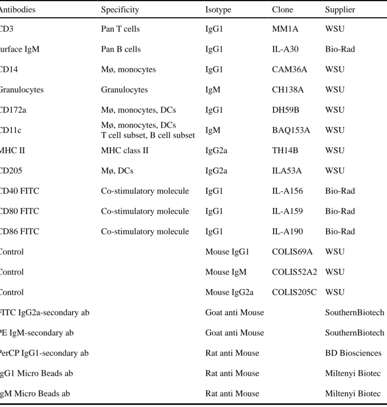
The anti-bovine antibodies in this study were purchased from WSU (Pullman, WA, 126
USA), Bio-Rad (Hercules, CA, USA), SouthernBiotech (Birmingham, AL, USA), BD 127
Biosciences (Franklin Lakes, NJ, USA) and Miltenyi Biotec (Bergisch Gladbach, 128
Germany)(Table 1).For the sorting of CD3−/sIgM−/CD14−/Granulocytes− cells, PBMC 129
were washed with PBS containing 0.5% bovine serum albumen (BSA), and incubated 130
with the mixture of mouse anti-bovine CD3 (diluted 1/50), mouse anti-bovine sIgM 131
(diluted 1/100), mouse anti-bovine CD14 (diluted 1/50), and mouse anti-bovine 132
Granulocytes (diluted 1/1000) antibodies for 30 min on ice, followed by the incubation 133
with rat anti-mouse IgG1 Micro Beads and rat anti-mouse IgM Micro Beads for 30 min 134
on ice, respectively. CD3−/sIgM−/CD14−/Granulocytes− cells containing DCs were 135
negatively selected using Auto MACS magnetic columns (Miltenyi Biotec, Bergisch 136
Gladbach, Germany). After negative selection, CD3−/sIgM−/CD14−/Granulocytes− cells 137
were incubated with mouse anti-bovine CD172a antibody (diluted 1/200) and rat 138
anti-mouse IgG1 Micro Beads for 30 min on ice, respectively. CD172a+ cells were 139
positively selected from CD3−/sIgM−/CD14−/Granulocytes− cells using Auto MACS 140 magnetic columns. 141 142 Flow cytometry 143
In order to detect bovine DCs, PBMC, CD3−/sIgM−/CD14−/Granulocytes− 144
negative-selected cells in MACS step 1 (negative-selected cells) and CD172a+
145
positive-selected cells in MACS step 2 (positive-selected cells) were stained with mouse 146
anti-bovine CD172a antibody and co-stained with mouse anti-bovine CD11c (diluted 147
1/500) and MHC class II (diluted 1/250) antibodies. PBMC and negative-selected cells 148
were incubated with anti-bovine CD3, sIgM, CD14 or Granulocytes antibody in order to 149
confirm the deletion of T cells, B cells, monocytes and granulocytes. Negative-selected 150
cells were incubated with mouse anti-bovine MHC class II, CD40, CD205, CD80 or 151
CD86 antibody, and treated with secondary fluorescent antibodies for 30 min on ice in 152
the dark. After the treatment of secondary fluorescent antibodies in Table 1, each cell 153
was subjected to the flow cytometry analysis using the Accuri C6 flow cytometer (BD 154
Biosciences) and the BD Accuri C6 software, Version 1.0.264.21 (BD Biosciences). In 155
each experiment, cells incubated with isotype-matched antibodies and secondary 156
fluorescent antibodies were selected as controls. 157
158
Immunocytochemical staining 159
Negative- and positive-selected cells were stained with mouse anti-bovine CD172a 160
antibody and co-stained with mouse anti-bovine CD11c and MHC class II antibodies, 161
and then stained with PerCP conjugated rat anti mouse-IgG1, PE conjugated goat anti 162
mouse IgM and FITC conjugated goat anti mouse-IgG2a fluorescent antibodies (Table 163
1). Cells were then centrifuged onto glass slides (Cytospin 2 Thermo Shandon, 164
Pittsburgh, PA, USA) at 600 × g for 5 minutes. After air drying for 5 min, cells were 165
counterstained with 4’,6-diamidino-2-phenylindole (DAPI) for 5 min at room 166
temperature in the dark, and were washed three times with PBS. Slide images were 167
viewed using a Laser Scanning Microscope 700 (Carl Zeiss, Jena, German), and 168
photographed at 400X with LSM software ZEN 2012, Version 8.0.0.273. 169
170
Quantitative real-time polymerase chain reaction (qRT-PCR) analysis 171
After the negative and positive selections, the purified bovine peripheral blood DCs 172
were stored at -80°C. Total RNA was extracted from them using ISOGEN II reagent 173
(Takara Bio Inc., Siga, Japan) following the manufacturer's instructions, and its 174
concentration was determined by the spectrophotometry at 260 nm. The reverse 175
transcription and cDNA synthesis were described as below. In brief, 2 μg of total RNA 176
was mixed with 500 ng oligo (DT)12-18 and 1 μL of 10 mM deoxynucleotide
177
triphosphates (dNTPs) (Invitrogen, Carlsbad, CA, USA). The mixture was heated to 178
65°C for 10 min in order to prepare for cDNA synthesis. Then the first-strand cDNA 179
was incubated with 200 units of Superscript RT III, 0.1M DTT and 5×First-Strand 180
Buffer (Invitrogen) at 50°C for 1 h, and then at 70°C for 15 min. 181
One μL cDNA sample, 7 μL SYBR Green Premix Taq (Takara Bio Inc.), 1 μL of 5pM 182
corresponding primer pair, and RNase-free water were added in a 20 μL final volume 183
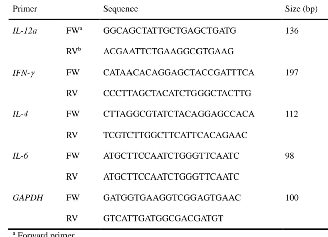
per well in 96-well plate. The primer sets of bovine cytokines were listed in Table 2 184
(Takara Bio Inc.). The transcripts using the bovine peripheral blood DCs cDNA were 185
amplified with the Thermal Cycler Dice Real Time System Single (Takara Bio Inc.): 1 186
cycle at 95°C for 30 sec; 40 cycles at 95°C for 5 sec, 60°C for 30 sec, then 95°C for 15 187
sec, 60°C for 30 sec, and finally 95°C for 15 sec. From template DNA, SYBR green 188
fluorescence was detected for the calculation of copy numbers. The specificity and the 189
integrity of PCR product were confirmed by the dissociation curve analysis. 190
GAPDH-specific primers were used as the internal controls, and the reactions without 191
template were used as negative control experiments. The results of target gene were 192
presented as the relative expression level to the expression of house-keeping GAPDH 193 gene. 194 195 Statistical Analysis 196
Values are reported as means ± SD. Statistical analyses were performed using the 197
software GraphPad 6.00 program (GraphPad software Inc., La Jolla, CA, USA). The 198
correlation between two parameters was analyzed by Pearson correlation coefficient test 199
(*: p<0.05, **: p<0.01).
Results 201
202
Purification of bovine peripheral blood DCs 203
We tried to purify bovine blood DCs from PBMC. Fig.1 shows the purification 204
process of bovine peripheral blood DC. The expression of the surface molecules such as 205
CD172a, CD11c, and MHC class II, such as specific markers of DC, were assessed by 206
three-color flow cytometry without any gate (Fig.1 A). Among the total PBMC, 14.8% 207
CD172a+CD11c+ cells were present and almost expressed a MHC class II molecule. 208
However, it is well known that CD11c is highly expressed on monocytes, macrophages 209
(Mø) and natural killer (NK) cells, and that CD172a+/CD11c+ cells possibly include a 210
subset of T cells, B cells, NK cells and monocyte/Mø. Therefore, we attempted to 211
remove these cell populations from PBMC using each specific monoclonal antibody. 212
After the negative selection, CD172a+/CD11c+ cells were found to represent about 6.5% 213
of the negative-collected cells and also expressed MHC class II on the cell surface. The 214
negative selection using MACS removed T cells (CD3+), B cells (surface IgM+),
215
monocytes (CD14+) and granulocytes from PBMC, and these populations in 216
negative-selected cells disappeared (Fig.1 B). Therefore, CD172a+/CD11c+ cells in the
217
negative-selected cells were considered as bovine peripheral blood DCs, which also 218
expressed MHC class II molecule. However, the negative-selected cells contained a 219
large amount of population of CD172a−/CD11c− non-DC cells. Next, we tried to purify 220
CD172a+/CD11c+ cells from the negative-selected cells. The positive selection with 221
CD172a antibody revealed that the purity of CD172a+/CD11c+ DCs was 84.8%, and that
222
they also expressed MHC class II strongly. 223
Photographs of peripheral blood DCs 225
Peripheral blood DCs after the negative and positive selections were stained with 226
anti-bovine CD172a (Red), CD11c (Green) and MHC class II (Green) antibodies. All 227
samples were counterstained with DAPI (Blue) (Fig.2). After the negative selection, 228
CD172a+/CD11c+ and CD172a+/MHC class II+ DCs were detected as a small population 229
in the photographs. Indeed, there was a plenty of CD172a−/CD11c−/MHC class II− 230
non-DC cells indicated with arrows. However, this cell population indicated with arrows 231
decreased after the positive selection with anti-CD172a antibody. Almost all the 232
positive-selected cells expressed CD172a, CD11c and MHC class II, which were 233
considered as the bovine peripheral blood DCs. These data suggest that the two-step 234
MACS method can purify highly DCs from bovine blood. 235
236
Phenotypic analysis and cytokine expression of bovine peripheral blood 237
CD172a+/CD11c+ DCs before parturition 238
Next, the surface expression of MHC class II, CD40, CD205, CD80 or CD86 was 239
analyzed on CD172a+/CD11c+ cells after the negative selection (Fig.3 A). The results 240
demonstrated that almost all the CD172a+/CD11c+ DCs expressed the molecules of
241
MHC class II (98.48±0.54%) and CD40 (94.98±0.88%). However, there were 242
individual differences in the expression of CD205, CD80 or CD86 in the 243
CD172a+/CD11c+ DCs. The percentages of CD205, CD80 and CD86 positive cells were
244
17.08±3.97, 29.68±4.23, and 23.50±6.02 of CD172a+/CD11c+ DCs, respectively. Before 245
parturition, there were significant correlations between the percentage of CD86 and the 246
percentages of CD80 or CD205 on CD172a+/CD11c+ DCs (Fig.3 B). 247
As the purity of bovine peripheral blood DC was more than 85% after positive 248
selection, it became available for the examination of the expression of T cell-modulation 249
cytokines in DCs (Fig.4). There were significant correlations in bovine peripheral DCs 250
with the activated molecule of CD205 and the mRNA expressions of IFN-γ and IL-6. In 251
addition, there were significant correlations between the co-stimulatory molecule CD80 252
and the expressions of IL-12a, IL-4, and IFN-γ, and between CD86 and the expressions 253
of IL-4, IFN-γ and IL-6. 254
Discussion 255
256
In this study, we have established a novel purification method for bovine peripheral 257
blood DCs. We have also characterized the phenotype and function of the DCs. A 258
previous study revealed that DCs were identified at 0.1-0.7% of PBMC (Renjifo et al., 259
1997). Because of the low percentage of DCs in the PBMC, it was necessary to deplete 260
the non-DC from bovine PBMC (Renjifo et al., 1997; Miyazawa et al., 2006; Gibson et 261
al., 2012; Sei et al., 2014). In this study, T cells, B cells, monocytes and granulocytes 262
were depleted from PBMC by negative selection. However, CD172a+/CD11c+ cells 263
with MHC class II molecule were detected at 6.5% of the negative-selected cells. This 264
cell fraction was revealed as DCs (Miyazawa et al., 2006; Gonzalez-Cano et al., 2014), 265
however, it was very difficult to investigate the functional and the genetic analysis of 266
bovine blood DCs using it. Using positive selection with anti-bovine CD172a antibody 267
and immunomagnetic microbeads, we were able to purify the CD172a+/CD11c+ DCs 268
with MHC class II molecule at 84.8% purity, and also confirm the purified cells as DCs 269
using the immunofluorescence photographs (Fig.2). 270
DCs are specialized antigen-presenting cells that regulate both immunity and 271
tolerance. DCs in the periphery play a key role in induction of T cell immunity, as well 272
as tolerance. DCs are phenotypically and functionally heterogeneous, and further 273
classified into several subsets depending on distinct marker expression and their 274
location. Co-stimulatory molecules were necessary to the T-cell responses and were 275
up-regulated during DC activation (Cools et al., 2007). The program of maturation of 276
DCs brings about the up-regulation of MHC II (Lanzavecchia & Sallusto, 2001) and 277
co-stimulatory molecules CD80 and CD86 (Mellman & Steinman, 2001). Bovine DCs 278
are characterized by the increased expression of MHC II, CD11c, CD80/CD86 and the 279
decreased expression of CD14 and CD21 surface markers (Denis & Buddle, 2008). 280
CD80 and CD86 on DCs and interact with the CD28 (stimulatory) and CTLA-4 281
(inhibitory) receptors of the T cell. The absence of CD80 and CD86 results in lack of 282
co-stimulatory signal delivery to T cells and leads to clonal anergy and lack of proper T 283
cell response (Schwartz, 1990). The signaling molecule CD40 is required to induce 284
immunogenic DCs and for the induction of IFNα (Martin et al., 2003; Le Bon et al., 285
2006). 286
The purified DCs from peripheral blood not only expressed CD172a, CD11c, and 287
MHC class II on the surface, but also expressed CD40, CD205, CD80 and CD86 (Fig.3). 288
The majority of the DCs expressed the molecules of MHC class II and CD40. It is well 289
known that CD205 has been expressed on many DCs in the T cell areas of lymphoid 290
tissues (Gliddon et al., 2004). It has been reported that CD205 can lead to tolerance in 291
the steady-state immunity after DC maturation (Bonifaz et al., 2002). Therefore, a part 292
of bovine peripheral blood DC before parturition might have been differentiated into 293
activated DCs with high CD205. In this study, before parturition there were strong 294
correlations in CD172a+/CD11c+ DCs between the CD86 expression and the
295
expressions of CD80, as well as CD205. Therefore, our phenotype analysis of DCs 296
revealed that there were both immature DCs and activated DCs in the peripheral blood, 297
and that the peripheral blood DCs might have the potential of regulation for T cell 298
lineage. 299
DCs collect and process antigens for presentation to T cells, and differ in the 300
regulatory signals they transmit, directing T cells to different types of immune response 301
or to tolerance (Shortman & Liu, 2002; Steinman, 1991). The priming with DCs was 302
strictly dependent on CD80 ⁄ CD86, and CD86 was well known to induce naive T cells 303
to become IL-4 producers (Debecker et al., 1994). DCs may determine the specificity, 304
the amplitude, and the character (Th1 ⁄ Th2) of the immune response. Therefore, we also 305
investigated the cytokine production of the DCs and the correlations between expression 306
of cytokine and co-stimulatory molecules.As the secretion of IL-2, IFN-γ and IL-4 from 307
DCs induced the development of T lymphocytes (Debecker et al., 1994), there were 308
great positive correlations between CD80/CD86 positivity and the expressions of IL-6, 309
IFN-γ and IL-4 (Fig. 4). IL-12 from DCs appeared as a potent and obligatory inducer of 310
Th1 priming (De Becker et al., 1998). In addition, IL-12 is produced by DCs and is able 311
to increase their stimulatory capacity of DCs (Kelleher & Knight, 1998). As CD80 312
high-positive DCs well induced IL-12a, there might be an autocrine effect of IL-12a on 313
DCs maturation (Fig. 4). In contrast, a half of cattle in this study showed the low 314
expressions of CD205, CD80 and CD86 with the low expressions of IL-12a, IL-4, IFN-γ 315
and IL-6. A previous study indicates that bovine DCs in late gestation have reduced 316
Th1-promoting cytokine production compared with regulatory cytokine production 317
(Pomeroy et al., 2015). Therefore, a half of bovine peripheral DCs before parturition 318
may be immature and tend to maintain tolerance based on the low cytokine production. 319
In addition, the other DCs with high CD205 and CD80/CD86 may already have the 320
ability of modulating the T-cell linage. Our purification method in this study was 321
considered as a useful tool to identify the capacity of DCs for activating T cell in vitro. 322
Further research should explore into the similar phenotype DCs in bovine after 323
parturition during the lactation period. 324
Acknowledgments 325
326
This research was supported by Grants-in-Aid for Scientific Research (24658224, 327
26660217) from the Ministry of Education, Culture, Sports, Science and Technology, 328
two grants (J120001170) from the Ministry of Agriculture, Forestry and Fisheries, and 329
two grants (J160000725, J170001750) from the Science and Technology Research 330
Promotion Program for Agriculture, Forestry, Fisheries and Food Industry. This work 331
was also financially supported by the Japan Society for the Promotion of Science (JSPS) 332
through JSPS Core-to-Core Program (Advanced Research Networks) entitled 333
“Establishment of international agricultural immunology research-core for a quantum 334
improvement in food safety”. 335
References 336
337
Banchereau, J. & Steinman, R. M. 1998. Dendritic cells and the control of immunity. 338
Nature, 392, 245-252. 339
Bonifaz, L., Bonnyay, D., Mahnke, K., Rivera, M., Nussenzweig, M. C. & Steinman, R. 340
M. 2002. Efficient targeting of protein antigen to the dendritic cell receptor 341
DEC-205 in the steady state leads to antigen presentation on major 342
histocompatibility complex class I products and peripheral CD8(+) T cell 343
tolerance. Journal of Experimental Medicine, 196, 1627-1638. 344
Cools, N., Ponsaerts, P., Van Tendeloo, V. F. I. & Berneman, Z. N. 2007. Balancing 345
between immunity and tolerance: An interplay between dendritic cells, 346
regulatory T cells, and effector T cells. Journal of Leukocyte Biology, 82, 347
1365-1374. 348
De Becker, G., Moulin, V., Tielemans, F., De Mattia, F., Urbain, J., Leo, O. & Moser, M. 349
1998. Regulation of T helper cell differentiation in vivo by soluble and 350
membrane proteins provided by antigen-presenting cells. European Journal of 351
Immunology, 28, 3161-3171. 352
Debecker, G., Sornasse, T., Nabavi, N., Bazin, H., Tielemans, F., Urbain, J., Leo, O. & 353
Moser, M. 1994. IMMUNOGLOBULIN ISOTYPE REGULATION BY 354
ANTIGEN-PRESENTING CELLS IN-VIVO. European Journal of Immunology, 355
24, 1523-1528. 356
Denis, M. & Buddle, B. M. 2008. Bovine dendritic cells are more permissive for 357
Mycobacterium bovis replication than macrophages, but release more IL-12 and 358
induce better immune T-cell proliferation. Immunology and Cell Biology, 86, 359
185-191. 360
Gibson, A., Miah, S., Griebel, P., Brownlie, J. & Werling, D. 2012. Identification of a 361
lineage negative cell population in bovine peripheral blood with the ability to 362
mount a strong type I interferon response. Developmental and Comparative 363
Immunology, 36, 332-341. 364
Gliddon, D. R., Hope, J. C., Brooke, G. P. & Howard, C. J. 2004. DEC-205 expression 365
on migrating dendritic cells in afferent lymph. Immunology, 111, 262-272. 366
Gonzalez-Cano, P., Arsic, N., Popowych, Y. I. & Griebel, P. J. 2014. Two functionally 367
distinct myeloid dendritic cell subpopulations are present in bovine blood. 368
Developmental and Comparative Immunology, 44, 378-388. 369
Green, M.J., Green, L.E., Medley, G.F., Schukken, Y.H. & Bradley, A.J. 2002. 370
Influenceof dry period bacterial intramammary infection on clinical mastitis in 371
dairy cows. Journal of Dairy Science, 85, 2589–2599. 372
Kelleher, P. & Knight, S. C. 1998. IL-12 increases CD80 expression and the stimulatory 373
capacity of bone marrow-derived dendritic cells. International Immunology, 10, 374
749-755. 375
Kimura, K., Goff, J. P., Kehrli, M. E. & Harp, J. A. 1999. Phenotype analysis of 376
peripheral blood mononuclear cells in periparturient dairy cows. Journal of 377
Dairy Science, 82, 315-319. 378
Kimura, K., Goff, J. P., Kehrli, M. E., Harp, J. A. & Nonnecke, B. J. 2002. Effects of 379
mastectomy on composition of peripheral blood mononuclear cell populations in 380
periparturient dairy cows. Journal of Dairy Science, 85, 1437-1444. 381
Lanzavecchia, A. & Sallusto, F. 2001. The instructive role of dendritic cells on T cell 382
responses: lineages, plasticity and kinetics. Current Opinion in Immunology, 13, 383
291-298. 384
Le Bon, A., Montoya, M., Edwards, M. J., Thompson, C., Burke, S. A., Ashton, M., Lo, 385
D., Tough, D. F. & Borrow, P. 2006. A role for the transcription factor RelB in 386
IFN-alpha production and in IFN-alpha-stimulated cross-priming. European 387
Journal of Immunology, 36, 2085-2093. 388
Lutz, M. B. & Schuler, G. 2002. Immature, semi-mature and fully mature dendritic 389
cells: which signals induce tolerance or immunity? Trends in Immunology, 23, 390
445-449. 391
MacDonald, K. P. A., Munster, D. J., Clark, G. J., Dzionek, A., Schmitz, J. & Hart, D. N. 392
J. 2002. Characterization of human blood dendritic cell subsets. Blood, 100, 393
4512-4520. 394
Maeda, Y., Ohtsuka, H., Tomioka, M. & Oikawa, M. 2013. Effect of progesterone on 395
Th1/Th2/Th17 and Regulatory T cell-related genes in peripheral blood 396
mononuclear cells during pregnancy in cows. Veterinary Research 397
Communications, 37, 43-49. 398
Martin, E., O'Sullivan, B., Low, P. & Thomas, R. 2003. Antigen-specific suppression of 399
a primed immune response by dendritic cells mediated by regulatory T cells 400
secreting interleukin-10. Immunity, 18, 155-167. 401
Mellman, I. & Steinman, R. M. 2001. Dendritic cells: Specialized and regulated antigen 402
processing machines. Cell, 106, 255-258. 403
Miyazawa, K., Aso, H., Honda, M., Kido, T., Minashima, T., Kanaya, T., Watanabe, K., 404
Ohwada, S., Rose, M. T. & Yamaguchi, T. 2006. Identification of bovine 405
dendritic cell phenotype from bovine peripheral blood. Research in Veterinary 406
Science, 81, 40-45. 407
Odoherty, U., Peng, M., Gezelter, S., Swiggard, W. J., Betjes, M., Bhardwaj, N. & 408
Steinman, R. M. 1994. HUMAN BLOOD CONTAINS 2 SUBSETS OF 409
DENDRITIC CELLS, ONE IMMUNOLOGICALLY MATURE AND THE 410
OTHER IMMATURE. Immunology, 82, 487-493. 411
Paibomesai, M., Hussey, B., Nino-Soto, M. & Mallard, B. A. 2013. Effects of 412
parturition and dexamethasone on DNA methylation patterns of IFN-gamma and 413
IL-4 promoters in CD4+ T-lymphocytes of Holstein dairy cows. Canadian 414
Journal of Veterinary Research-Revue Canadienne De Recherche Veterinaire, 77, 415
54-62. 416
Pomeroy, B., Sipka, A., Klaessig, S. & Schukken, Y. 2015. Monocyte-derived dendritic 417
cells from late gestation cows have an impaired ability to mature in response to 418
E-coli stimulation in a receptor and cytokine-mediated fashion. Veterinary 419
Immunology and Immunopathology, 167, 22-29. 420
Renjifo, X., Howard, C., Kerkhofs, P., Denis, M., Urbain, J., Moser, M. & Pastoret, P. P. 421
1997. Purification and characterization of bovine dendritic cells from peripheral 422
blood. Veterinary Immunology and Immunopathology, 60, 77-88. 423
Robertson, S. A., Mau, V. J., Tremellen, K. P. & Seamark, R. F. 1996. Role of high 424
molecular weight seminal vesicle proteins in eliciting the uterine inflammatory 425
response to semen in mice. Journal of Reproduction and Fertility, 107, 265-277. 426
Sakaguchi, S., Sakaguchi, N., Asano, M., Itoh, M. & Toda, M. 1995. 427
IMMUNOLOGICAL SELF-TOLERANCE MAINTAINED BY ACTIVATED 428
T-CELLS EXPRESSING IL-2 RECEPTOR ALPHA-CHAINS (CD25) - 429
BREAKDOWN OF A SINGLE MECHANISM OF SELF-TOLERANCE 430
CAUSES VARIOUS AUTOIMMUNE-DISEASES. Journal of Immunology, 155, 431
1151-1164. 432
Schumacher, A., Wafula, P. O., Teles, A., El-Mousleh, T., Linzke, N., Zenclussen, M. L., 433
Langwisch, S., Heinze, K., Wollenberg, I., Casalis, P. A., Volk, H. D., Fest, S. & 434
Zenclussen, A. C. 2012. Blockage of Heme Oxygenase-1 Abrogates the 435
Protective Effect of Regulatory T Cells on Murine Pregnancy and Promotes the 436
Maturation of Dendritic Cells. Plos One, 7, 13. 437
Schwartz, R. H. 1990. A CELL-CULTURE MODEL FOR LYMPHOCYTE-T 438
CLONAL ANERGY. Science, 248, 1349-1356. 439
Sei, J. J., Ochoa, A. S., Bishop, E., Barlow, J. W. & Golde, W. T. 2014. Phenotypic, 440
Ultra-Structural, and Functional Characterization of Bovine Peripheral Blood 441
Dendritic Cell Subsets. Plos One, 9, 17. 442
Shortman, K. & Liu, Y. J. 2002. Mouse and human dendritic cell subtypes. Nature 443
Reviews Immunology, 2, 151-161. 444
Steinman, R. M. 1991. THE DENDRITIC CELL SYSTEM AND ITS ROLE IN 445
IMMUNOGENICITY. Annual Review of Immunology, 9, 271-296. 446
Steinman, R. M. & Cohn, Z. A. 1973. IDENTIFICATION OF A NOVEL CELL TYPE 447
IN PERIPHERAL LYMPHOID ORGANS OF MICE. Journal of Experimental 448
Medicine, 137, 1142-1162. 449
Tafuri, A., Alferink, J., Moller, P., Hammerling, G. J. & Arnold, B. 1995. T-CELL 450
AWARENESS OF PATERNAL ALLOANTIGENS DURING PREGNANCY. 451
Science, 270, 630-633. 452
Thomas, R., Davis, L. S. & Lipsky, P. E. 1993. ISOLATION AND 453
CHARACTERIZATION OF HUMAN PERIPHERAL-BLOOD DENDRITIC 454
CELLS. Journal of Immunology, 150, 821-834. 455
VanGool, S. W., Vandenberghe, P., DeBoer, M. & Ceuppens, J. L. 1996. CD80, CD86 456
and CD40 provide accessory signals in a multiple-step T-cell activation model. 457
Immunological Reviews, 153, 47-83. 458
Williams, D.J., Guy, C.S., Mcgarry, J.W., Guy, F., Tasker, L., Smith, R.F., Maceachern, 459
K., Cripps, P.J., Kelly, D.F. & Trees, A.J. 2000. Neospora
460
caninum-associatedabortion in cattle: the time of experimentally-induced 461
parasitaemia during gestation determines foetal survival. Parasitology, 121, 462
347–358. 463
Zenclussen, A. C. 2013. Adaptive Immune Responses During Pregnancy. American 464
Journal of Reproductive Immunology, 69, 291-303. 465
Figure Legends 466
467
Fig.1 Purification of bovine peripheral blood DCs. 468
T cells, B cells, monocytes and granulocytes were removed from PBMC by the 469
negative selection using magnetic-activated cell sorting (MACS) with anti-bovine CD3, 470
sIgM, CD14 and Granulocytes antibodies. After the negative selection, peripheral DCs 471
were purified from the negative-selected cells by the positive selection using MACS 472
with anti-bovine CD172a antibody. The size (FSC), complexity (SSC), and expression 473
of surface molecule CD172a, CD11c and MHC class II were analyzed on PBMC, the 474
negative-selected cells and the purified DC by flow cytometry (A). The flow cytometry 475
histograms show the expression of CD3, sIgM, CD14 or Granulocytes in PBMC and the 476
negative-selected cells (B). Data are representative from six independent experiments. 477
478
Fig.2 Photographs of peripheral blood DCs. 479
Peripheral blood DCs after the negative selection and the positive selection were 480
stained by CD172a (Red), CD11c (Green) and MHC class II (Green). All samples were 481
counterstained with DAPI (Blue). Arrows show the unstained cells by CD172a and 482
CD11c. Bars: 50 μm. 483
484
Fig.3 Phenotypic characterization of bovine peripheral blood CD172a+CD11c+ DCs 485
before parturition. 486
After the negative selection, the surface expression of MHC class II, CD40, CD205, 487
CD80 or CD86 on DCs was analyzed on CD172a+CD11c+ DCs (A). Figure B showed 488
the correlations between the percentage of CD86 and the percentage of CD80 and 489
CD205 on DCs. **: p<0.01 490
491
Fig.4 Relationship between expression of cytokines and surface molecule positivity 492
in bovine peripheral blood DCs before parturition. 493
The correlations between the expression of IL-12a, IL-4, IFN-γ, and IL-6 and the 494
percentages of surface molecules CD205, CD80 or CD86 were shown in DCs after the 495
negative and positive selections. *: p<0.05, **: p<0.01 496
Tables 498
499
Table 1 Antibodies used in this study 500
Antibodies Specificity Isotype Clone Supplier
CD3 Pan T cells IgG1 MM1A WSU
surface IgM Pan B cells IgG1 IL-A30 Bio-Rad
CD14 Mø, monocytes IgG1 CAM36A WSU
Granulocytes Granulocytes IgM CH138A WSU
CD172a Mø, monocytes, DCs IgG1 DH59B WSU
CD11c Mø, monocytes, DCs
T cell subset, B cell subset IgM BAQ153A WSU
MHC II MHC class II IgG2a TH14B WSU
CD205 Mø, DCs IgG2a ILA53A WSU
CD40 FITC Co-stimulatory molecule IgG1 IL-A156 Bio-Rad
CD80 FITC Co-stimulatory molecule IgG1 IL-A159 Bio-Rad
CD86 FITC Co-stimulatory molecule IgG1 IL-A190 Bio-Rad
Control Mouse IgG1 COLIS69A WSU
Control Mouse IgM COLIS52A2 WSU
Control Mouse IgG2a COLIS205C WSU
FITC IgG2a-secondary ab Goat anti Mouse SouthernBiotech
PE IgM-secondary ab Goat anti Mouse SouthernBiotech
PerCP IgG1-secondary ab Rat anti Mouse BD Biosciences
IgG1 Micro Beads ab Rat anti Mouse Miltenyi Biotec
IgM Micro Beads ab Rat anti Mouse Miltenyi Biotec
Table 2 Primer information for quantitative real-time PCR in this study 502
Primer Sequence Size (bp)
IL-12a FWa GGCAGCTATTGCTGAGCTGATG 136 RVb ACGAATTCTGAAGGCGTGAAG IFN-γ FW CATAACACAGGAGCTACCGATTTCA 197 RV CCCTTAGCTACATCTGGGCTACTTG IL-4 FW CTTAGGCGTATCTACAGGAGCCACA 112 RV TCGTCTTGGCTTCATTCACAGAAC IL-6 FW ATGCTTCCAATCTGGGTTCAATC 98 RV ATGCTTCCAATCTGGGTTCAATC GAPDH FW GATGGTGAAGGTCGGAGTGAAC 100 RV GTCATTGATGGCGACGATGT a Forward primer. 503 b Reverse primer. 504
Figure 1 505
Figure 2 507
Figure 3 509
Figure 4 511
和文抄録 513 514 新規精製法によるウシ末梢血樹状細胞の分娩前における表現型と機能の解析 515 516 庄 涛1,2・浦川めぐみ1,2・佐藤秀俊3・佐藤佑子3・田口輝明1,2・海野 剛1,2・ 517 甲藤士郎1,2・田中孝太郎1,2・吉村 梢1,2・高田直和3・小林宏子3・伊藤 愛3・ 518 Michael T. Rose4・菊 佳男5・長澤裕哉5・北澤春樹6・渡邊康一1,2・野地智法 519 1,2・林 智人5・麻生 久1,2 520 521 1機能形態学分野,2食と農免疫国際教育研究センター,東北大学大学院農学研究 522 科,仙台市青葉区,980-0845 523 3宮城県畜産試験場,大崎市,989-6445 524
4Institute of Biological, Environmental and Rural Sciences, Aberystwyth 525
University, Cardiganshire, United Kingdom 526 5農研機構動物衛生研究部門寒地酪農衛生ユニット,札幌市,062-0045 527 6 動物資源化学分野,東北大学大学院農学研究科,仙台市青葉区,980-0845 528 529 樹状細胞(DC)は抗原提示細胞であり、自然免疫および適応免疫応答において 530 重要な役割を果たす。ウシ末梢血樹状細胞は、CD172a+/CD11c+/MHC class II+を 531 発現するが、末梢血単核球中に 0.1〜0.7%しか存在しないために、妊娠中の免 532 疫寛容性維持に関する表現型および機能は未だ解明されていないのが現状であ 533 る。本研究では、磁気細胞分離装置(MACS)を用いてウシ末梢血単核球から樹 534
状細胞の精製を試み、純度 84.8%の MHC class II および CD40 を高発現する 535 CD172a+/CD11c+樹状細胞を精製する新規法を確立した。分娩前末梢血中樹状細胞 536 では、抗原取込みに関わる CD205 および共刺激分子 CD80 と CD86 の発現には多 537 様性があったが、共刺激分子の発現強度とサイトカイン発現には正の相関があ 538 ることを発見した。また、共刺激分子およびサイトカインの発現が低い樹状細 539 胞も存在して免疫寛容の維持に関わると考えられた。本研究成果によって、分 540 娩前のウシ末梢血中では、低サイトカイン産生能の未成熟な樹状細胞と、高い 541 共刺激分子発現して T 細胞を調節する能力を持つ成熟した樹状細胞が混在する 542 ことにより、免疫寛容性を維持していることが示唆された。 543