IRUCAA@TDC : Effect of combination of trigger point injection and stellate ganglion block on non-odontogenic mandibular molar pain referred from masseter muscle: a case report.
6
0
0
全文
(2) Bull Tokyo Dent Coll (2013) 54(3): 171–175. Case Report. Effect of Combination of Trigger Point Injection and Stellate Ganglion Block on Non-odontogenic Mandibular Molar Pain Referred from Masseter Muscle: A Case Report Toshiyuki Handa, Ken-ichi Fukuda and Tatsuya Ichinohe* Department of Clinical Health and Oral Science, Division of Dental Anesthesiology (Suidobashi Hospital Dental Anesthesia/Orofacial Pain Center), Tokyo Dental College, 2-9-18 Misaki-cho, Chiyoda-ku, Tokyo 101-0061, Japan * Department of Dental Anesthesiology, Tokyo Dental College, 1-2-2 Masago, Mihama-ku, Chiba 261-8502, Japan Received 20 February, 2013/Accepted for publication 15 April, 2013. Abstract We report a case of myofascial pain syndrome (MPS), manifested as nonodonto genic mandibular molar pain referred from the masseter muscle, relieved by a combi nation of trigger point injection (TPI) and stellate ganglion block (SGB). The patient was a 32-year-old woman who had experienced cold hypersensitivity in the right third mandibular molar 2 months prior to visiting our department. Subsequently, she had visited a family dentist and undergone pulpectomy under local anesthesia. She eventually visited our clinic because there was no marked change in her symptoms. On the first visit, no tooth abnormality was found and the patient was neither anxious nor depressive. Tender points were found in the right masseter and temporal muscles during muscle palpation. Referred pain radiating to the right mandibular molars was observed when pressure was applied to the central portion of the right masseter muscle. As a result, we diagnosed MPS based on evidence of nonodontogenic tooth pain caused by referred pain from the masseter muscle. We performed TPI with 2% lidocaine hydrochloride to the tender point in the masseter muscle. Although the visual analog scale (VAS) pain score dropped from 97 to 36, complete pain relief was not achieved. The TPI was effective for approximately 7 hrs, after which severe throbbing pain returned. The sustained nature of the tooth pain suggested that it was sympathetic nerve-dependent. Subse quently, we performed SGB, resulting in a reduction in the VAS pain score from 90 to 32. Therefore, we performed another TPI and the VAS pain score dropped to 0. We continued SGB and TPI for the next 3 days and the symptoms disappeared. Thus, a combination of TPI and SGB controlled MPS manifested as masseter muscle-mediated nonodontogenic tooth pain. Key words:. Myofascial pain syndrome — Referred pain — Trigger point injection — Stellate ganglion block. 171.
(3) 172. Handa T et al.. Introduction Myofascial pain syndrome (MPS) occur ring in the head and neck region often manifests as referred pain in the teeth1,11), and is sometimes too severe to be alleviated. Although MPS is believed to be related to sympathetic nerve activation, to our knowl edge, no studies to date have investigated the effectiveness of stellate ganglion block (SGB) for MPS-related tooth pain. Here, we report a case of severe mandibular molar pain caused by referred pain from the masseter muscle in MPS that was relieved using a combination of trigger point injection (TPI) and SGB.. Case This case report was approved by the Tokyo Dental College Human Research Ethics Board and informed consent to publish obtained from the patient. The patient was a 32-yearold woman. She had visited another hospital complaining of dull pain and cold hypersen sitivity in the right third mandibular molar 2 months prior to presenting at our department. Subsequently, she had undergone pulpectomy under local anesthesia. Although the cold hypersensitivity was resolved, the dull pain persisted around the right third mandi bular molar. Although the patient subsequently underwent repetitive root canal treatment, these symptoms persisted, even after admin istration of oral loxoprofen sodium hydrate. One week prior to visiting our clinic, the dull pain became severe and the patient was unable to sleep. The patient also experienced hypoesthesia, mainly in the right corner of the oral cavity. Although the patient’s family doctor advised a wait-and-see approach, the patient decided to visit our clinic as there was no change in her symptoms. On her first visit to our clinic, X-ray imag ing revealed no specific findings. Percussion examination also revealed no abnormal find ings in the first, second, or third mandibular molars. Mouth opening distance was >30 mm,. and there was no clicking sound when the mouth was opened and closed. The patient gave the following answers to the McGill Pain Questionnaire: throbbing, pricking, pressing, tingling, pulling, taut, numb, splitting, pound ing, fearful, and unbearable. The visual ana log scale (VAS) pain score was 90; the pain started upon rising in the morning and was most severe at approximately 8 pm. The pain disturbed the patient when she was going to sleep, and she woke repeatedly during sleep. The State-Trait Anxiety Inventory (STAI) and Hospital Anxiety and Depression (HAD) scales were used to assess psychological factors. On STAI, the score was 30 for trait anxiety (level II: moderately low) and 38 for state anxiety (level III: average). In contrast, on HAD, the score was 2 for anxiety and 1 for depression. This indicated that the patient was neither anxious nor depressive. The SemmesWeinstein test around the corner of the oral cavity yielded a score of 1.65–3.65, indicating mild hypoesthesia. Palpation of the mastica tory muscles showed markedly tender points at the right masseter and temporal muscles accompanied by a taut band at these loca tions. The patient exhibited the jump sign9) when pressure was applied to the central por tion of the right masseter muscle, and there was a myofascial trigger point (TP) that caused referred pain, radiating to the right second and third mandibular molars. Although we attempted a regional block using 2% lido caine hydrochloride at the second and third mandibular molars, there was no change in the severity of the pain. We suspected that the tooth pain was caused by referred pain due to MPS and therefore performed TPI with 2% lidocaine hydrochloride at a dose of 1 ml into the masseter TP. The VAS score for the tooth pain dropped from 97 to 36. Complete pain relief, however, was not achieved. The next day, the patient visited our clinic again, still complaining of severe throbbing pain. The TPI administered the previous day had been effective for approxi mately 7 hrs. We speculated that the patient might have hyperalgesia due to sympathetic nerve hyperactivity because the tooth pain.
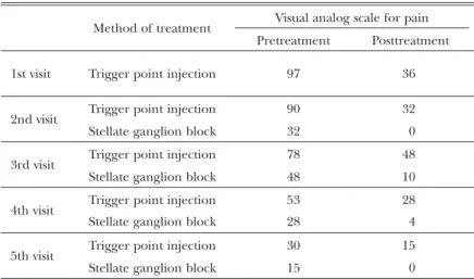
(4) 173. Effect of Combination of TPI and SGB. Teble 1 Process of treatment Method of treatment 1st visit 2nd visit 3rd visit 4th visit 5th visit. Visual analog scale for pain Pretreatment. Posttreatment. Trigger point injection. 97. 36. Trigger point injection. 90. 32. Stellate ganglion block. 32. 0. Trigger point injection. 78. 48. Stellate ganglion block. 48. 10. Trigger point injection. 53. 28. Stellate ganglion block. 28. 4. Trigger point injection. 30. 15. Stellate ganglion block. 15. 0. Fig. 1 Trigger point injection for the masseter muscle. Fig. 2 Stellate ganglion block. had lasted for >1 week. Therefore, we injected SGB at the transverse process of the 6th left cervical vertebra using 2% lidocaine hydro chloride at a dose of 5 ml. The VAS score for tooth pain subsequently dropped from 90 to 32. We then performed another TPI at the masseter tender point, and the VAS score dropped from 32 to 0. Because a combination of masseter TPI and SGB achieved complete pain relief, we diagnosed MPS manifest as referred pain from the masseter muscle, com plicated by sympathetic nerve-dependent pain. We continued masseter TPI and SGB over the following 3 days and the symptoms of both pain and hypoesthesia disappeared.. Discussion It has been reported that the etiology of tooth pain in 3% of all patients is nonodon togenic and that in 9% of all patients it is mixed in nature8). Tooth pain of myofascial origin is the most common among patients with nonodontogenic pain. Another study3) reported that 11% of patients with MPS experienced nonodontogenic tooth pain. Studies suggest that the main masticatory muscles involved are the masseter and tem poral muscles5,7). Observation of pain relief after TPI is useful for diagnostic examina tion5). Therefore, in patients showing tooth.
(5) 174. Handa T et al.. pain with no abnormalities in the tooth or periodontal tissues, detailed examination of the head and neck muscles is key in achieving a diagnosis. In tooth pain of myofascial origin, referred pain arises from TPs formed in fatigued muscles, as described in the classic theory of myofascial pain by Travell and Simmons9). Although Mascia et al. reported that this type of tooth pain disappeared when TPI was administered5), only a few articles have provided scientific evidence for this method10). In routine clinical practice, however, TPI is undoubtedly a useful method for the diag nosis and treatment of myofascial pain. Cord-like indurations and localized tender ness, a localized twitch response, and a jump sign were observed in the present case. These symptoms were partly relieved by TPI, and the addition of SGB resulted in complete pain relief. Travell and Simmons9) reported that the origin of TPs might lie in the sympa thetic nervous system. Although the mecha nism remains to be elucidated, it may be related to a failure of peripheral blood flow and pain-producing substances such as nor epinephrine. Hubbard and Berkoff 2) recorded spontaneous electrical activity in myofascial TPs in patients with temporomandibular pain and observed muscle spindles in biopsies from these sites. This was attributable to intrafusal muscle fibers because the electrical activity was eliminated using the sympathetic blocker phentolamine. Furthermore, the involvement of the sympathetic nerves is important in the development of MPS. Fibrous C polymodal nociceptors distributed densely in the muscle can form a tender point with ischemia, lead ing to the accumulation of endogenous painproducing substances and stimulation of the sympathetic nervous system, thereby establish ing a vicious cycle of reflexes4). The present results suggest that the sympathetic nerves are always involved where there is myofascial pain. This case demonstrates the effectiveness of SGB for pain that cannot be completely resolved with TPI. Perry et al.6) reported that MPS was caused not only by excessive movement, but also by. sympathetic nerve hyperactivity due to emo tional stress such as anxiety or nervousness. In this case, the patient did not complain of daily or social activities that caused emotional stress, and the results of the HAD and STAI psychological tests provided no evidence sup porting the involvement of emotional factors. Therefore, it seems unlikely that emotional factors were involved in the pain reported by this patient. Referred pain from the masticatory muscles in MPS is one of the most common presenta tions in a clinical setting. However, referred pain is often overlooked, leading to pointless repetition of cycles of dental treatment. In the present case, referred pain from the masseter muscle caused sustained mandibular molar pain, resulting in sympathetic nerve hyperac tivity and severe pain. There are various theo ries on the mechanism underlying referred pain. This report reconfirms that in cases of sustained pain, sympathetic nerve activa tion could amplify symptoms, and that SGB in combination with TPI can be effective in resolving such symptoms.. References 1) Farella M, Michelotti A, Gargano A, Cimino R, Ramaglia L (2002) Myofascial pain syn drome misdiagnosed as odontogenic pain: a case report. Cranio 20:307–311. 2) Hubbard DR, Berkoff GM (1993) Myofascial trigger points show spontaneous needle EMG activity. Spine 18:1803–1907. 3) Kim ST (2005) Myofascial pain and toothaches. Aust Endod J 31:106–110. 4) Livingston WK (1943) Pain Mechanisms: A Physiological Interpretation of Causalgia and Its Related States, pp. 83–127, Macmillan Press, New York. 5) Mascia P, Brown BR, Friedman S (2003) Toothache of nonodontogenic origin: a case report. J Endod 29:608–610. 6) Perry F, Heller PH, Kamiya J, Levine JD (1989) Altered autonomic function in patients with arthritis or with chronic myofascial pain. Pain 39:77–84. 7) Reeh ES, elDeeb ME (1991) Referred pain of muscular origin resembling endodontic involvement. Case report. Oral Surg Oral Med.
(6) Effect of Combination of TPI and SGB. Oral Pathol 71:223–227. 8) Sanner F (2010) Acute right-sided facial pain: a case report. Int Endod J 43:154–162. 9) Travell JG, Simmons DG (1999) Myofascial Pain and Dysfunction: The Trigger Point Manual — Upper Half of Body, pp. 5–36, Williams & Wilkins, Baltimore. 10) Türp JC, Komine F, Hugger A (2004) Efficacy of stabilization splints for the management of patients with masticatory muscle pain: a qualitative systematic review. Clin Oral Investig 8:179–195. 11) Wright EF (2000) Referred craniofacial pain patterns in patients with temporomandibular disorder. J Am Dent Assoc 131:1307–1315.. Reprint requests to: Dr. Toshiyuki Handa Division of Dental Anesthesiology, Department of Clinical Health and Oral Science, Tokyo Dental College Suidobashi Hospital, 2-9-18 Misaki-cho, Chiyoda-ku, Tokyo 101-0061, Japan E-mail: [email protected]. 175.
(7)
図
関連したドキュメント
Standard domino tableaux have already been considered by many authors [33], [6], [34], [8], [1], but, to the best of our knowledge, the expression of the
By using the first order averaging method and some mathematical technique on estimating the number of the zeros, we show that under a class of piecewise smooth quartic
The inclusion of the cell shedding mechanism leads to modification of the boundary conditions employed in the model of Ward and King (199910) and it will be
In fact in order to show that Condorcet rankings are medians we are going to use a more general notion of median namely the notion of metric median in a metric space.. I begin by
We show that a discrete fixed point theorem of Eilenberg is equivalent to the restriction of the contraction principle to the class of non-Archimedean bounded metric spaces.. We
Includes some proper curves, contrary to the quasi-Belyi type result.. Sketch of
We will later see that non-crossing and non-nesting set partitions can be seen as the type A instances of more general constructions:.. ▸ non-crossing partitions NC ( W ) , attached
In this paper, we establish the boundedness of Littlewood- Paley g-functions on Lebesgue spaces, BMO-type spaces, and Hardy spaces over non-homogeneous metric measure spaces
