• 検索結果がありません。

Peripheral Organ Dose Evaluation using a Human Body Phantom in Intensity Modulated Radiation Therapy for Lung Cancer with Helical Type Accelerator

N/A
N/A
Protected

Academic year: 2021

シェア "Peripheral Organ Dose Evaluation using a Human Body Phantom in Intensity Modulated Radiation Therapy for Lung Cancer with Helical Type Accelerator"

Copied!
8
0
0

読み込み中.... (全文を見る)

全文

(1)

Introduction

Intensity modulated radiation therapy (IMRT) is a treat- ment that concentrates a radiation dosage in a target vol- ume

1

. The normal tissue dose can be reduced to the greatest extent by optimization calculations according to the treat- ment plan

2

. Simulations need to be performed to investigate the effect of secondary radiation on these optimization cal- culations, with corrections based on differences in pene- trated organ density. For this purpose, highly complex cal- culations and verification experiments using a phantom are

necessary prior to the treatment by a method called inverse planning

3

.

The exposure time in IMRT (150 to 300 seconds or more) is generally longer than that in conventional radiation ther- apy because IMRT employs many narrow photon beams

7

. Moreover, according to the algorithm used for the optimi- zation calculations, the dose in the vicinity of the body sur- face, such as the breasts and eyes is affected by factors such as the airspace surrounding the body; thus, the optimized calculation value for the treatment plan and the actual ab- sorbed dose might not be in agreement. The likelihood of Address correspondence: Department of Radiological Sciences, Nagasaki University Graduate School of Biomedical Science, 1-7-1 Sakamoto, Nagasaki 852-8501, Japan

TEL: +81-03-3816-2129, FAX: +81-03-5803-1990, E-mail: [email protected] Received Fabruary 2, 2010; Accepted November 30, 2010

MS#AMN 07076

Peripheral Organ Dose Evaluation using a Human Body Phantom in Intensity Modulated Radiation Therapy for Lung Cancer with Helical Type Accelerator

Shinichi G OTOH ,

1

Yasuhiro K AWAHARADA ,

2

Satoshi S UDA ,

3

Masataka U ETANI

1

1

Department of Radiological Sciences, Nagasaki University Graduate School of Biomedical Sciences, 1-7-1 Sakamoto, Nagasaki 852-8501, Japan

2

Department of Radiological Technology, Gunma Prefectural College of Health Sciences, 323-1 Kamioki-cho, Maebashi-shi, Gunma 371-0052, Japan

3

Oncology Center of Hidaka Hospital, 886 Nakao-cyo, Takasaki-shi, Gunma 370-0001, Japan

Purpose: Intensity modulated radiation therapy (IMRT) is characterized by a relatively long period for beam exposure and consequently the risk for unnecessary exposure to non-targeted organs, mainly due to the scattered radiation, should be con- sidered. The puposes of this study are to measure the absorbed dose of the peripheral organs during helical IMRT using a fluorescent glass dosimeter.

Materials and Methods: In this research, we took lung cancer as a model and measured the absorbed dose of the periph- eral organs during helical IMRT using a fluorescent glass dosimeter. The planning target volume (PTV) dose of 95% was set to be 5 Gy in the phantom.

Results and Discussion: The highest exposure dose was observed for the breasts, which were on the PTV trajectory, with the left and right breasts receiving doses of 227.94 mGy and 371.90 mGy, respectively. The exposure doses of the left and right lenses were 3.13 mGy for the left lens and 3.22 mGy for the right lens. An exponential dose reduction to the distance from PTV was confirmed. Our data suggest that the doses for peripheral organs were acceptable in lung cancer case based on past literature search. However, the use of custom blocks for the eyes should be considered to prevent possible late occurance of cataract.

ACTA MEDICA NAGASAKIENSIA 55: 61−67, 2011

Keywords: helical tomotherapy, intensity modulated radiation therapy, peripheral organ dose, glass dosimeter

(2)

errors is especially high in the low-dose region because the measurement performance of the ionization chamber detec- tor is relatively poor at such doses.

The purpose of this study is to measure the absorbed dose of the risk organs during helical tomotherapy IMRT using a fluorescent glass dosimeter.

Materials and Methods Helical Tomotherapy

Helical tomotherapy is one of the most advanced IMRT equipment, which has computerized tomography (CT) based helical type exposure functions

4-6

. In this system, radiation therapy is executed as the couch moves through the gantry, which rotates in the same manner as a CT apparatus for di- agnostic use. The beam used for exposure is a slit shaped narrow beam called a beamlet. As the apparatus rotates, the beam is modulated with exposure time, and thereby IMRT can be effectively performed

9-11

. We smployed TomoTherapy Hi-Art System (TomoTherapy, Wisconsin, USA) for heli- cal tomotherapy. , The treatment beam energy was applied via a 6 MV convex beam without use of a flattening filter.

The radiation source to isocenter separation was 85 cm and the maximum irradiation field was 5×40 cm

2

at the isocenter.

Fluorescent Glass Dosimeter

The beam generated in helical tomotherapy is a slit nar- row beam, which can be as small as 0.6×1.25 cm

2

. For this reason, we used a fluorescent glass dosimeter, which is suitable for measurements in the low-dose region for ex- tremely narrow beams

8

. A glass dosimeter (Asahi Glass, Japn) was used to measure the absorbed dose values in various parts of the phantom (RAN-100, THE PHANTOM LABORATORIES,INC., USA) (Fig.1). This apparatus util- izes radiophotoluminescence, a luminescent phenomenon which occurs when certain types of glass are struck by ul- traviolet rays after exposure to radiation. Fluorescent glass dosimeters exploit the chemical transition of silver ions in silver-activated phosphate glass; because the fluorescence center produced by the divalent silver ion (Ag

2+

) or silver particles (Ag

0

) due to the radiation is exceedingly stable, the loss of dose information, called fading, is extremely small at less than 1% per year. Moreover, as the fluores- cence center does not vanish when measured and can be re- peatedly read many times, the statistical accuracy of the measurements is enhanced and a stable measurement value can be acquired

12,13

. We used GD-352 for the phosphate

glass element. In addition, we used it as a bare element without an additional filter for low energy and calibrated it with 6MV X-ray. As a result, the calibration constant we obtained was 1.16.

Experimental Measurements

We carried out absorbed dose measurements by estab- lishing a virtual target in the chest of a human-body phan- tom. Measurements were carried out at the locations corre- sponding to the left and right breasts on the exposure trajectory and at locations corresponding to the thyroid, pelvic region, and left and right crystalline lenses outside the exposure trajectory. We calibrated each detection ele- ment of the florescent glass dosimeter by using a standard- ized linear accelerator. Exposure was carried out with an ir- radiation field of 15 cm×15 cm and a radiation source to ionization chamber detector separation of 100 cm accord- ing to the 6 MV X-rays acquired from a standard linear ac- celerator (Varian Torilogy, USA). A conventional standard ionization chamber was used in this study (M30013, PTW, Germany).

For the treatment plan, we adopted a right lung cancer as

a model and set the PTV at 15 cm below the thyroid gland

(Fig.2). We assumed spheres of 2 cm in diameter for the

PTV. The exposure dose was established such that the dose

at 95% volume of the PTV curve in DVH for the treatment

plan optimized calculation was 5 Gy. We verified the model

for this treatment plan using an ionization chamber dosime-

ter (A1Sl, Standard Imaging Inc., USA) and a cylindrical

solid phantom (cheese phantom, TomoTherapy Inc., USA)

(Fig.3). This phantom is a cylinder of 30 cm in diameter

Fig. 1. Human body Rando-phantom on the Helical type accelera-

tor treatment couch.

(3)

and 18 cm in height. The phantom can be split in half from the central part, and the beam profile can be measured using EDR film. Also, there are holes for point dosimetry measurement, and the absorbed dose can be measured with an ionization chamber dosimeter. The absorbed dose as de- termined from actual measurements and the values from the optimized calculations for the treatment plan were com- pared in the region where the dose distribution inside the PTV was flat.

The absorbed dose of the peripheral organ was measured with a fluorescent glass dosimeter in the location of the left and right eyes, the thyroid, the left and right breasts, and the pelvic region. For this purpose, the florescent glass do- simeter was used in conjunction with an MIX-R

14

of 2 mm in thickness for the left and right eyes and a MIX-R of 1 cm in thickness for all other locations. The basic ingredients of MIX-R were polyisoprene, sulfur, zinc oxide, stearic acid and Nocceler CZ. The florescent glass dosimeters were used in a set of five, and evaluations were carried out using the mean value. The X-rays used in helical tomotherapy had energy of 6 MV, which approximates 1.25 MV cobalt gamma rays. Thus, a conversion was carried out for the ac- quired absorbed dose by using the following equation, where 1 Gy = 1 Sv:

H = D・Q・N.

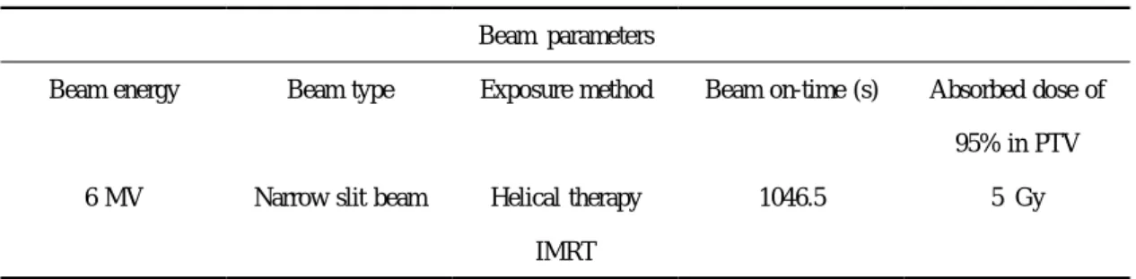
Here, H is the patient dose [Sv], D is the absorbed dose [Gy], Q is the beam quality factor [Q = 1 MV], and N is a correction factor [N = 1.0]. The treatment beam parameters at time of exposure are shown in Table 1. In addition, to in- vestigate the dose due to the change of dosimetry distance, we used a glass dosimeter to measure the relationship be- tween the distance and dose. This measurement was per- formed in the same manner as the measurement of the ab sorbed dose at the phantom body. Dosimetry was performed at the points along the central axis at 10, 20, 30, 40 and 50 cm from the axial plane including PTV center.

Fig. 2. A PTV position in the Rando-Phantom. (a) an axial and (b) an coronal CT images.

Fig. 3. Dedicated QA phantom used to verify the absorbed dose values acquired through the treatment plan optimization calcula- tions for IMRT and the beam profile. The dose profile on the coronal plane is measured by EDR-2 film. Then, point absorbed dose is measured by a small ionization chamber detector. We start radiotherapy when the actual measurement value shows variance within 3% of the calculated value in the treatment plan.

Beam parameters

Beam energy Beam type Exposure method Beam on-time (s) Absorbed dose of 95% in PTV

6 MV Narrow slit beam Helical therapy 1046.5 5 Gy

IMRT

Table 1. Exposure parameters used in optimization calculation for lung cancer treatment plan simulations.

(4)

Results

The measured doses were 3.13 mGy for the left lens and 3.22 mGy for the right lens. These doses were less than 0.1% of the established dose of 95% PTV at 5 Gy. The thy- roid received a dose of 13.26 mGy (0.27% of the PTV dose), which was an exposure dose of approximately 4 times higher than the dose received by the lenses, owing to the proximity of the thyroid to the PTV.

The left and right breasts, which were on the trajectory of the helical tomotherapy, received doses of 227.94 mGy and 371.90 mGy, respectively. This confirmed that the left breast on the non-treated side received an equivalent of 4.6% of the PTV dose and the right breast on the treated side an equivalent of 7.4%. The pelvic region received a dose of 1.47 mGy, which was the lowest exposure dose among the target measurements, owing to its distance from the PTV. This represented less than 0.05% of the PTV dose.

The relation between the distance from the PTV and the dose is shown in Fig. 3. The dose is exponentially attenu ated with increasing the distance from the PTV, 20 to 30

mGy at 10 cm, less than 10 mGy at 20 cm and around 2 mGy at 50 cm (Fig.4).

Fig. 4. Absorbed dose relation to the distance from the axis plane including PTV center.

organ No. 1

rod

No. 2 rod

No. 3 rod

No. 4 rod

No. 5 rod

Mean dose

a

(mGy) SD

b

Right eye 3.08 3.17 3.20 3.29 3.34 3.22 0.10

Left eye 3.15 3.16 3.09 3.14 3.12 3.13 0.03

Thyroid 13.35 12.75 13.16 13.17 13.85 13.26 0.40

Right breast

c

292.90 350.40 395.50 389.90 430.80 371.90 52.57

Left breast

c

235.60 243.20 239.60 202.50 218.80 227.94 17.02

Pelvic area 1.44 1.57 1.44 1.46 1.43 1.47 0.06

Table 2. Absorbed dose for each position during helical tomotherapy treatment as measured with a fluorescent glass dosimeter.

a

A group of five fluorescent glass dosimeters was used for each position of the phantom and the absorbed dose (mGy) was cal- culated as the mean.

b

SD: standard deviation

c

Right and left breast are in direct radiation field areas.

(5)

Discussion

This study confirmed that the off-trajectory dose in heli- cal tomotherapy can be sufficiently low. However, for pe- ripheral organs on the trajectory of the helical tomotherapy, caution and validation are necessary in accordance with the exposure dose, although this also depends on the type and position of the peripheral organ and whether the treatment is single-dose or total dose.

Concerning the measurement of off-axis doses in helical tomotherapy, Ramsey et al. have reported results measured using a human-body phantom and a Thermo Luminescence Dosimeter (TLD)

7

. They consider an off-axis increase of leakage dose to be possible because the exposure time for treatment is from 5 to 15 minutes for IMRT by helical tomotherapy, which is longer than treatment using conven- tional linear accelerators. For that reason, they used an identical human-body phantom and carried out measure- ments of absorbed dose with a TLD under the same condi- tions using helical tomotherapy and a general linear accel- erator. In that study, they found the highest dose to be 1×10

-4

Sv/s and the lowest dose to be 1×10

-6

Sv/s for treatment times of 120 to 600 seconds. Although dependent on factors such as the measurement location and phantom, this maximum value is similar to the breast dose (maximum value) that we measured. In comparison with IMRT using a general linear accelerator, the off-axis dose of helical tomotherapy was equal or lower, irrespective of its longer exposure time. They attributed this to the special construc- tion of the shielding structure of the helical tomotherapy system.

In the present study, the actual measurement was 13.26 mGy when PTV (planning target volume) was 5Gy. Therefore, it becomes 132.6mGy, 10 times as much as the measure- ment, if the total dosage for radiotherapy is 50Gy; however, it is not clear from reports to date whether this dose can definitely increase the risk of thyroid cancer. Recent stud- ies, showed that thyroid cancer can be induced by relatively low dose of radiation. In a follow-up survey of the atomic bomb survivors at Hiroshima and Nagasaki, thyroid cancer was more likely to occur in younger subjects, which was not observed in subjects over the age of 40. However, a causal relation with radiation exposure was not established

15

. Pacini et al. reported that thyroid cancer is clearly increas- ing at a greater rate in individuals exposed to radioactivated iodine in childhood due to the fallout of the Chernobyl in- cident in comparison with non-exposed individuals. Taking into account the possibility of increased risk of thyroid can- cer in young children, the dose to the thyroid should be

minimized by implementing an optimal treatment plan

16

. Day et al. investigated ophthalmologic disorders in indi- viduals exposed to the fallout of the Chernobyl incident, to evaluate the relation between contamination due to the inci- dent and cataracts. The participants in the study were ap- proximately 1000 people who lived in the contaminated re- gion and were under the age of 12 at the time of the incident and approximately 800 people who lived in a scarcely contaminated region. Based on the results of this study, posterior capsule opacification was found to be sig- nificantly increased in the contaminated region

17

.

In the research of Worgel et al., ophthalmological examina- tions were carried out in 1998 and 2000, targeting approxi- mately 8000 cleanup workers living in five provinces in Ukraine. The odds ratio for posterior capsule opacification and type 1 cortical opacity increased with contamination with a threshold to be between 0.3 and 0.5 Gy, suggesting a far lower likelihood than values previously considered

18

. Chumak et al. studied problems relating to the validity of the reported doses used in the research of Worgel et al. and the evaluation of the beta rays. As a result, the dose to the crystalline lens for the majority of workers was found to be 100-200 mGy with a small proportion exceeding 500 mGy

19

. Moreover, as a result of re-examining 730 survivors of the Hiroshima and Nagasaki atomic bombs, Neriishi et al. re- ported that the point estimation for the threshold dose was 0.6 Sv for cortical cataracts (95% confidence interval [CI],

<0.0-1.2 Sv), and 0.8 Sv for posterior capsule opacification (95% confidence interval [CI], <0.0-2.8 Sv)

20

. This falls far under the 5 Gy threshold for cataracts proposed in the 60th publication of the International Commission on Radiological Protection (ICRP)

21

.

Considering that the lower limit of the lens dose reported by Worgel et al. was 0.3 Sv, the lens dose measured in the present research was far lower than this threshold. However, in the case that the distance between the PTV and the lens was less than 5 cm, the dose to the lens became approxi- mately 0.2-0.3 Sv; therefore, a custom block should be used to reduce exposure to the lens.

In the present research, the dose to the pelvic region was 1.72 mGy. This value became 17.2 mGy in the case that the total dose of the PTV was 50 Gy. This value is exceedingly low in comparison with the threshold dose at which sterility or an effect on embryos arises (ICRP recommended val- ues).

Mammary glands are within the path of the therapeutic beams, and the dose in this study is 227.94 to 371.90 mGy.

Regarding this point, when the total radiotherapy dosage is

50 Gy, the dose to mammary glands is approximately 2 to

(6)

4 Gy. Increased breast cancer was recognized more than ten years after the exposure to radiation. It has been shown that its risk is higher in females exposed to radiation at young ages than in those exposed at the age of 40 or more. In par- ticular, exposure at the age of less than 20 has the highest risk

22,23

. The age of onset agrees with the age of high inci- dence of usual breast cancer, and differences in tissue types and so on that characterize radiation-induced breast cancer are not recognized between high-dose exposure, low-dose exposure, and no exposure. Although significant increase in incidence risk of bilateral breast cancer is not recognized, it is suggested that the risk may be high when the age of ex- posure is less than 20

24

. The dose to mammary gland in atomic bomb victims is estimated to be 0 to 6 Gy (0 to 6.08 Sv, average 0.276 Sv), and it is shown that incidence of breast cancer increases almost linearly with the exposed dose

25

.

On the other hand, influence of exposure to radiation in the cases of a low dose, a low dose rate, or a low dose rate with a high dose is not clarified scientifically. Therefore, for influence of low-dose exposure, the risk is estimated by model analyses based on the assumption that no threshold exists and that there exists a dose-response relation similar to that in the high-dose exposure, namely, cancer occurs stochastically in correlation with the total dose. However, we need to notice that the estimation methods are not well established and the results contain uncertainty. In fact, the linear dose-response relation is not confirmed even in atomic bomb victims below 0.25 Gy (average 0.17 Sv), and in- crease in incidence risk of contralateral breast cancer due to breast irradiation after breast-conserving surgery is also not recognized

26,27

.

From these facts, it is necessary to reduce the dose to mammary gland as much as possible. To realize it, reme- dies such as setting direction of radiation beam to avoid mammary gland tissues should be examined.

Conclusions

In this research, we measured the exposure dose of the peripheral organs during helical tomotherapy IMRT with a fluorescent glass dosimeter. It was confirmed that the expo- sure dose reached a maximum value on the PTV trajectory and decreased in proportion to distance. Using the lung cancer model, the exposure dose to the left and right lenses was confirmed to be acceptable. However, when the distance from the PTV is less than 5 cm, a custom block should be used in order to reduce the dose to the lens.

References

1. Burman C, Chui CS, Kutcher G et al. Planning, delivery, and quality assurance of intensity-modulated radiotherapy using dynamic multileaf collimator: a strategy for large-scale implementation for the treatment of carcinoma of the prostate. Int J Radiat Oncol Biol Phys 39(4):863- 73, 1997

2. Sauer OA, Shepard DM, Mackie TR. Application of constrained opti- mization to radiotherapy planning. Med Phys 26(11):2359-66, 1999 3. Watanabe Y. Point dose calculations using an analytical pencil beam

kernel for IMRT plan checking. Phys Med Biol 46(4):1031-8, 2001 4. Low DA, Chao KS, Mutic S, Gerber RL, Perez CA, Purdy JA. Quality

assurance of serial tomotherapy for head and neck patient treatments.

Int J Radiat Oncol Biol Phys 42(3):681-92, 1998

5. Low DA, Mutic S, Dempsey JF et al. Quantitative dosimetric verifica- tion of an IMRT planning and delivery system. Radiother Oncol 49(3):305-16, 1998

6. Kapulsky A, Gejerman G, Hanley J. A clinical application of an auto- mated phantom-film QA procedure for validation of IMRT treatment planning and delivery. Med Dosim 29(4):279-84, 2004

7. Ramsey C, Seibert R, Mahan SL, Desai D, Chase D. Out-of-field dosimetry measurements for a helical tomotherapy system. J Appl Clin Med Phys 24;7(3):1-11, 2006

8. Araki F, Moribe N, Shimonobou T, Yoshiura T, Ikegami T, Ishidoya T. A study for narrow beam dosimetry using a radiophotoluminescent glass rod dosimeter [in Japanese]. Japanese Journal of Radiological Technology 60(7):939-947, 2004

9. Mackie TR, Balog J, Ruchala K et al. Tomotherapy. Semin Radiat Oncol 9(1):108-17, 1999

10. Mavroidis P, Stathakis S, Gutierrez A, Esquivel C, Shi C, Papanikolaou N. Expected clinical impact of the differences between planned and delivered dose distributions in helical tomotherapy for treating head and neck cancer using helical megavoltage CT images. J Appl Clin Med Phys 21;10(3):2969, 2009

11. Kissick MW, Mackie TR, Jeraj R. A delivery transfer function (DTF) analysis for helical tomotherapy. Phys Med Biol 7;52(9):2355-65, 2007

12. Kron T. Thermoluminescence dosimetry and its applications in medi- cine--Part 2: History and applications. Australas Phys Eng Sci Med 18(1):1-25, 1995

13. Thilmann C, Adamietz IA, Ramm U et al. In vivo dose increase in the presence of dental alloys during 60Co-gamma-ray therapy of the oral cavity. Med Dosim 21(3):149-54,1996

14. Ichirou Y. A New Flexible Water-Equivalent Material. Jpn J Radiol 36(2):143-7, 1975

15. Imaizumi M, Usa T, Tominaga T et al. Radiation dose-response rela- tionships for thyroid nodules and autoimmune thyroid diseases in Hiroshima and Nagasaki atomic bomb survivors 55-58 years after ra- diation exposure. JAMA 1;295(9):1011-22, 2006

16. Pacini F, Vorontsova T, Demidchik EP et al. Post-Chernobyl thyroid carcinoma in Belarus children and adolescents: comparison with natu- rally occurring thyroid carcinoma in Italy and France. J Clin Endocrinol Metab 82(11):3563-9,1997

17. Day R, Gorin MB, Eller AW. Prevalence of lens changes in Ukrainian children residing around Chernobyl. Health Phys 68(5):632-42, 1995 18. Worgul BV, Kundiyev YI, Sergiyenko NM et al. Cataracts among

Chernobyl clean-up workers: implications regarding permissible eye exposures. Radiat Res 167(2):233-43, 2007

19. Chumak VV, Worgul BV, Kundiyev YI et al. Dosimetry for a study of low-dose radiation cataracts among Chernobyl clean-up workers.

Radiat Res 167(5):606-14, 2007

20. Neriishi K, Nakashima E, Minamoto A et al. Postoperative cataract cases among atomic bomb survivors: radiation dose response and threshold. Radiat Res 168(4):404-8, 2007

21. Mountford PJ, Temperton DH. Recommendations of the International

(7)

Commission on Radiological Protection (ICRP) 1990. Eur J Nucl Med 19(2):77-9, 1997

22. Tokunaga M, Land CE, Yamamoto T, Asano M, Tokuoka S, Ezaki H, et al. Incidence of female breast cancer among atomic bomb survivors, Hiroshima and Nagasaki, 1950-1980. Radiat Res. 112:243-72, 1987 23. Tokunaga M, Land CE, Tokuoka S, Nishimori I, Soda M and Akiba S.

Incidence of female breast cancer among atomic bomb survivors, 1950-1985. Radiat Res. 138:209-23. 1994

24. Land CE, Tokunaga M, Koyama K, Soda M, Preston DL, Nishimori I, et al. Incidence of female breast cancer among atomic bomb survi- vors, Hiroshima and Nagasaki, 1950-1990. Radiat Res. 160:707-17.

2003

25. Thompson DE, Mabuchi K, Ron E, Soda M, Tokunaga M, Ochikubo S, et al. Cancer incidence in atomic bomb survivors. Part II:Solid tu- mors, 1958-1987. Radiat Res. 137:S17-67. 1994

26. Veronesi U, Cascinelli N, Mariani L, Greco M, Saccozzi R, Luini A, et al. Twenty-year follow-up of a randomized study comparing breast- conserving surgery with radical mastectomy for early breast cancer. N Engl J Med. 347:1227-32. 2002

27. Poggi MM, Danforth DN, Sciuto LC, Smith SL, Steinberg SM, Liewehr DJ, et al. Eighteen-year results in the treatment of early breast carcinoma with mastectomy versus breast conservation therapy:the National Cancer Institute Randomized Trial. Cancer 98:697-702.

2003

(8)

Table 1. Exposure parameters used in optimization calculation for lung cancer treatment plan simulations.
Table 2. Absorbed dose for each position during helical tomotherapy treatment as measured with a fluorescent glass dosimeter.

参照

関連したドキュメント

6 have reported mean breast thickness and average glandular dose (AGD) in their study popula- tion to be 6.1 and 1.36 mGy per view, respectively, while mean ESAK recorded by one

Intensification Therapy with Anti-Parathyroid Hormone-related Protein Antibody Plus Zoledronic Acid for Bone Metastases of Small Cell Lung Cancer Cells in SCID Mice.. Tadaaki

16 By combining the tissue clearing method CUBIC, melanin bleaching, and immunostaining, we succeeded in making the eye transparent and acquiring images of the retina from outside

The impact of rotational error regarding the treatment of prostate cancer with VMAT has not yet been evaluated, and our preliminary study showed that pitch angle error affected

We used this software package to estimate percentage dose reduction values of the average organ dose (indicated as 'Average dose in total body' in PCXMC) and effective dose for

Meta-analysis: comparison of F-18 fluorodeoxyglucose- positron emission tomography and bone scintigraphy in the detection of bone metastasis in patients with lung cancer..

1) Aberle DR, Adams AM, Berg CD, Black WC, Clapp JD, Fagerstrom RM, Gareen IF, Gatsonis C, Marcus PM, Sicks JD. Reduced lung -cancer mortality with low-dose computed tomographic

Standard domino tableaux have already been considered by many authors [33], [6], [34], [8], [1], but, to the best of our knowledge, the expression of the