Acta Med. Nagasaki 40: 13-17
A Monoclonal Antibody Assessment of the Beta-Microseminoprotein Expression in Prostatic Carcinoma as a Prognostic Indicator
Yasuo YOGI
Department of Urology, Nagasaki University School of Medicine, 1-7-1 Sakamoto, Nagasaki 852, Japan
Using a newly generated monoclonal antibody for human
beta‑microseminoprotein(β‑MSP), which is among the
proteins secreted by the prostatic gland, this study has
investigated whetherβ‑MSP immunoreactivity can be a
prognostic indicator of prostatic carcinama. To accomplish
this,β‑MSP immunoreactivity was. evaluated in needle
biopsy specimens of 116 patients with prostatic carcinoma.
Results have indicated a higher cancer-specific survival
rate in patients with a greater response to β‑MSP immuno‑
staining (p = 0.04). Further, a multivariate analysis of possible prognostic factors, i. e., the patient age, the clinical stage, the histological grade (Gleason's score), the serum
PAP content, the β‑MSP immunoreactivity, and the initial treatment received, has confirmed that β‑MSP immuno‑
reactivity is a significant prognostic indicator of prostatic carcinoma (p = 0.05).
Thus, the use of a monoclonal antibody to evaluate β‑MSP
immunoreactivity has been found to be a useful indicator for assessing the activity of prostatic carcinoma.
Key words : /3-microseminoprotein, monoclonal antibody, immunohistochemistry, prostatic carcinoma,
multivariate analysis, prognosis
Introduction
Although the beta-microseminoprotein (/3-MSP), a 10.7 kDa protein secreted by the normal human prostatic gland [1-4], has been thought to be a protein specific to only prostatic gland, in recent years secretions of 8 -MSP by other mucus producing cells, such as those of the tracheal epithelium, have also been detected by immunohisto- chemical analysis and/or northern blot analysis [5, 6].
Thus /3 -MSP is no longer considered to be a prostate- specific protein. However, since the /3-MSP concentration in seminal plasma is far higher than in other human body fluids, /3 -MSP may play a somewhat greater role in prostate and/or semen.
Prostatic acid phosphatase (PAP) and the prostate- specific antigen (PSA or r-Sm), which are also secreted by
the prostate, are known to be serum markers of prostatic
carcinoma, and the use of immunohistochemical methods to determine their localization is a well established proce- dure [7-11]. To the best of our knowledge, however, only a few reports have appeared with regard to /3-MSP localiza- tion in prostatic carcinoma [2, 12, 13]. Previously using a
/3-MSP polyclonal antibody, we have studied the response in 96 prostatic carcinoma patients, so as to assess the
relationship between the /3 -MSP immunoreactivity and cancer death, and have found that differences in /3 -MSP immunoreactivity to be an important factor for prog- nosing the outcome of prostatic carcinoma [14].
This success has led us to speculate that if a highly specific monoclonal /3 -MSP antibody were to be used to assess the /3 -MSP immunoreactivity in prostatic carci- noma patients, an even more accurate prognosis could be achieved. Therefore, using newly produced monoclonal /3 - MSP antibody, this study has investigated the correlation between /3 -MSP immunoreactivity and the prognosis of prostatic carcinoma.
Materials and methods
Patients and Tissues
Studied were 116 new prostatic carcinoma patients, diagnosed between March 1980 and July 1992 at the Department of Urology of Nagasaki University Hospital.
Their average age at time of diagnosis was 72.4 years (range : 50 to 91 years). As for their cancer status, 19 patients were in stage B (T2NOMO ; TNM classification [15]), 32 in stage C (T3-4NOMO), and 65 in stage D (N1-3 or M1). Initially, all patients had received endocrine therapy ; the majority were given estrogen with or with- out a bilateral orchiectomy and the remainder received a luteinizing hormone-releasing hormone agonist. In addi- tion, 76 patients had received chemotherapy ; the majority were given cisplatin plus doxorubicin and/or 5- fluorouracil, and the remainder were given other agents.
For the immunohistochemical study, formalin-fixed,
paraffin-embedded, tissue specimens of each neoplastic prostatic gland were retrieved from the files of the Pathology Division of the Central Diagnostic Laboratory.
All specimens were prepared from transperineal or transrectal needle biopsies that had been performed before treatment. All hematoxylin and eosin-stained sections of these cases were reviewed to assess the histological grade according to the Gleason grading system [161. For the controls similarly fixed, paraffinembedded, tissue speci- mens of normal prostatic glands were stained immuno- histochemically, these tissue specimens having been obtained from patients with a bladder cancer at the time they had received a total cystectomy.
Preparation of the Monoclonal Antibody for /3-MSP The monoclonal antibody for /3 -MSP was prepared by conventional technique used for creating a hybridoma [171. Purified /3 -MSP, isolated from human seminal plasma, was used as the immunogen in BALB/c mice, after which the immune splenocytes were fused with murine myeloma cells P3X63Ag8.653. The hybridomas producing the anti-,8 -MSP antibody were cloned by limiting dilution.
The immunoglobulin class and subclass of the clones were determined by using a monoclonal antibody isotyping kit (Zymed Laboratories, Inc., San Francisco). An IgG1 (i )- producing clone was selected for this study. The purifica- tion of the monoclonal antibody from the hybridoma supernatants was performed by affinity chromatography on Affi-gel Protein A (Bio-Rad Laboratories, Richmond, CA). The specificity of the monoclonal antibody for /-3- MSP was demonstrated by an immunoblotting analysis with extracts from normal prostatic tissues, seminal plasma, and purified /3-MSP.
Immunohistochemical Procedure and Evaluation of /3 - MSP Immunoreactivity
Paraf f fined sections were stained immunohistochemi- cally by using the biotin-streptavidin method with horse- radish peroxidase. The sections then were deparaffinized in xylene-alcohol and incubated for 30 min in methanol containing 0.3% hydrogen peroxide to block endogenous peroxidase, which was followed by another incubation for 60 min in a Block Ace solution (Dainippon Pharmaceutical Co., Ltd., Osaka) containing 2% bovine serum albumin and 10% goat serum to decrease the nonspecific back- ground staining. After these procedures, the specimens were incubated for 120 min with the monoclonal /3 -MSP antibody, 15 ug/ml, in phosphate-buffered saline (PBS), pH 7.2, then incubated for 60 min with rabbit biotinylated anti-mouse IgG (Zymed Laboratories, Inc., San Francisco, CA) 1 : 100 in PBS, pH 7.2, followed by a final incubation for 30 min with peroxidase-conjugated streptavidin (Zym- ed Laboratories, Inc., San Francisco, CA) 1 : 500 in PBS, pH 7.2. After this final incubation the sections were subjected to a diaminobenzidine reaction in a 0.05 M Tris
buffer, pH 7.2, with 0.05% hydrogen peroxide for 5 min, followed by counterstaining with hematoxylin, dehydra- tion, and mounting in Permount (Fisher Scientific, Inc., Fair Lawn, NJ).
Grading of the /3 -MSP immunoreactivity was based on the number (%) of tumor cells that stained positively. The grading parameters follow : -, < 5% ; 1 +, 5-25% ; 2+, 25-50% ; 3+, 50-75% ; and 4+, >75%. All specimens were examined and classified without knowledge of the clinical outcomes.
Statistical Analysis
Cancer-specific survival curves were calculated accord- ing to the Kaplan-Meier method and indications of a statistical significance were calculated by the generalized Wilcoxon method. In the cancer-specific survival analysis, deaths from causes unrelated to the prostate cancer were treated as withdrawals in the same manner as those lost to follow-up.
Results
Although the /3-MSP immunostaining of the epithelial cells in the normal prostatic glands was strong and diffuse, in the prostatic carcinoma patients the number of immunoreactive cells was extremely reduced. The grades of /3 -MSP immunoreactivity were as follows : - in 93 cases (80.2%), 1+ in 10 cases (8.6%), 2+ in 6 cases (5.2%), 3+
in 3 cases (2.6%), and 4+ in 4 cases (3.4%). Since only 23 patients had grades of from 1+ to 4+, the grades of /3- MSP immunoreactivity were grouped into two sets : those with a negative immunoreactivity (-) and those with a positive immunoreactivity (1 + to 4+ ).
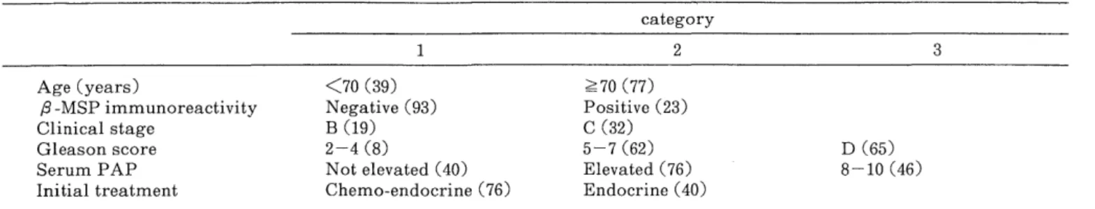
Table 1 lists the 6 parameters and their categories evaluated in a multivariate analysis. The chi-square test was used to investigate the correlation between the /3 -M SP immunoreactivity and the other parameters shown in Table 1. Results revealed that the /3 -MSP immunoreacti- vity showed no significant correlation with the Gleason score or with the other four parameters, i. e., the patient's age, clinical stage, serum PAP, and the initial treatment received (Table 2).
Of the 116 patients studied, 38 died of their prostatic carcinoma. The cancer-specific survival curves, based on the 6 -MSP immunoreactivity, age, clinical stage, Gleason score, serum PAP and the initial treatment received are shown in Figure 3. A significantly higher survival rate was noted in patients with a positive /3 -MSP immunoreacti- vity (p = 0.04), and the results of a generalized Wilco- xon's test analysis showed a significant prognostic differ- ence in survival, depending on the clinical stage and the Gleason score.
Table 3 shows the results of the multivariate analysis of
Table 1 Six parameters and Their Categories Studied in Cox's Regression Model
category
1 2 3
Age (years) <70 (39) z70 (77)
,8 -MSP immunoreactivity Negative (93) Positive (23)
Clinical stage B (19) C (32)
Gleason score 2-4 (8) 5-7 (62) D (65)
Serum PAP Not elevated (40) Elevated (76) 8-10 (46)
Initial treatment Chemo-endocrine (76) Endocrine (40)
Numbers in parentheses indicate number of patients.
Table 3 Multivariate Analysis of Possible Prognostic Parameters in Cox's Regression Model for Patients with Prostatic Carcinoma
Parameter Regression coefficient Standard error t value* p value
Age 0.151 0.385 0.392 0.70
/3-MSP immunoreactivity -1.080 0.554 -1.948 0.05
Clinical stage 1.468 0.406 3.618 0.0005
Gleason score 0.817 0.340 2.403 0.02
Serum PAP -0.502 0.460 -1.092 0.28
Initialtretment -0.192 0.476 -0.403 0.69
*t = regression coefficient/standard error .
Table 2 Correlation between /3-MSP immunoreactivity and Other Parameters
/3 -MSP immunoreactivity Chi-square
Negative Positive test
Age (years)
<70 30 9
p=0.53
? 70 63 14
Clinical stage
B 15 4
C 24 8 p=0.64
D 54 11
Gleason score
2-4 6 2
5-7 52 10 p=0.56
8-10 35 11
Serum PAP
Not elevated 32 8
p=0.97
Elevated 61 15
Initial treatment
Chemo-endocrine 62 14
p-0.60
Endocrine 31 9
Fig. 1 The acini of normal prostate show strong staining for /3-microseminoprotein, X200.
6 parameters. As can be noted, the clinical stage (p <
0.01), the Gleason score (p = 0.02), and the -/3 -MSP immunoreactivity (p = 0.05) appear to have a statisti- cally significant effect on cancer-specific survival.
carcinoma, since only 23 patients (20%) showed a positive /3 -MSP immunoreactivity.
Discussion
This study has found that /3 -MSP immunostaining was strong and diffuse in the epithelial cells of normal prostatic glands but that the /3 -MSP immunoreactivity was extremely weak in the prostatic carcinoma patients. In this regards many reports have advocated the usefulness of immunohistochemical staining to determine the PAP and PSA expression for identifying a metastatic prostatic carcinoma. Therefore, although 8-MSP is secreted by the normal prostatic gland in addition to PAP and PSA, our results appear to indicate that /3 -MSP cannot be consid- ered a useful immunohistochemical marker of prostatic
Fig. 2 No 8 -microseminoprotein reactive cells are seen in a Gleason grade 2 adenocarcinoma, X 100.
Fig. 3 Cancer-specific survival curves according to the a-MSP immunoreactivity (A), age (B), clinical stage (C), Gleason score (D), serum PAP (S)and initial treatment (F).
Using a polyclonal antibody, Okabe et al. have studied the distribution of /3 -MSP in 40 prostatic carcinoma patients [12], and found that those showing a positive /3- MSP expression amounted to 22 patients (55%), and that a higher differentiated tumor showed a greater expression of /3-MSP immunoreactivity. Abrahamsson et al., using a polyclonal antibody, have also conducted an immunohisto- chemical investigation of 40 prostatic carcinoma patients [2], and found the number of patients in whom the immunoreactive cells in each specimen were more than one third of the total number of tumor cells counted, amounted to 30 patients (75%). Further, in our previous study using a /3-MSP polyclonal antibody, the incidence of a positive response to immunostaining was 38.5% (37/
96) [14].
In this study, the incidence of a /3-MSP-positive cellular response in our prostatic carcinoma cases was extremely low in comparison to the results of other studies.
However, this discrepancy may be due to differences between the quality of the polyclonal antibodies that were used in these previous studies and our monoclonal anti- body.
In general, assessing the tumor grade by the various criteria being used is still considered to be the most reliable indicator at present to predict the biological behavior of prostatic carcinoma. In an effort to estimate the biological activity of prostatic carcinoma more precisely, researchers have studied other possible indica- tors, such as the nuclear androgen receptor content, the DNA ploidy, the nuclear roundness factor, neuro-endocrine differentiations and many.others. In this regard Sakai et al. have investigated the prognostic significance of PAP and PSA immunoreactivity in patients with advanced prostatic carcinoma [7, 81, and found that the PAP immunoreactivity is a far better prognostic indicator for assessing the ultimate outcome than the tumor grade.
Similarly, our multivariate analysis of 6 parameters has revealed that /3 -MSP immunoreactivity is a significant prognostic indicator of prostatic carcinoma, although it follows the clinical stage and Gleason score.
In conclusion, this study has found that prostatic carcinoma patients who lack a strong /3 -MSP expression appear to have a high malignant potential, and that investigation of /3 -MSP immunoreactivity with our monoclonal antibody has been found to be a useful indica- tor for evaluating the prognosis of prostatic carcinoma.
Acknowledgement
The author is grateful to Professor Yutaka Saito and Dr. Hideki Sakai for their advice and helpful comments throughout this study. Thanks are also due to Dr.
Ryouichi Tsuda for providing the purified /3 -microserriin- oprotein.
References
1) Tsuda R, Inoue T, Hara M : A seminal plasma specific antigen of prostate gland. Jpn J Leg Mod 36 : 703-709, 1982.
2) Abrahamsson PA, Lilja II, Falkmer S, \Vadstrom LB : Immunohisto-
chmical distribution of the three predominant secretory proteins in the parenchyma of hyparplastic and neoplastic prostate glands. Prostate
12 : 39-46, 1988.
3) Lilja H, Abrahamsson PA : Three predominant proteins secreted by the human prostate gland. Prostate 12 : 29-38, 1988.
4) Dube JY, Pelletier G, Gagnon P, Tremblay RR : Immunohistochemical
localization of a prostatic secretory protein of 94 amino acids in normal prostatic tissue, in primaly prostatic tumors and in their
metastases. J Urol 138: 883-887, 1987.
5) Ulvsbil.ck M, Lindstrom C, Weiber II, Abrahamsson PA, Lilja 1-1, Lundwall A : Molecular cloning of a small prostate protein, known as
b-microseminoprotein, PSP94 or b-inhibin, and demonstration of
transcripts in non-genital tissues. Biochem Biophys Res Commun 164 1310-1315, 1989.
6) Weiber H, Andersson C, Murne A, Rannevik G, Lindstrom C, Lilja 1-1, Fernlund P : 6 microseminoprotein is not a prostetespecific protein.
Am J Path 137: 593-603, 1990.
7) Sakai H, Shiraishi K, Minami Y, Yushita Y, Kanetake H, Saito Y Immunohistochemical prostatic acid phosphatase level as a prognostic
factor of prostatic carcinoma. Prostate 19 : 265-272, 1991.
8) Sakai H, Yogi Y, Minami Y, Yushita Y, .Kanetake H, Saito Y Prostate specific antigen and prostatic acid phosphatase immuno-
reactivity as prognostic indicators of advanced prostatic carcinoma. J
Urol 149: 1020-1023, 1993.
9) Epstein JT, Eggleston JC : Immunohistochemical localization of prostate-specific acid phosphatase and prostate-specific antigen in
stage A2 adenocarcinoma of the prostate : Prognostic implications.
Hum Pathol 15 : 853-859, 1984.
10) Shinha AA, Gleason DF, Wilson MJ, Wick MR, Reddy PK, Blackard CE : Relationship of prostatic acid phosphatase localization in human
prostate by a monoclonal antibody with the Gleason grading system.
The Prostate 13 : 1-15, 1988.
11) Lam KW, Li CY, Yam LT, Sun T, Lee G, Ziesmer S : Improved immunohistochemical detection of prostatic acid phosphatase by a
monoclonal antibody. The Prostate 15 : 13-21, 1989.
12) Okabe T, Eto K : Clinical studies of prostatic antigens (r -seminopro tein, S-microseminoprotein). Jpn J Urol 74 : 1313-1319, 1983.
13) Tsuda R, Hara M, Inoue T, Okabe T : Immunochemical localization of r-seminoprotein and /3-microseminoprotein in prostatic glands. Jpn J Leg Med 37 : 16-19, 1983.
14) Hyakutake II, Sakai H, Yogi Y, Tsuda R, Minami Y, Yushita Y, Kanetake 1-I, Nakazono I, Saito Y : Beta- microseminoprotein immuno-
reactivity as a new prognostic indicator of prostatic carcinoma.
Prostate 22 : 347-355, 1993.
15) Schrdder FH, Hermanek P, Denis L, Fair WR, Gospodarowicz MK, Pavone-Macaluso M : The TMN classification of prostate cancer.
Prostate 4 (Suppl) : 129-138, 1992.
16) Gleason DF, The Veterans Administration Cooperative Urological Resarch Group : Histologic and clinical staging of prostatic carcinoma.
In Tannenbaum M (ed) : "Urologic Pathology : The Prostate."
Philadelphia : Lea & Febiger, 1977, pp 171-198.
17) Harlow E, Lane D : Antibodies-A laboratory manual. Cold Spring Harbor Laboratory, 234, 1988.